Method of preparing basement membrane, method of constructing basement membrane specimen, reconstituted artificial tissue using the basement membrane specimen and process for producing the same

a technology of basement membrane and reconstituted artificial tissue, which is applied in the field of preparing basement membrane, reconstituted artificial tissue using the basement membrane specimen and process for producing the same, can solve the problems of complex culture protocol for the formation of a basement membrane itself, the complex wash procedure of such reagents, and the inability of epithelial tissue to maintain itself or achieve original performan

Inactive Publication Date: 2007-03-22
JAPAN SCI & TECH CORP +1
View PDF0 Cites 2 Cited by
  • Summary
  • Abstract
  • Description
  • Claims
  • Application Information

AI Technical Summary

Benefits of technology

[0027] The present invention 4 also relates to a process for producing a basement membrane specimen or an artificial tissue which can be transplanted while maintaining the structure of a basement membrane wherein a protein support structure is temporarily adhered to plastic surface through an adsorptive polymer by hydrophobic bonding having a hydrophobic linear carbon skeleton and a functional group which can react with protein in a molecule, and a basement membrane specimen or an artificial tissue is formed thereo

Problems solved by technology

In other words, without a basement membrane, an epithelial tissue cannot maintain itself or achieve its original performance.
However, it was also confirmed that type II alveolar epithelial cells were unable to constitute a basement membrane without the supply of, for example, a fibroblast, exogenous basement membrane components from Matrigel or a growth factor TGF-β, since the endogenous basement membrane components from type II alveolar epithelial cells are not enough for the cells to assemble a basement membrane structure effectively.
However, following problems still remained: a problem of frequent troubles that, when fibroblasts secreting basement membrane components and a growth factor of TGF-β are used, gel contraction occurs during the culture, and the fibroblasts are peeled off from plastic membrane together with alveolar epithelial cells; a problem that reagents used to peel alveolar epithelial cells for the preparation of basement membrane specimen tend to remain inside the gel if fibroblasts a

Method used


Image

  • Method of preparing basement membrane, method of constructing basement membrane specimen, reconstituted artificial tissue using the basement membrane specimen and process for producing the same
  • Method of preparing basement membrane, method of constructing basement membrane specimen, reconstituted artificial tissue using the basement membrane specimen and process for producing the same
  • Method of preparing basement membrane, method of constructing basement membrane specimen, reconstituted artificial tissue using the basement membrane specimen and process for producing the same

Examples


example 1

(Epithelial Cells, Endothelial Cells and the Like Forming a Basement Membrane)

[0075] As for epithelial cells, type II alveolar epithelial cells (obtained from rats transfected with SV40-large T antigen genes; T2 cells) which were provided by Dr. A. Clement, H{circle around (o)}pital Armand Trousseau, Paris (Clement et al., Exp. Cell Res., 196:198-205, 1991) were cultured in DMEM (Dulbecco's modified Eagle medium) wherein 10 mM of 2-[4-(2-hydroxyethyl) -1-piperazinyl] ethanesulfonic acid (HEPES) (pH 7.2), 10% fetal bovine serum (FBS; Hyclone Laboratories Inc., Logan, Utah), penicillin, and streptomycin are added, in the atmospheric condition of air 95% / CO2 5%, and used. As for endothelial cells, human pulmonary arterial vascular endothelial cells (HPAE cells) purchased from Clonetics were cultured in culture medium of MCDB 131 alone wherein 10 mM of HEPES (pH 7.2), 2% FBS, growth factor, penicillin, and streptomycin are added, or culture medium of equal mixture of MCDB 131 and DMEM,...

example 2

(Preparation of Fibrous Collagen Gel)

[0076] Collagen gel fiber was prepared on the model of dense matrix of collagen gel usually constituted by fibroblasts. Type I neutral collagen solution in DMEM (pH 7.2) (0.3-0.5 mg / ml of type I collagen obtained from 0.42 ml of bovine dermis by acid extraction; Koken Co., Tokyo) were added to 4.3 cm2 of cultured fibroblast layer together with polyethylene terephthalate ester membrane of 6-well culture plate (Becton Dickinson Labware, Franklin Lakes, N.J.), and incubated in CO2 incubator for a few hours—24 hours, then allowed to gelling. This gel was air-dried and compressed at room temperature for 24-48 hours, and used as high-density collagen fiber (fib). As for the above-mentioned fibroblasts, pulmonary fibroblasts derived from male rats Jcl: Escher 344 were prepared according to the method previously described (CELL STRUCTURE AND FUNCTION 22: 603-614, 1997).

example 3

(Constitution of Tissue Model 1)

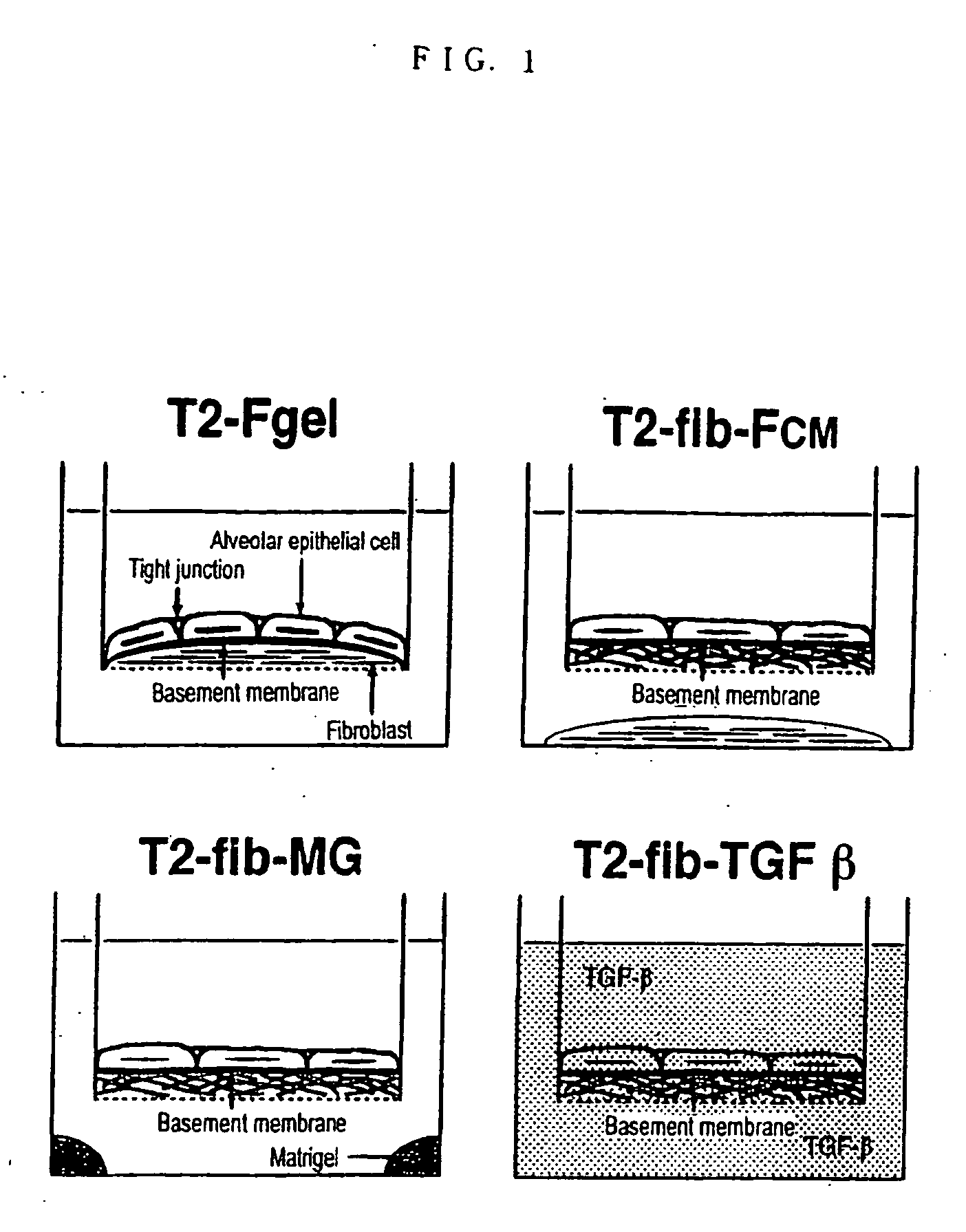
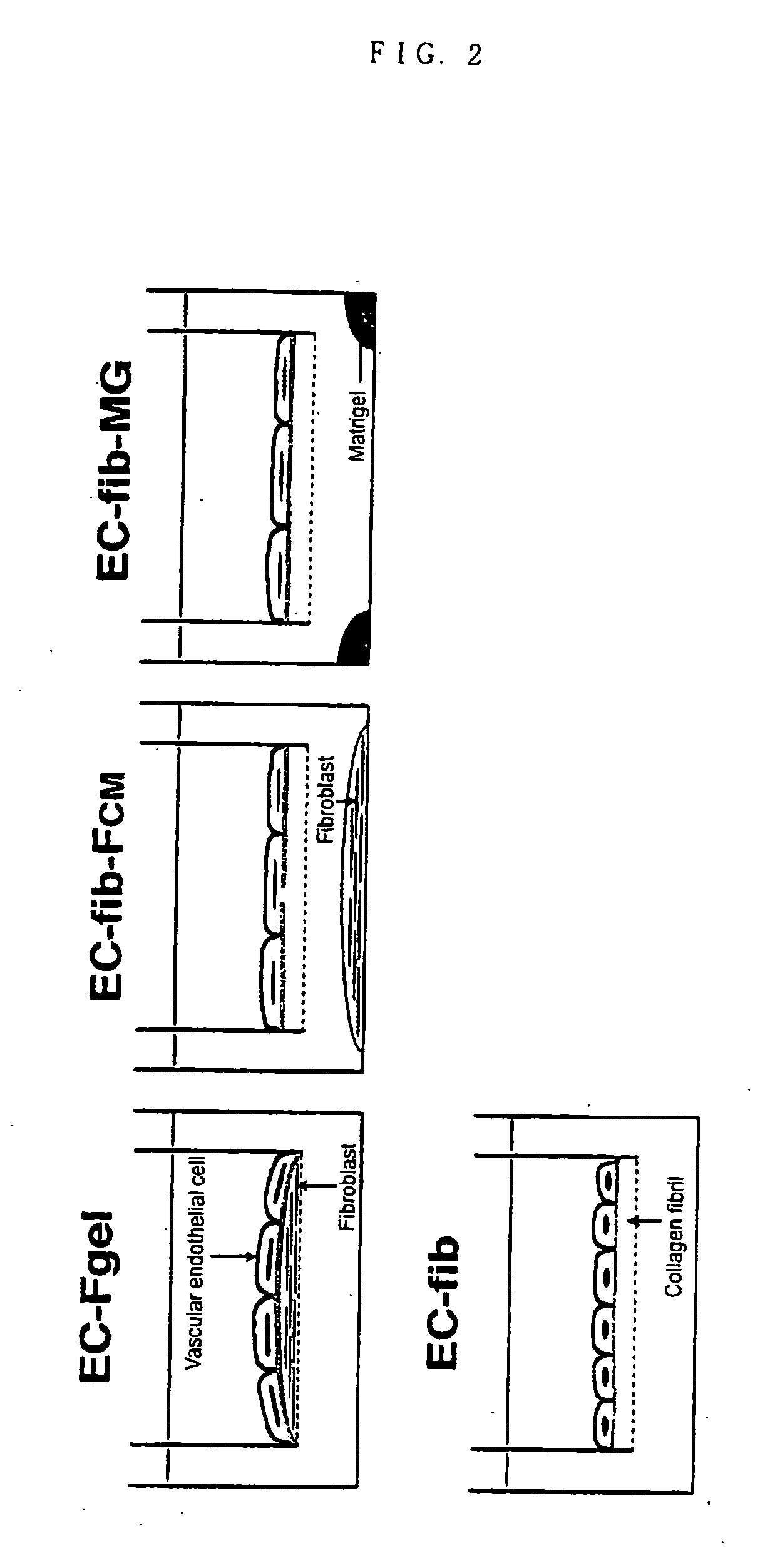
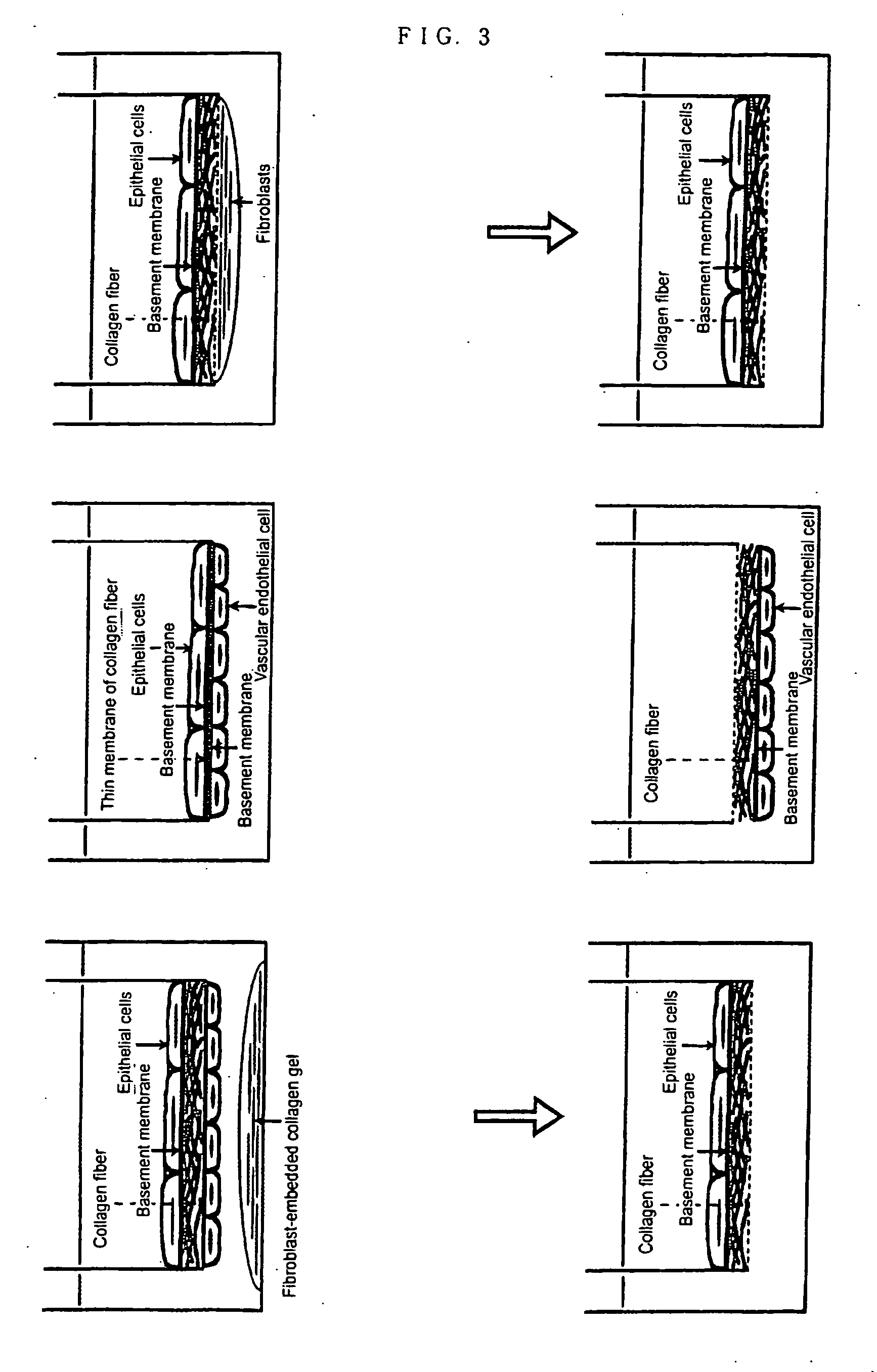
[0077] The methods for forming epithelial tissues and endothelial tissues having a basement membrane beneath the type II alveolar epithelial cells and vascular endothelial cell layers are shown in FIG. 1 and FIG. 2 respectively. In order to constitute most basic tissue model, the above-mentioned epithelial cells (T2) or endothelial cells (HPAEC) were seeded directly on the collagen gel wherein fibroblasts were embedded, and cultured for 2 weeks (T2-Fgel in FIG. 1, EC-Fgel in FIG. 2). Further, in order to form epithelial tissue and endothelial tissue having a basement membrane using the above-mentioned fib as culture matrix, epithelial cells (T2) or endothelial cells (HPAEC) were directly seeded on fib and cultured for 2 weeks (T2-fib-Fcm, T2-fib-MG and T2-fib-TGF in FIG. 1). In FIG. 1, Fcm shows the coculture with collagen gel wherein fibroblasts are embedded (Fgel), MG shows the culture wherein the bottom of culture plate is coated with Matrigel 200...


PUM

PropertyMeasurementUnit
Structureaaaaaaaaaa
Hydrophobicityaaaaaaaaaa

Abstract

The object of the present invention is to provide the following: a method for preparing a basement membrane which is extracellular matrices having a function to control morphology, differentiation, proliferation, motility, function expression and the like of cells; a method for constructing a specimen of a basement membrane; a process for producing a reconstituted artificial tissue; a basement membrane specimen or an artificial tissue which can be transplanted while maintaining the structure of a basement membrane. A basement membrane having a barrier function is formed by culturing alveolar epithelial cells or vascular endothelial cells on a fibrous collagen matrix coated with a polymer having a sugar chain which can localize a receptor having an activity to accumulate a basement membrane component on the basal surface of the cells having an ability to form a basement membrane. A reconstructed artificial tissue is obtained by seeding and culturing desired homogeneous or heterogeneous cells having an ability to form a basement membrane on the basement membrane specimen constructed by the following process: the cells having an ability to form a basement membrane adhered onto a support structure through a basement membrane are treated with a surface active agent; the lipid component of cells is lysed; the mixture of an alkaline solution and a protease inhibitor is used to lyse the protein remained on the surface of the basement membrane of the cells. A protein support structure is temporarily adhered to plastic surface through an adsorptive polymer by hydrophobic bonding, such as an alternating copolymer of methyl vinyl ether and maleic anhydride, which has a hydrophobic linear carbon skeleton and a functional group which can react with protein in a molecule and a basement membrane specimen or an artificial tissue is formed thereon, then the protein support structure supporting a basement membrane specimen or an artificial tissue is physically exfoliated from plastic surface when desired.

Description

REFERENCE TO RELATED APPLICATIONS [0001] This application is a divisional of U.S. application Ser. No. 10,809,218 filed on Mar. 25, 2004, which is a continuation-in-part of International Patent Application PCT / JP02 / 09841 filed Sep. 25, 2002 and published as WO 03 / 026712 on Apr. 3, 2003, which claims priority to Japanese Patent Application Numbers 2001-292675, 2001-292676, 2001-292677 and 2001-292510, all of which were filed Sep. 25, 2001. Each of the above applications, and each document cited in this text and in each of the above applications (“application cited documents”) and each document cited or referenced in each of the application cited documents, and any manufacturer's specifications or instructions for any products mentioned in this text and in any document incorporated into this text, are hereby incorporated herein by reference; and, technology in each of the documents incorporated herein by reference can be used in the practice of this invention. [0002] It is noted that ...

Claims


Application Information

Patent Timeline
IPC IPC(8): C12N5/08A61L27/20A61L27/24A61L27/38C12N5/071
CPCA61L27/20A61L27/24C12N2533/00C12N5/0697A61L27/3895A61L27/3804
Inventor MOCHITATE, KATSUMI
Owner JAPAN SCI & TECH CORP
Why Eureka
Try Eureka