Transcutaneous localization of arterial bleeding by two-dimensional ultrasonic imaging of tissue vibrations

a tissue vibration and ultrasound imaging technology, applied in the field of tissue vibration identification, can solve the problems of insufficient sensitivity for internal bleeding diagnosis, inability to suggest how to detect a bleeding site, and inability to image a limited area of interest, so as to improve the visualization of internal bleeding sites, localize bleeding in patients, and localize bleeding.

Inactive Publication Date: 2007-03-22
UNIV OF WASHINGTON
View PDF21 Cites 23 Cited by
  • Summary
  • Abstract
  • Description
  • Claims
  • Application Information

AI Technical Summary

Benefits of technology

[0010] Real-time tissue vibration imaging has been implemented at frame rates, for example, of 10 frames/second, on an ultrasound system with a software-programmable signal and image processing back-end. The preliminary results confirm that vibrations produced as a result of arterial bleeding can be detected and imaged with this invention. The vibration amplitude is expected to be the largest near the site of the bleeding, and this fact can be used to localize a bleeding site quickly and non-invasively. The strong backscattered ultrasonic echoes from tissue vibrations can improve visualization of internal bleeding sites that are otherwise hard to image due to weak scattering from blood.
[0011] Potentially,

Problems solved by technology

However, this technique suffers from the disadvantage of imaging a limited region of interest.
Although color-flow ultrasound can image a large region of interest, currently it lacks sufficient sensitivity for diagnosing internal bleeding, due to weak scattering from blood and the slow flow velocity of blood bleeding from a wound, especially in the case of deep bleeds and organ bleeds.
However this paper does not suggest how to detect a bleeding site with acoustic streaming.
Yet, the use of contrast agents is time-consuming and can sometimes be dangerous.
Each of these prior art techniques for detecting bleeding i

Method used


Image

  • Transcutaneous localization of arterial bleeding by two-dimensional ultrasonic imaging of tissue vibrations
  • Transcutaneous localization of arterial bleeding by two-dimensional ultrasonic imaging of tissue vibrations
  • Transcutaneous localization of arterial bleeding by two-dimensional ultrasonic imaging of tissue vibrations

Examples


Embodiment Construction

Tissue Vibration Imaging System

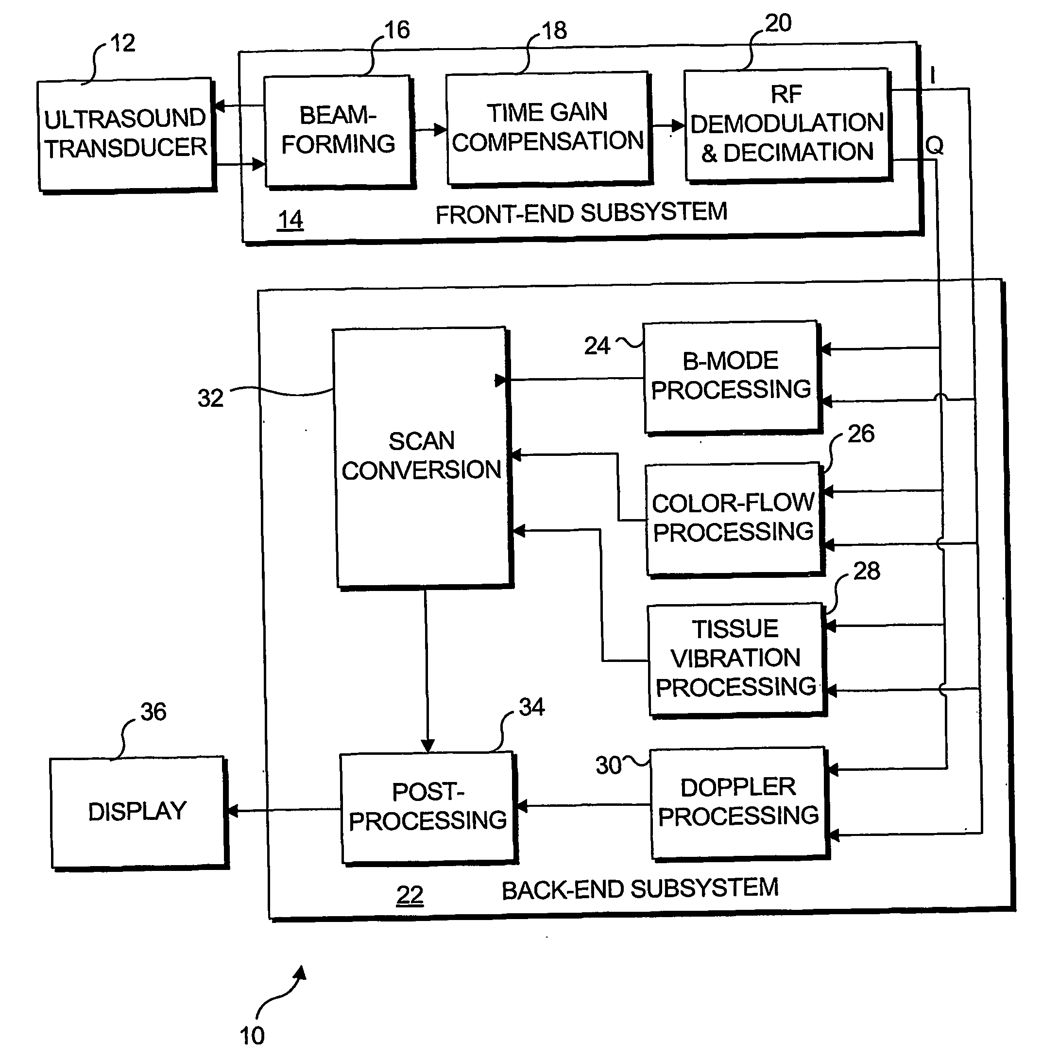
[0031]FIG. 1 is a block diagram illustrating an ultrasound system 10, which is generally similar to a conventional ultrasound system, but which has been modified to include tissue vibration imaging and is thus usable in practicing the present invention. Ultrasound system 10 includes an ultrasound transducer 12 that transmits a signal, which is modulated with a carrier frequency, typically 1 MHz-15 MHz, using multiple cycles (i.e., 2-20 cycles). The transmitted signal is reflected by scatterers (not shown) along the beam path and is received after a time delay, which depends upon the distance of the scatter from the transducer. In an acquisition stage, the acoustic echoes received from the tissue are converted to electrical signals by the transducer, and these signals are digitized by analog-to-digital converters (not separately shown). A front-end subsystem 14 includes a beam former 16 that performs dynamic focusing, apodization, and steering of both...


PUM


Abstract

An ultrasound based technique for detecting and imaging vibrations in tissue caused by eddies produced during bleeding through punctured arteries or from organs. A clutter signal, normally suppressed in conventional color flow imaging, is employed to detect and characterize local tissue vibrations, to detect internal bleeding in an image, or as an audible or palpable signal, or a readout. Using a tissue vibration image, the origin and extent of vibrations relative to the underlying anatomy and blood flow can be visualized in real time, enabling measurements of vibration amplitude, frequency, and spatial distribution. Bleeding rate can be determined from the frequency and amplitude of the vibrations. Signal processing algorithms usable to identify tissue vibrations from an ensemble of 2D ultrasound data include those based on phase decomposition, spectral estimation using eigendecomposition, and spectral estimation using autoregressive modeling for isolating vibrations from clutter, blood flow, and noise.

Description

GOVERNMENT RIGHTS [0001] This invention was funded at least in part with grants from the U.S. Office of Naval Research (ONR) (Award Nos. N00014-96-1-0630, N00014-01-G-0460, and N00014-99-1-0982), and grants from the U.S. Department of Defense (DoD) (ARMY MRMC Award Nos. DAMD17-02-2-0014, and DAMD17-00-2-0063), and the U.S. government may have certain rights in this invention.FIELD OF THE INVENTION [0002] This invention generally relates to a method for identifying tissue vibrations using an ensemble of two-dimensional (2D) ultrasound data acquired with an ultrasound transducer, and more specifically, to a method and system for identifying bleeding sites within a body of a patient in real time, based upon the tissue vibration that occurs at the sites, so that arterial bleeding and bleeding from organs can readily be localized in an image. BACKGROUND OF THE INVENTION [0003] Internal bleeding is a significant cause of death in cases of trauma, and rapid and effective diagnosis of patie...

Claims


Application Information

Patent Timeline
IPC IPC(8): A61B8/00A61BA61B8/06
CPCA61B8/06G01S15/8981A61B8/488A61B8/13
Inventor SIKDAR, SIDDHARTHAKIM, YONGMINBEACH, KIRK
Owner UNIV OF WASHINGTON
Why Eureka
Try Eureka