
Methods and compositions for labeling nucleic acids

a nucleic acid and composition technology, applied in the field of methods and compositions for labeling nucleic acids, can solve the problems of labor-intensive and time-consuming autoradiography, several limitations of the method, and the complications of using radioactivity

Active Publication Date: 2007-05-03
PRESIDENT & FELLOWS OF HARVARD COLLEGE +1
View PDF11 Cites 50 Cited by
  • Summary
  • Abstract
  • Description
  • Claims
  • Application Information

AI Technical Summary

Benefits of technology

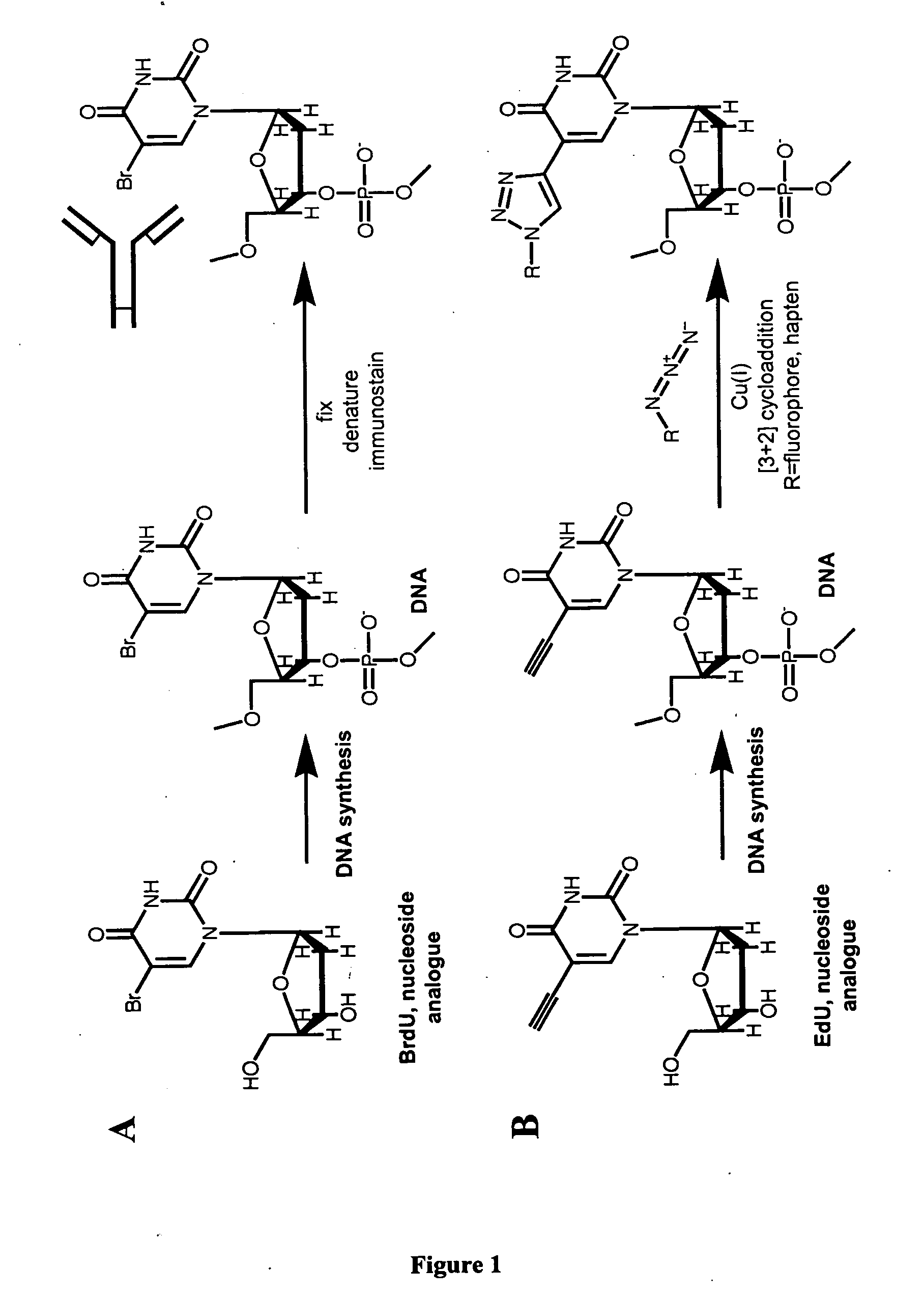
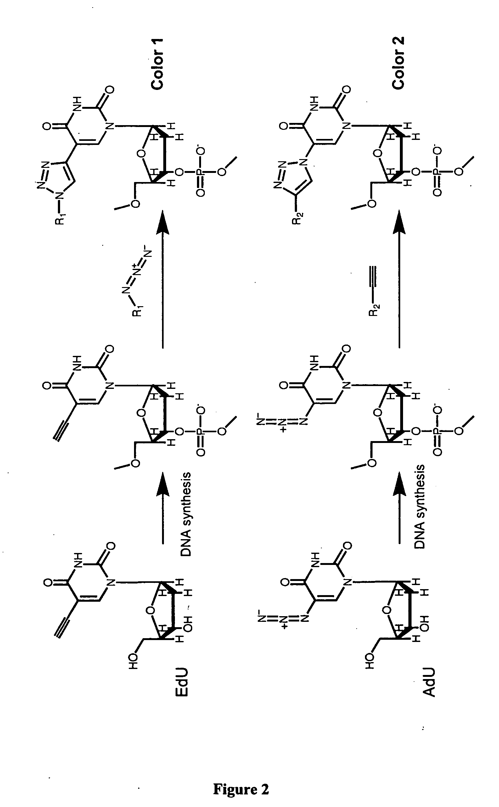
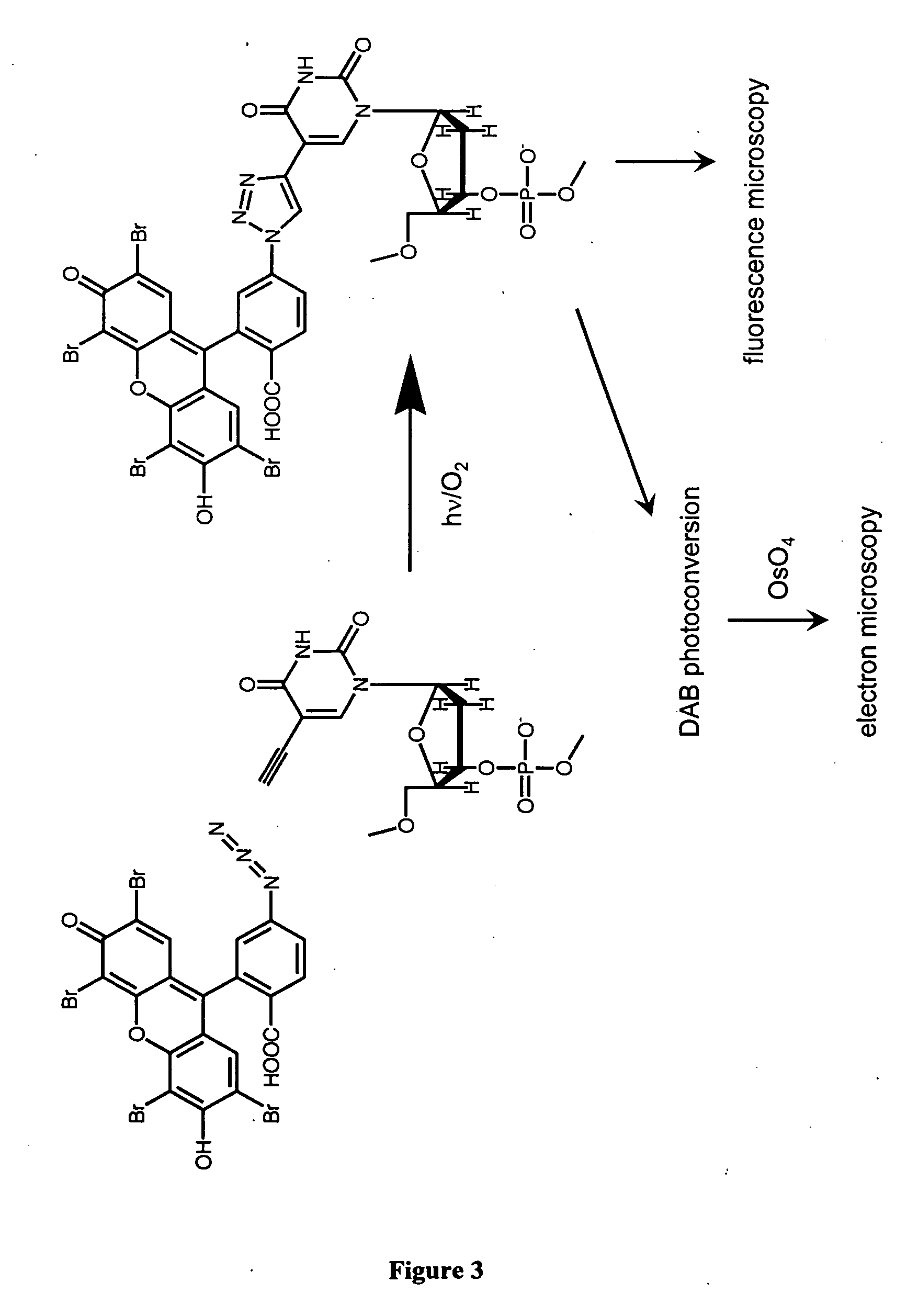
[0008] The present invention is directed to improved systems and strategies for studying cell division. More specifically, the present invention provides methods and compositions useful for labeling nucleic acid molecules and for measuring cellular proliferation in vitro and in vivo. In general, the inventive methods include a chemical reaction between a nucleotide analogue incorporated into a nucleic acid polymer and a reagent comprising a label, wherein the nucleotide analogue contains a first reactive group and the reagent contains a second reactive group such the reaction between the first and second reactive groups

Problems solved by technology

Although [3H]-thymidine and BrdU incorporation labeling methods have proven valuable for studying cell cycle kinetics, DNA synthesis and sister chromatid exchange, as well as for assessing cell proliferation of normal or pathological cells or tissues under different conditions, these methods exhibit several limitations.
The most notable disadvantage of [3H]-thymidine incorporation results from the complications and risks of using radioactivity.
In addition, autoradiography is labor-intensive and time-consuming.
Furthermore, because both methods are sample destructive, quantification can be performed at only one predetermined time point, and continuous monitoring of a single sample is not possible.
Although this method has several advantages including being safe for use in humans, it has disadvantages including that it involves a lengthy and destructive processing of the sample prior to detection.

Method used


Image

  • Methods and compositions for labeling nucleic acids
  • Methods and compositions for labeling nucleic acids
  • Methods and compositions for labeling nucleic acids

Examples


example 1

General Protocols for Labeling of Cells with Ethynyl-dU

[0263] Ethynyl-dU (EdU) was used in tissue culture media (DMEM complemented with penicillin, streptomycin, and fetal calf serum (FCS)) ranging from 10 nM to 1 82 M depending on the length of the labeling pulse. For example, if cells to be labeled were synchronized in the S phase, 100 nM to 1 μM of EdU was used for 1 to 2 hours. After labeling, the cells were washed 3 or 4 times with PBS and then tissue culture media was added.

Staining of Living Cells:

[0264] Staining Solution: 100 mM Tris pH 8.5 (from 2 M stock in water), 0.5 to 1 mM CuSO4 (from 1 M stock in water), 0.5 to 1 μM TMR-propyl-azide (from approximately 100 mM stock in DMF), and water (as required) were mixed together. Ascorbic acid (from a 0.5 mM stock in water) was then added to this solution to a final concentration of 50 mM, and the resulting staining solution is mixed thoroughly.

[0265] For staining cells alive without permeabilization (e.g., when the staining...

example 2

Labeling of HeLa Cells with Ethynyl-dU

[0271] HeLa cells were labeled or not with 1 μM EdU as described in Example 1. They were then fixed / permeabilized and stained with an Xrhodamine-azide reagent (which is not cell permeable and therefore requires permeabilization in order to perform the click chemistry).

[0272] Images of both populations of cells (i.e., labeled or not with EdU) are presented on FIG. 5. A higher resolution view of these cells is presented in FIG. 11 (right), along with images obtained from EdU-labeled HeLa cells stained with OliGreen (FIG. 11 left). Note the speckled appearance of the EdU stain in the bottom cell which is undergoing anaphase. The speckles are due to the fact that the labeling pulse of EdU was shorter than the time required for the cell to replicate its DNA; thus only the DNA replicating during the pulse was labeled.

example 3

Time-Lapse Fluorescence Imaging of Labeled Live Cells

[0273] To stain DNA under conditions as close to the native state as possible, a cell-permeable TMR-azide reagent was developed. TMR-propyl-azide was synthesized by first reacting TMR-carboxy-NHS ester with 3-bromo-propylamine to form bromo-propyl-TMR. The latter compound was then reacted with sodium azide to form TMR-propyl-azide, which was found to be cell-permeable and thus suitable for labeling cells without the need for permeabilization and / or fixation.

[0274] To demonstrate staining of cells using TMR-propyl-azide, HeLa cells were first labeled with EdU. To facilitate live microscopic imaging, EdU-labeled cells were plated in coverslip chambers which were mounted on the heated stage (37° C.) of an inverted Nikon TE200)U microscope equipped for wide-field fluorescence microscopy as well as spinning-disk confocal fluorescence microscopy (Yokogama spinning disk confocal head from Perkin-Elmer). The media covering the cells was...


PUM

PropertyMeasurementUnit
Timeaaaaaaaaaa
Fluorescenceaaaaaaaaaa

Abstract

The present invention relates to methods for the labeling of nucleic acid polymers in vitro and in vivo. Certain methods are provided that include a [3+2] cycloaddition between a nucleotide analogue incorporated into a nucleic acid polymer and a reagent attached to a label. Other methods are provided that include a Staudinger ligation between a nucleotide analogue incorporated into a nucleic acid polymer and a reagent comprising a substituted triarylphosphine attached to a label. Such methods do not require fixation and denaturation and therefore can be applied to the labeling of nucleic acid polymers in living cells and in organisms. Also provided are methods for measuring cellular proliferation. In these methods, the amount of label incorporated into the DNA is measured as an indication of cellular proliferation. The methods of the invention can be used in a wide variety of applications including clinical diagnosis of diseases and disorders in which cellular proliferation is involved, toxicity assays, and as a tool for the study of chromosomes' ultrastructures.

Description

RELATED APPLICATIONS [0001] The present invention claims priority from Provisional Application No. 60 / 730,745, filed on Oct. 27, 2005 and entitled “Methods and Compositions for Labeling Nucleic Acids”. The Provisional Application is incorporated herein by reference in its entirety.GOVERNMENT SUPPORT [0002] Part of the work described herein was funded by the National Institutes of Health (Grant No. GM039565).BACKGROUND OF THE INVENTION [0003] Cell division and cell death play central roles in the proper development of multi-cellular organisms and in the homeostatic maintenance of tissues. Loss or reduction of cell proliferative capability and dysregulation of cell death are among the most important phenomena that characterize the aging process (D. Monti et al., Am. J. Clin. Nutr., 1992, 55 (6 Suppl): 1208S-1214S; H. R. Warner et al., J. Am. Geriatr. Soc., 1997, 45: 1140-1146; L. Ginaldi et al., Immunol. Res., 2000, 21: 31-38). Disruption of normal control of cell proliferation and ce...

Claims


Application Information

Patent Timeline
IPC IPC(8): C12Q1/68C07H21/04
CPCC12Q1/68C12Q1/6816C12Q1/6841G01N33/5011G01N33/582C12Q1/6806C12Q2525/101C12Q2563/107
Inventor GEE, KYLE R.AGNEW, BRIAN
Owner PRESIDENT & FELLOWS OF HARVARD COLLEGE
Learn More