Method and apparatus for method for viewing and analyzing of one or more biological samples with progressively increasing resolutions

a biological sample and resolution technology, applied in the field of viewing and analyzing one or more biological samples with progressively increasing resolution, can solve the problems of limiting the options for screening for pre-neoplastic conditions, significant challenges in identification and diagnosis, and removal of tissue from patients

Inactive Publication Date: 2007-05-31
THE GENERAL HOSPITAL CORP
View PDF99 Cites 16 Cited by
  • Summary
  • Abstract
  • Description
  • Claims
  • Application Information

AI Technical Summary

Benefits of technology

[0009] One of the objects of the present invention is to overcome certain deficiencies and shortcomings of the prior art systems (including those described herein above), and provide exemplary embodiments of methods and arrangement for viewing and analyzing of one or more biological samples and anatomic structures with progressively increasing resolutions. Such exemplary methods and arrangements can be used along with a visual inspection of the data or by automatic processing procedures of the data to guide the visualization of areas that are most likely to contain abnormal and / or unhealthy tissue.

Problems solved by technology

Because precancerous growth and early stage cancers often arise on a microscopic scale, they can present significant challenges for identification and diagnosis.
Although this approach may be regarded as a current standard for microscopic diagnosis, it requires the removal of tissue from the patient and significant processing time to generate slides.
However, the lack of an appropriate imaging technology can greatly limits options for screening for pre-neoplastic conditions (e.g. metaplasia) and dysplasia.
In addition, an inability to identify areas of dysplasia and carcinoma in situ has led to screening procedures such as, e.g., random biopsy of the prostate, colon, esophagus, and bladder, etc., which can be highly undesirable and indiscriminate.
Resolutions approaching 20 μm have been achieved by these technologies, but fundamental physical limitations can still prevent their application in patients.
High NA imaging, however, can be particularly sensitive to aberrations that arise as light propagates through inhomogeneous tissue.
Also, high-resolution imaging with RCM is typically limited to a depth of about 100-400 μm.
Development of endoscopic confocal microscopy systems has been more difficult, owing at least in part to the substantial technical challenges involved in miniaturizing a scanning microscope.
One major obstacle to direct application of the concepts of confocal microscopy to endoscopy is the engineering of a mechanism for rapidly rastering a focused beam at the distal end of a small-diameter, flexible probe.
As currently implemented, point sampling can be inherent to RCM because it has a limited field of view, which may be comparable to or less than that of an excisional biopsy, and the imaging rate can be too slow for comprehensive large field microscopy.
Another challenge in adapting confocal microscopy to endoscopic applications can include miniaturization of high NA objectives that may be used for optical sectioning.
Indeed, the amount of this data can be large and difficult to view at one time such data, and thus such methods and arrangements would be beneficial for viewing and analysis thereof.

Method used


Image

  • Method and apparatus for method for viewing and analyzing of one or more biological samples with progressively increasing resolutions
  • Method and apparatus for method for viewing and analyzing of one or more biological samples with progressively increasing resolutions
  • Method and apparatus for method for viewing and analyzing of one or more biological samples with progressively increasing resolutions

Examples


Embodiment Construction

[0043] In accordance with exemplary embodiments of the present invention, methods and arrangement according to exemplary embodiments of the present invention can be provided for viewing and analyzing of one or more biological samples and anatomic structures with progressively increasing resolutions. Such exemplary methods and arrangements can be used along with a visual inspection of the data or by automatic processing procedures of the data to guide the visualization of areas that are most likely to contain abnormal and / or unhealthy tissue.

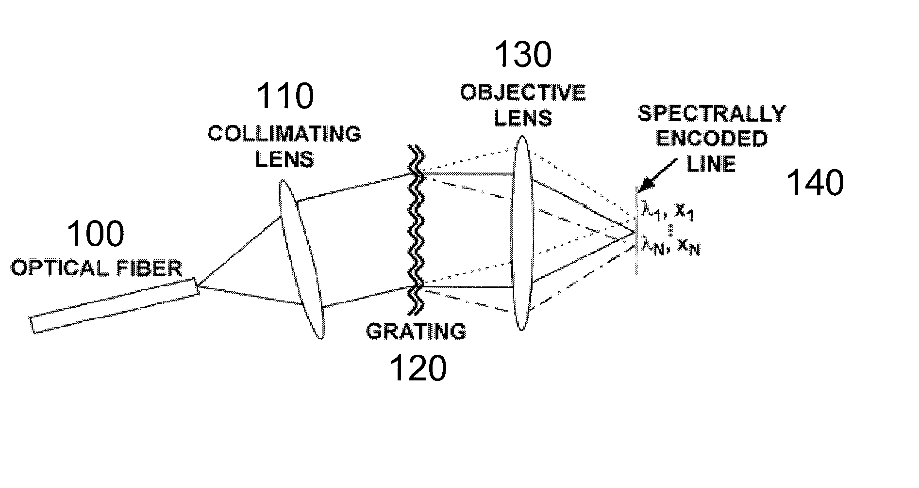
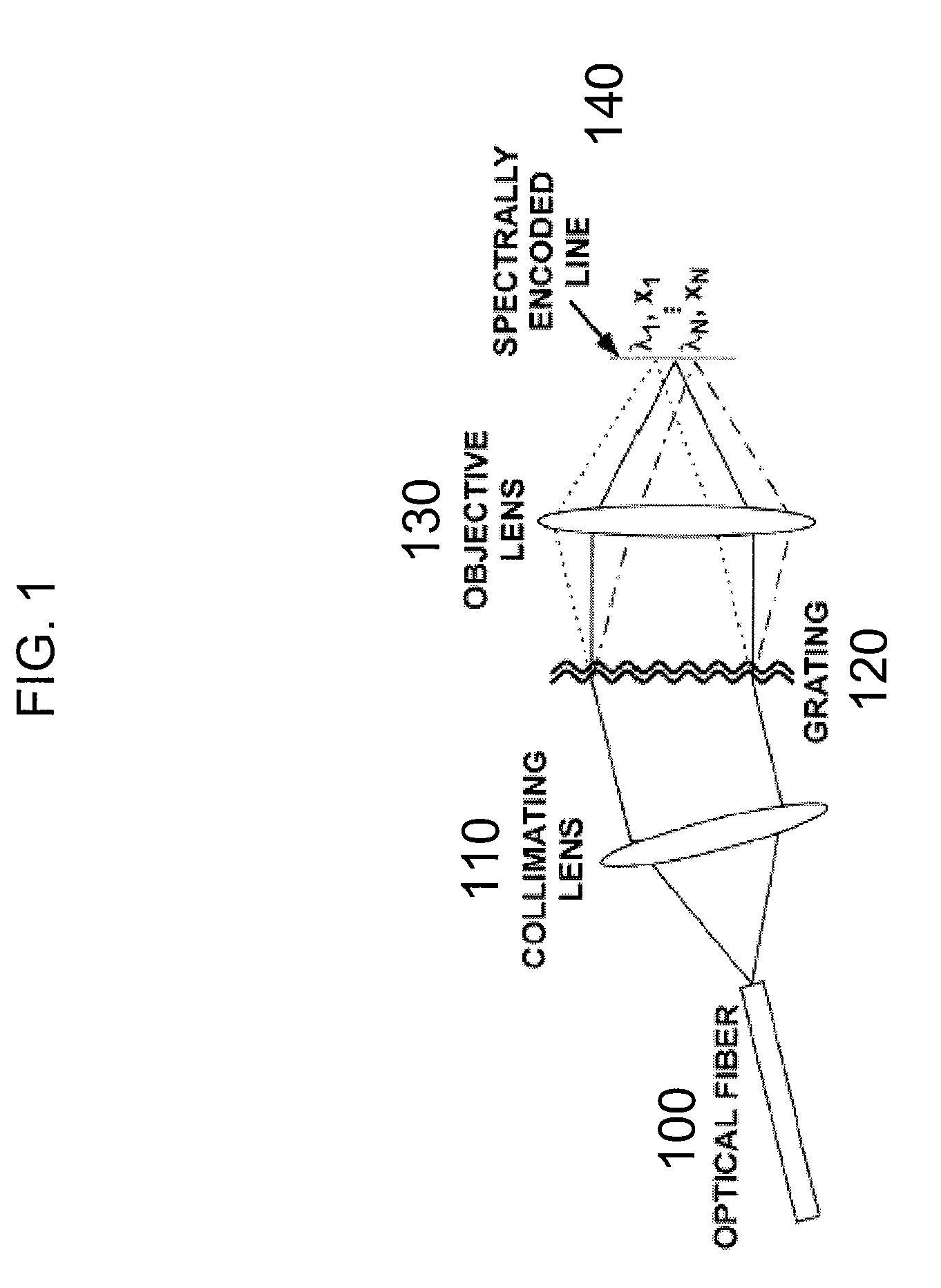
[0044] An exemplary SECM technique is shown in FIG. 1. The output from a single-mode optical fiber 100, which may be located at a distal end of a probe, can be collimated by a collimating lens 110, and then illuminate a dispersive optical element (such as, e.g., a transmission diffraction grating 120). An objective lens 130 can then focus each diffracted wavelength to a distinct spatial location within the specimen, resulting in a transverse lin...


PUM


Abstract

Method, apparatus and arrangement according an exemplary embodiment of the present invention can be provided for analyzing and / or illustrating at least one portion of an anatomical structure. For example, light can be forwarded to such portion so as to generate first information which is related to the portion. For example, the light can be provided on or within a subject. The first information can be received, and at least one section of the portion may be selected based on the first information so as to generate second information. A magnification of a display of the portion may be progressively modified as a function of the second information.

Description

CROSS-REFERENCE TO RELATED APPLICATION(S) [0001] This application is based upon and claims the benefit of priority from U.S. Patent Application Ser. No. 60 / 721,802, filed Sep. 29, 2005, the entire disclosure of which is incorporated herein by reference.FIELD OF THE INVENTION [0002] The present invention relates to relates to methods and arrangement for viewing and analyzing of one or more biological samples and anatomic structures with progressively increasing resolutions. BACKGROUND OF THE INVENTION [0003] Radiological techniques such as X-ray computed tomography (“CT”), magnetic resonance imaging (“MRI”), and ultrasound can enable noninvasive visualization of human pathology at the organ level. Although these modalities may be capable of identifying large-scale pathology, the diagnosis of cancer can require the evaluation of microscopic structures that is beyond the resolution of conventional imaging techniques. Consequently, biopsy and histopathologic examination may be required ...

Claims


Application Information

Patent Timeline
Patent Type & Authority Applications(United States)
IPC IPC(8): H01S3/00
CPCA61B5/0062G01N2021/1765A61B5/0068A61B5/0073A61B5/0075A61B5/0084A61B5/6852G01N21/4795G01N21/6458G01N23/046G02B21/0028G02B23/2423G02B23/243G02B23/2461G02B23/2476G01B9/02G01B9/02064G01B9/02027G01B9/02091G01B9/02049G01B9/02087G01B9/04G01N21/25G01N21/6486G01N2223/419G01N21/27G01N33/4833A61B5/0066G01N33/48
Inventor TEARNEY, GUILLERMO J.YELIN, DVIRVAKOC, BENJAMIN J.SUTER, MELISSABOUMA, BRETT EUGENE
Owner THE GENERAL HOSPITAL CORP
Why Eureka
Try Eureka