Apparatus, system, and method for treatment of posterior leaflet prolapse

a technology of posterior leaflet and appendix, applied in the field of medical devices, can solve the problems of chordae tendineae rupture, severe debilitating and even fatal valve disease, and inability to repair posterior leaflet prolapse, so as to prevent the heart valve annulus from further and undesired deformation

Active Publication Date: 2007-05-31
EDWARDS LIFESCIENCES CORP
View PDF98 Cites 165 Cited by
  • Summary
  • Abstract
  • Description
  • Claims
  • Application Information

AI Technical Summary

Benefits of technology

[0015] The invention can include application of a heart valve annuloplasty ring. The annuloplasty ring can reshape the heart valve annulus to a desired shape,

Problems solved by technology

Diseased heart valves may be categorized as either stenotic, wherein the valve does not open sufficiently to allow adequate forward flow of blood through the valve, and/or incompetent, wherein the valve does not close completely, causing excessive backward flow of blood through the valve when the valve is closed.
Valve disease can be severely debilitating and even fatal if left untreated.
In many diseased valves, the chordae tendineae are either ruptured, otherwise damaged, or of an improper length.
When chordae tendineae are too long, too short, or otherwise damaged, the corresponding tricuspid or mitral valve to which they are attached typically may fail to close properly.
This can lead to regurgitation, which is the unwanted backflow of blood from a ventricle to

Method used


Image

  • Apparatus, system, and method for treatment of posterior leaflet prolapse
  • Apparatus, system, and method for treatment of posterior leaflet prolapse
  • Apparatus, system, and method for treatment of posterior leaflet prolapse

Examples


Embodiment Construction

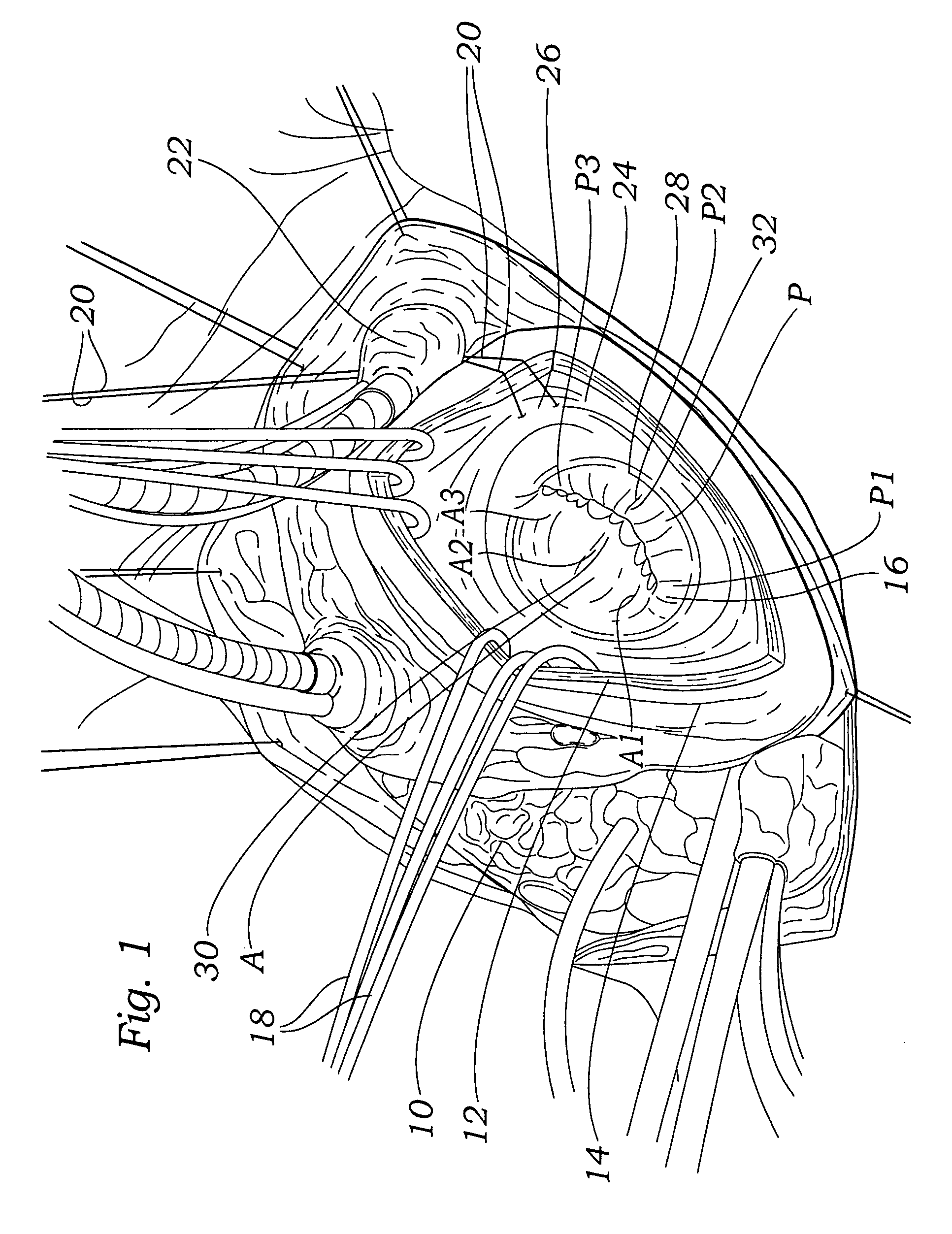
[0038]FIG. 1 depicts a heart 10 with an incision 12 in the left atrial wall 14 through which the mitral valve 16 is exposed for viewing during a surgical proceeding. The atrial wall incision 12 is held open with one or more retractors 18, giving the surgeon a full view for analysis of the mitral valve 16. Note that the viewing can be achieved directly a shown, as is typically the case for open chest and / or open heart surgical methods, or indirectly through an endoscope or other visualization devices, as may be used for minimally invasive procedures. In the exposure depicted in FIG. 1, a suture 20 (such as a 3-0 suture) is passed around and below the inferior vena cava 22, then makes a shallow pass through (i.e., takes a superficial bite of) the left atrial endothelium 24 at a position 26 about 1.5 cm behind the mitral valve annulus 28. Note that in the particular embodiment depicted, the suture is passed through the left atrial endothelium 18 at approximately the 5 o'clock position ...


PUM


Abstract

The invention is an apparatus, system, and method for repairing heart valves. A suture line is secured to a papillary muscle, and then passed through a portion of a heart valve leaflet. A reference element is provided at a desired distance from a plane defined by the heart valve annulus. The suture line is secured to the heart valve leaflet at a position adjacent the reference element. The reference element may part of a device configured for placement on or in a heart valve annulus. The reference element may be slidingly secured to the device so that the distance of the reference element from the main body of the device can be varied by a surgeon or other user. The reference element may be a line of suture, which may be pre-installed during manufacture of the device or may be installed by the surgeon or other user.

Description

CROSS-REFERENCES TO RELATED APPLICATIONS [0001] This application claims priority to commonly assigned U.S. provisional patent application No. 60 / 694,479 filed Jun. 27, 2005, which is incorporated herein by reference in its entirety for all purposes.FIELD OF THE INVENTION [0002] The present invention relates generally to medical devices and particularly to repairing posterior leaflet prolapse in a mitral valve. BACKGROUND OF THE INVENTION [0003] In vertebrate animals, the heart is a hollow muscular organ having four pumping chambers: the left and right atria and the left and right ventricles, each provided with its own one-way valve. The natural heart valves are identified as the aortic, mitral (or bicuspid), tricuspid, and pulmonary, and are each mounted in an annulus comprising dense fibrous rings. The mitral and tricuspid valves have thread-like bands of fibrous tissue that attach to the valve at one end and to the papillary muscles at the other end. [0004] Heart valve disease is ...

Claims


Application Information

Patent Timeline
IPC IPC(8): A61F2/24
CPCA61B17/02A61B17/0469A61F2/2466A61B2017/06057A61F2/2457A61B2017/00243
Inventor PERIER, PATRICKADZICH, VASO
Owner EDWARDS LIFESCIENCES CORP
Why Eureka
Try Eureka