Device for cell separation and analysis and method of using

a cell and analysis technology, applied in the field of cell separation and analysis, can solve the problems of method risks and associated significant challenges, and achieve the effect of facilitating separation

Active Publication Date: 2007-07-12
BIOCEPT INC
View PDF70 Cites 94 Cited by
  • Summary
  • Abstract
  • Description
  • Claims
  • Application Information

AI Technical Summary

Benefits of technology

[0009] The invention provides a microflow device for recovering rare cells or other target biomolecules from relatively small amounts of bodily fluids or the like, which device incorporates a microchannel arrangement wherein there is a collection region that is formed with a plurality of transverse fixed posts extending from a base surface of the collection region. The posts are arranged in a particular irregular array pattern to disrupt straight-line flow therethrough and importantly to break-up regular streamlined flow through the array, thereby assuring collisions with the posts and promoting swirling eddies in a bodily fluid or other liquid that is being caused to travel along this flow path through the collection region. Sequestering agents for the desired target biomolecules are appropriately attached to the surfaces of the transverse posts and throughout the entire collection region.
[0010] The collection region is preferably molded as a part of a cavity in the bottom surface of a body wherein a flow path is provided having an inlet and an outlet. The cavity is closed by a flat bottom plate of rigid material, and the body preferably contains a tab extending from one longitudinal end thereof which facilitates separation, i.e. peeling, of the body from the plate after the target biomolecules have been recovered from the sample. A top cap having openings leading to the inlet and outlet is disposed atop the body, and a polymeric wrap, preferably a heat-shrunk sleeve, sandwiches the body between the two plates and assures fluidtight sealing between the bottom surface of the body and the top surface of the plate. Slitting of the polymeric sleeve allows the body to be readily peeled from the bottom plate and exposes the captured target biomolecules on the upper surface of the plate for ready microscopic observation and/or analysis.
[0011] In one particular aspect, the invention provides a microflow device for separating biomolecules, such as cells, from a sample of a bodily fluid or other liquid, which device comprises a body having a flow path formed therein through which such a sample containing target biomolecules can be caused to flow, the flow path having inlet means, outlet means, and a microchannel arrangement which includes a collection region extending between said inlet and outlet means, which collection region is formed as a cavity in a flat bottom surface of said body and includes a plurality of transverse separator posts that protrude from a base surface of said cavity, a flat, rigid closure plate having a top surface that is in abutting contact with said flat bottom surface of said body and closes said flow path cavity, said posts being integral with said base surface of said collection region and extending to the top surface of said closure plate, said posts being arranged in ...

Problems solved by technology

Fetal cells have been obtained by methods such as amniocentesis and chorionic villus sampling; however, such methods can pose risks, especially to the fetus.
Th...

Method used


Image

  • Device for cell separation and analysis and method of using
  • Device for cell separation and analysis and method of using
  • Device for cell separation and analysis and method of using

Examples


example 1

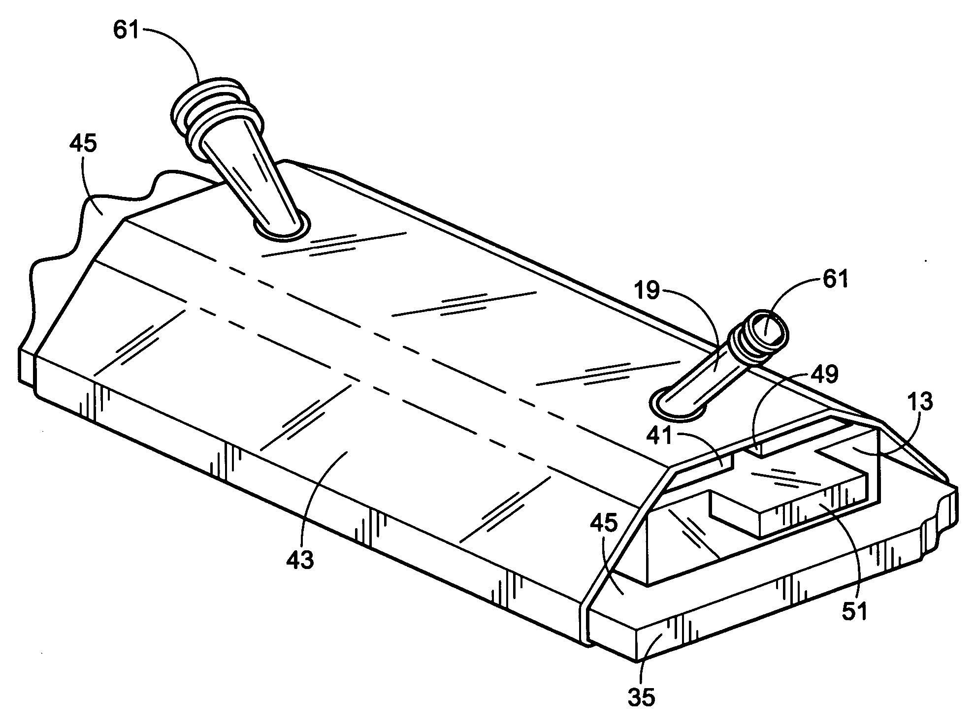
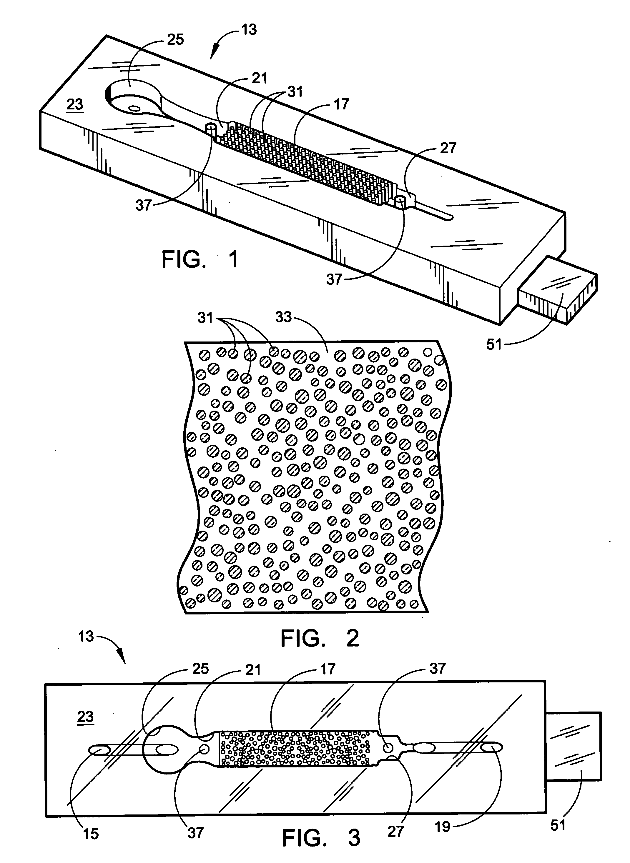
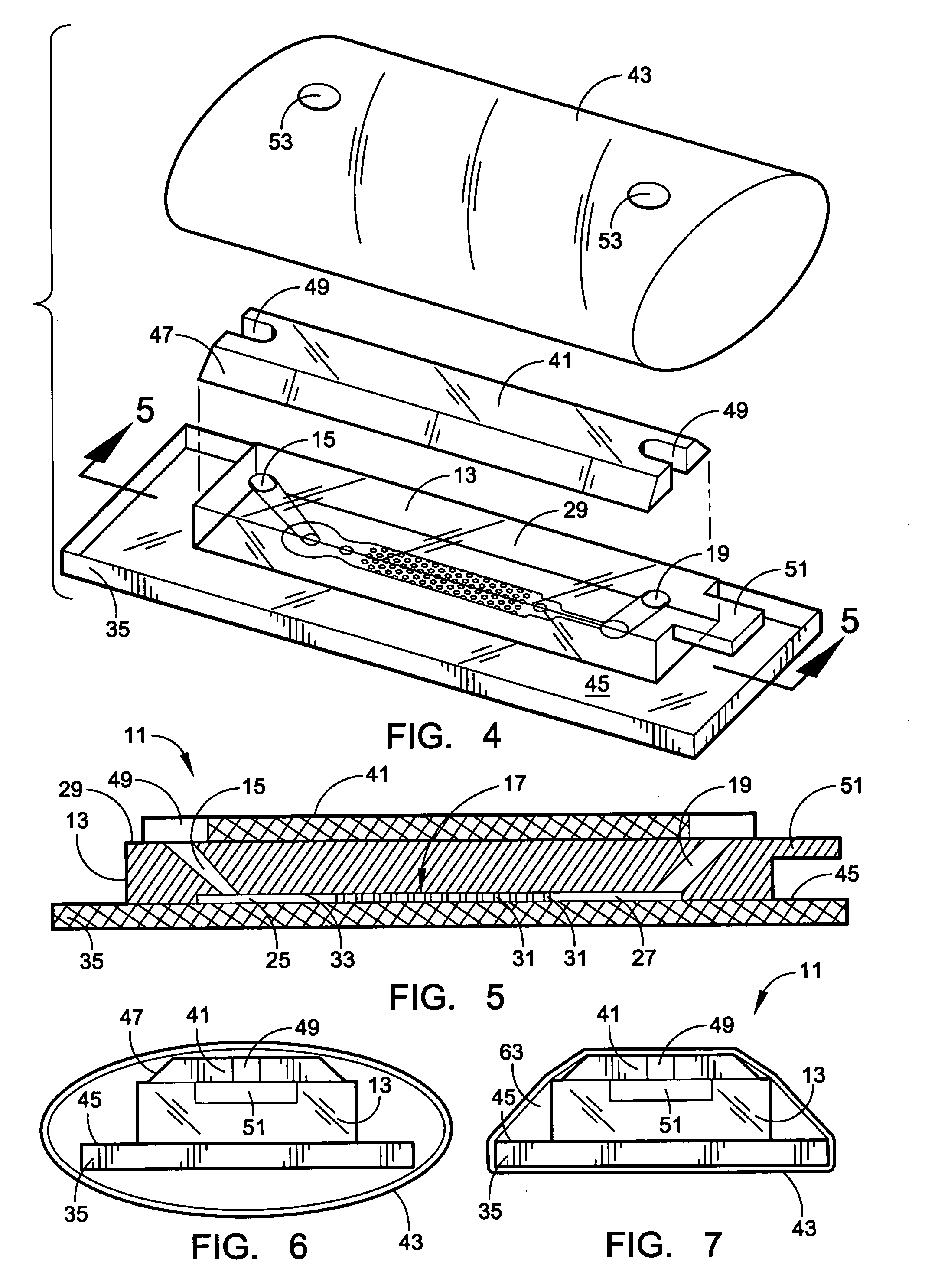
[0051] A microflow device for separating biomolecules is constructed to provide a prototype device as in the drawings. The body 13 is formed from PDMS, and with a cap plate 41 in place, it is pressed against a flat glass plate 35 by a heat-shrunken sleeve 43 of dimensionally oriented polymer to close the flow channel. The interior surfaces throughout the collection region 17 are derivatized by incubating for 30 minutes at room temperature with a 10 volume % solution of Dow Corning Z-6020 or Z-6011. After washing with ethanol, they are treated with nonfat milk at room temperature for about one hour to produce a thin casein coating. Following washing with 10% ethanol in water, a treatment is effected to coat all the interior surface with a permeable hydrogel that is based on isocyanate-capped PEG triols having an average MW of about 6000. A prepolymer solution is made using 1 part by weight polymer to 6 parts of organic solvent, i.e. acetonitrile and DMF, and it is mixed with an 1 mg / ...

example 2

[0055] Another microflow device of the same construction is similarly coated with a permeable hydrogel which carries Trop-1 and Trop-2 Abs. Cervical mucus from expectant mothers (8-12 weeks gestation) is diluted to 10 ml with HAM's media (InVitrogen) and treated with DNAse (120 units) at 37° C. for 30 minutes. After filtering through a 100 μm cell strainer, the cells are spun at 1500 RPM for 30 minutes. The cell pellet is resuspended in HAM's media (100 μl) and passed through the Trop-1 and Trop-2 coated microflow device by connecting the outlet tubing to a vacuum pump and supplying about 50 microliters of this cell suspension of cervical mucus extract to a vertically oriented conical reservoir. The pump is operated to produce a slow continuous flow of the sample liquid through the microflow device at room temperature and preferably at a rate of about 3-5 μl / min. During this period, the Trop-1 and Trop-2 Abs that are attached to the surfaces in the collection region, capture trophob...


PUM

PropertyMeasurementUnit
Fractionaaaaaaaaaa
Fractionaaaaaaaaaa
Fractionaaaaaaaaaa

Abstract

A microflow device for separating or isolating cells from a bodily fluid or other liquid sample uses a flow path where straight-line flow is interrupted by a pattern of transverse posts which are arranged across the width of a collection region in an irregular or set random pattern so as to disrupt streamlined flow. Sequestering agents, such as Abs, are attached to all surfaces in the collection region via a hydrophilic permeable hydrogel coating. The collection region is formed as a cavity in a body molded from PDMS, which flexible body is sandwiched between a glass slide or comparable flat plate and a rigid top cap plate, both of which are pressed into abutting relation with the PDMS body by a heat-shrunk polymeric sleeve. Following cell separation and washing, cells can be released from the sequestering agents and the device centrifuged to force said cells to collect adjacent the hydrogel-coated slide or plate. Slitting the polymeric sleeve allows the body to then be peeled from the slide or plate, using an integral tab, to expose the separated cells on the top surface thereof for ready microscopic examination.

Description

INTRODUCTION [0001] This invention relates to separation or isolation of target biomolecules from feed liquids and more particularly to an improved device for separating desired target human cells from bodily fluids or the like. BACKGROUND OF THE INVENTION [0002] Effective isolation and collection of rare cells from a heterogeneous cell population remains of high interest, due to the increasing demand for isolated cell populations for use in disease diagnosis and treatment, e.g. gene therapy, as well as for basic scientific research. For example, pathologically changed cells, such as cancerous cells, can be separated from a larger normal cell population, and the cleaned cell populations may then be transplanted back into the patient. [0003] One prominent demand is for the isolation of particular fetal cells from heterogeneous maternal cell populations to permit early fetus diagnosis, such as early screening of potential chromosomal disorders during pregnancy. Fetal cells have been o...

Claims


Application Information

Patent Timeline
IPC IPC(8): G01N33/567C12M1/34C12N5/07C12N5/071
CPCB01L3/5027G01N1/40G01N1/2813C12M41/36
Inventor TSINBERG, PAVELTANG, ZHONGLIANG
Owner BIOCEPT INC
Why Eureka
Try Eureka