Methods of monitoring cellular death using low frequency ultrasound

a low-frequency ultrasound and cellular death technology, applied in ultrasonic/sonic/infrasonic diagnostics, applications, therapy, etc., can solve the problems of time-consuming and inconvenient monitoring, and the methods are not only invasiv

Active Publication Date: 2007-07-19
SUNNYBROOK HEALTH SCI CENT
View PDF11 Cites 13 Cited by
  • Summary
  • Abstract
  • Description
  • Claims
  • Application Information

AI Technical Summary

Benefits of technology

[0004] A method of detecting cellular damage within a subject can comprise transmitting low frequency ultrasound (20 MHz or below) into a selected site within the subject wherein the selected site has been exposed to a stress capable of causing cellular death at the selected site. At least a portion of ultrasound backscattered from the ultrasound transmitted into the selected site can be received. The received backscattered ultrasound can be compared to a control backscatter measurement. An increase or a decrease in intensity or spectral slope of the received backscattered ultrasound when compared to the control backscatter measurement can indicate cellular death and / or damage at the selected site within the subject. Also provided herein are systems for performing the disclosed methods.

Problems solved by technology

These methods, however, are not only invasive, but are also time-consuming, requiring processing of a cell or tissue sample before data relating to apoptosis can be obtained.

Method used


Image

  • Methods of monitoring cellular death using low frequency ultrasound
  • Methods of monitoring cellular death using low frequency ultrasound
  • Methods of monitoring cellular death using low frequency ultrasound

Examples


example 1

[0123] A backscatter signal can be composed of contributions from many individual scatterers. Each scatterer contributes an amplitude and a phase to the received signal. This can be modeled by equation 1 (Raju and Srinivasan, 2002): r⁢ ⁢ⅇjΨ=∑i=1n⁢ri⁢ⅇjθi.(1)

[0124] The amplitude and the phase (ri and θi) contribution from each scatterer can be determined by its position, size and acoustic properties. As there can be a statistical distribution of these properties, the amplitude and phase (r and Ψ) of the received signal can also have a probability distribution. Numerous probability density functions (PDFs) have been proposed to describe the statistics of ultrasonic backscatter (Dutt and Greenleaf, 1994; Narayanan et al., 1994; Shankar et al., 1996; Raju and Srinivasan, 2002). Two of these distributions, the Rayleigh distribution (Strutt, 1880) and the generalized gamma (GG) distribution (Stacy, 1962) can be used in the described methods.

[0125] The Rayleigh PDF, often used as a simpl...

example 2

[0127] The response of the HEP-2 cell line (a Human Caucasian larynx carcinoma cell line) to the chemotherapeutic drug camptothesin, shown to induce apoptosis in this cell-line was determined.

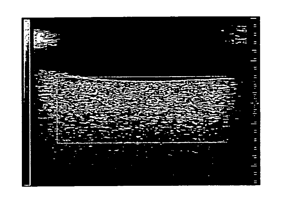
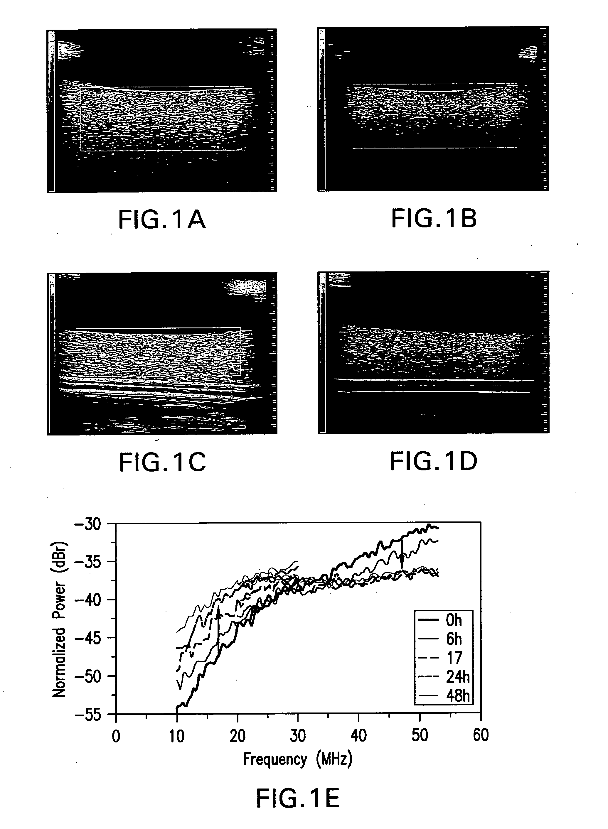
[0128] HEP-2 cell death induction: For the induction of apoptosis, cells were incubated at 37° C. and 5.0% CO2 in 30 μg / mL of camptothecin for various timepoints up to 48 hrs. Pipetting an appropriate amount of the reagent and dissolving it in MEM with 0.1% gentamycin prepared the camptothecin solution. The camptothecin solution was prepared fresh for each experiment. After the treatment, cells were washed with phosphate buffered saline (PBS) and detached using trypsin. These were then pelleted in flatbottom cryotubes at 1500 g on a desktop swinging bucket centrifuge with a diameter and height of approximately 1 cm. Pellets were imaged with a 20 or 40 MHz transducer, and RF data were collected.

[0129]FIG. 1 shows exposure of HEP2 cells to the chemotherapeutic camptothesin. Pellets of unexposed...

example 3

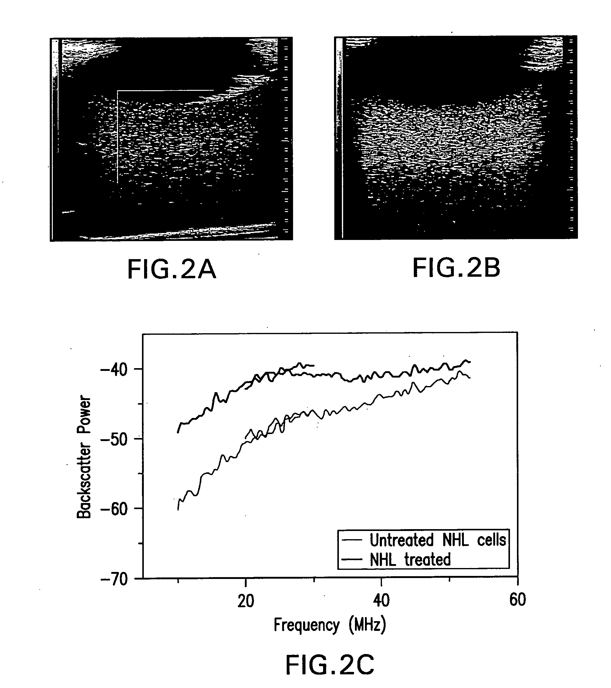
[0130] The response of NHL cells (Non-Hodgkins Lymphoma, human B cell lymphoma established from the pleural effusion of a 46-year-old woman with nodular histiocytic lymphoma) to CHOP chemotherapy are demonstrated, both in-vitro (Daunorubicin 0.012 mM, Cyclophosphamide 0.35 mM, Vincristine 0.2 nM, Prednisone 0.037 mM) and in-vivo in an animal model (table 4.1).

[0131] NHL cell death induction: Non-Hodgkin's Lymphoma cells (CRL2261, American Tissue Culture Collection) were cultured, then a concentration of 106 cells in 50 μl of media was injected intra-dermally into the left hind leg of severe combined immunodeficiency disease (SCID) mice, or were grown to form pellets. Primary tumors were allowed to develop for approximately 4 weeks, until they reached a diameter of 8-10 mm. The mice / cells were treated with the CHOP chemotherapy regimen, as is used clinically to treat aggressive NHL. In this regimen four drugs (Cyclophosphamide, Hydroxydoxorubicin, Vincristine (Oncovin) and Prednison...


PUM


Abstract

A method of detecting cellular damage within a subject comprises transmitting low frequency ultrasound (20 MHz or below) into a selected site within the subject wherein the selected site has been exposed to a stress capable of causing cellular death at the selected site. At least a portion of ultrasound backscattered from the ultrasound transmitted into the selected site is received. The received backscattered ultrasound is compared to a control backscatter measurement. An increase or a decrease in intensity or spectral slope of the received backscattered ultrasound when compared to the control backscatter measurement indicates cellular death or damage at the selected site within the subject.

Description

CROSS-REFERENCE TO RELATED APPLICATIONS [0001] This application claims priority to U.S. Provisional Application No. 60 / 691,577, filed on Jun. 16, 2005. The aforementioned application is herein incorporated by reference in its entirety.BACKGROUND OF THE INVENTION [0002] It is useful in both experimental and clinical applications to know whether cells are undergoing cellular death. Cells can undergo death by apoptosis or non-apoptotic cell death. Cell death plays a significant role in both normal and disease-related biological processes. Cells undergo apoptosis in response to a variety of stresses including chemotherapy, radiation therapy, photodynamic therapy and heat. Whether a cell is undergoing apoptosis is currently determined by taking samples of cells or tissues of interest and observing, using histological and DNA measurement methods, whether the cells exhibit the morphological changes that are indicative of apoptosis. These changes include membrane blebbing, DNA condensation ...

Claims


Application Information

Patent Timeline
Patent Type & Authority Applications(United States)
IPC IPC(8): A61B8/00
CPCA61B8/08A61B2019/5276A61N5/10A61N5/062A61F7/00A61B2090/378
Inventor KOLIOS, MICHAELCZARNOTA, GREGORYSHERAR, MICHAELTUNIS, ADAMHUNT, JOHN
Owner SUNNYBROOK HEALTH SCI CENT
Why Eureka
Try Eureka