Method and diagnostic tests based on flow cytometric analysis of antigen-specific t lymphocytes

a flow cytometric and antigen-specific technology, applied in the direction of material testing goods, measurement devices, instruments, etc., can solve the problems of increasing the total number of cases, increasing the cost of intervention, and remaining concerned about orthopoxvirus

Inactive Publication Date: 2007-08-02
INST NAT PER LE MALATTIE INFETTIVE LAZZARO SPALLANZANI IRCCS
View PDF0 Cites 14 Cited by
  • Summary
  • Abstract
  • Description
  • Claims
  • Application Information

AI Technical Summary

Benefits of technology

[0012] The present invention provides a method for the set-up of a specific kit allowing the immune diagnosis, in particular of bio-terrorism agent exposure by measuring the immune response to antigens associated with all those pathologies that generate a T cell response. Specifically, the method is focused on the use of pathogen-discriminating epitopes that have been selected between commercially available recombinant proteins, and designed as a set of synthetic peptides (peptide composition) efficiently and, in general, promiscuously inducing the stimulation of T-lymphocytes specific for pathogenic and nonpathogenic variants of the biological threat agent. The quantitative determination of antigen-specific T lymphocytes (referred as Ag-Sp), was analysed by using these newly devised pathology-specific antigen or epitope compositions which represent a further embodiment of the invention (Patho-tope arrays). After the stimulation, the test uses a rapid method for the selective measurement of the Ag-Sp T lymphocytes that are identified through: A) monoclonal antibodies recognizing membrane structures of T lymphocytes and of their sub-populations; B) monoclonal antibodies binding to cytokines accumulating at intracellular level after the stimulation with the antigen; and C) mixtures of A) and B). The flow cytometric detection of the presence of markers of differentiation on T lymphocytes and of intracytoplasmic cytokines allows the acquisition of both qualitative and quantitative results. The diagnostic test described in the present invention is performed using venous blood, is composed by a simple reagent kit, and uses a flow cytometer for read-out—commonly used in laboratories of clinical pathology for the quantification of the T, B and NK lymphocytes. The availability of mobile flow-cytometer units may allow the use of this assay under field investigation conditions.

Problems solved by technology

Although the last natural case of smallpox was reported in Somalia in 1977, this orthopoxvirus remains a source of concern.
Assuming an average of 15 days needed for infected persons to become infectious, delay in intervention will be costly, increasing the total number of cases.
Moreover, the intentional spread of envelopes containing Anthrax spore in the Unites States has determined a great alarm and some deaths.
However, these tests based on B lymphocytes fail to give positive results until two weeks post-antigen exposure, allowing for the minimal time necessary to activate the B lymphocytes.
Both of these approaches give only qualitative results and provide information about the ability of immune cell to recognize the antigen although they do not allow the measurement of the frequency of the cells responding to the antigen stimulation.
However, these approaches were restricted to the M.tuberculosis-specific response, required a long stimulation time and were poorly sensitive.
However, these methods do not allow to discriminate pathogenic from nonpathogenic species of the same family of microorganism which is crucial for the detection of biological threat agents (for example to distinguish the highly pathogenic variola virus from the safe administration of a vaccine or to discriminate the dangerous SARS coronavirus from the common cold OC43 or E229 coronavirus).

Method used


Image

  • Method and diagnostic tests based on flow cytometric analysis of antigen-specific t lymphocytes
  • Method and diagnostic tests based on flow cytometric analysis of antigen-specific t lymphocytes
  • Method and diagnostic tests based on flow cytometric analysis of antigen-specific t lymphocytes

Examples


example 1

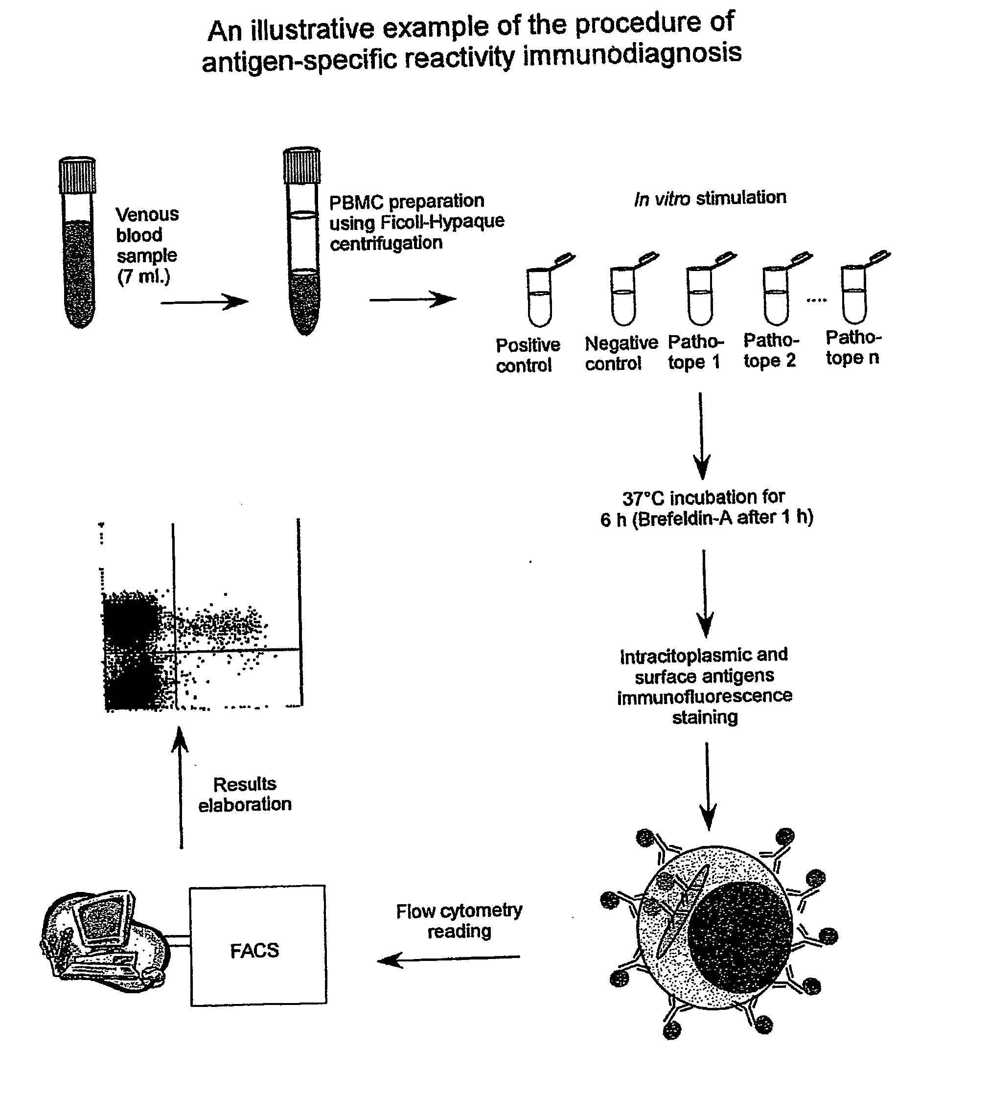
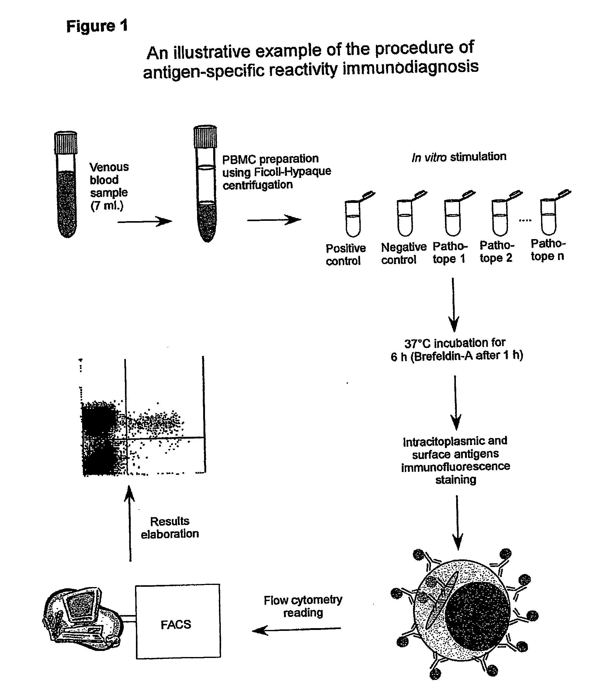
Procedure of Execution of the Patho-tope Arrays T Lymphocytes Immuno-diagnosis Test by Flow-cytometry.

[0265] The following specific monoclonal antibodies for human antigens were used for the execution of the test: purified anti-CD28e anti-CD49d as co-stimulator factors during the cellular cultures; anti-IFN-gamma conjugated with fluorescein (FITC);

[0266] anti-CD3 conjugated with phycoerythrin (PE); anti-CD45 conjugated with phycoerythrin-cyanin-5(Cy-5) and a isotypic control (IgG1) conjugated with FITC.

[0267] The antibodies are used at the concentration of 0,25 μg / ml. Each new batch of antibodies was tested, and the antibodies mixtures (mix) were set-up ready for use in 1 mL microcentrifuge tubes. Specifically, each antibody was used in saturating conditions to exclude differences in the samples during the staining. The tubes were then placed in a Speedvac freeze-dryer until complete evaporation of the solvent (20 min). At the moment of the use each mix was reconstituted by addin...

example 2

Elaboration of Results and Formulation of a Diagnostic Response

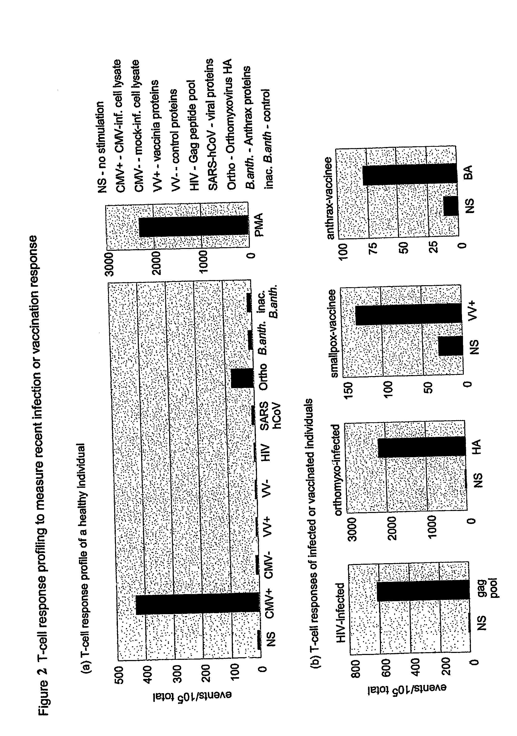
[0269] A T-cell response profile was developed for several individuals (see FIG. 1a). A marked, specific response to CMV antigens was seen in each of the healthy donor panels. There was a large individual variability, but sample duplicates confirmed specificity. Neither of these results were unexpected, as the prevalence of seropositivity for CMV in Italy is quite high and the response levels were expected to vary, depending on the individual. Pathogen-infected or recently vaccinated individuals were used as controls to confirm the reactivity of the antigen mixes for the response panel. As shown in FIG. 1b, a robust response was observed in infected or vaccinated individuals for their respective pathogens.

[0270] A small, but reproducible response was seen to the recombinant SARS protein pool in a number of healthy donors (FIG. 2a). Selected epitopes in our preparation are not unique to the class IV coronavirus (SARS-hC...

example 3

Description of the Procedure using as Ag-Sp Formulations a Raw Antigenic Extract, Recombinant Proteins, or Mixtures of Peptides

[0271] In this experiment different antigenic preparations were used: (a) raw protein extract, (b) purified or recombinant proteins, (c) mixtures of peptides. [0272] (a) As an example, the methodology of purification of antigenic extract from fibroblast or VERO cells infected with the vaccinia virus is reported. The cells susceptible to the infection were transferred in a tube containing vaccinia virus to a multiplicity of infection (MOI)=100. The incubation was performed at 37° C. until 50% of the cytopathic effect was found. At that time, a centrifugation to 850 g×15 min was performed followed by fixation in PFA 2% for 10 min. at 4° C. After three washes in PBS, the cell pellets were sonicated for 20 min at 4° C. in PBS, centrifuged at 850 ×g for 15 min at 4° C. and aliquoted at −20° C. Each new antigenic preparation batch must be checked to find the best...


PUM

PropertyMeasurementUnit
concentrationaaaaaaaaaa
frequencyaaaaaaaaaa
densityaaaaaaaaaa

Abstract

The present invention provides a method for the immuno-diagnosis of diseases with different aetiology (infectious diseases, tumors etc) by measurement of the T cell response J, B and NK lymphocytes) induced by a set of diseasespecific antigens. The method is based on the quantitative determination of antigenspecific T lymphocytes (referred as Ag-Sp), stimulated by using a newly devised pathology-specific antigen or epitope compositions which represent further embodiments of the invention. After stimulation, the selective measurement of the Ag-Sp T lymphocytes is performed by: A) monoclonal antibodies recognizing membrane structures of T lymphocytes and of their sub-populations; B) monoclonal antibodies binding to cytokines accumulating at intracellular level after the stimulation with the antigen; or C) mixtures of A) and B). The flow cytometric detection of the presence of markers of differentiation on T lymphocytes and of intracytoplasmic cytokines allows the acquisition of both qualitative and quantitative results. The invention also provides diagnostic kits for performing the method of the invention.

Description

FIELD OF THE INVENTION [0001] The present invention refers to a method and corresponding diagnostic tests to assay immune responses to antigens associated with pathologies that generate T cell responses. The test is based on the flow-cytometry analysis of the antigen-specific T lymphocytes (referred as Ag-Sp T lymphocytes). [0002] More particularly the invention refers to a method and corresponding diagnostic tests to simultaneously assay the exposure to antigens associated with biological threat agents. The method can also be applied to other clusters of disease and may allow to determine at once the occurrence of respiratory infections, sexually transmitted diseases, in utero infections, post-transplant infection, blood borne infections or neoplastic diseases with known tumor associated antigens. BACKGROUND [0003] Although the last natural case of smallpox was reported in Somalia in 1977, this orthopoxvirus remains a source of concern. No evidence exists that smallpox will recur a...

Claims


Application Information

Patent Timeline
Patent Type & Authority Applications(United States)
IPC IPC(8): G01N33/567G01N33/50G01N33/569G01N33/571G01N33/576
CPCG01N33/505G01N33/56911G01N33/56972G01N33/576G01N33/56983G01N33/571G01N33/56977
Inventor POCCIA, FABRIZIOGIOIA, CHRISTIANAAGRATI, CHIARAMONTESANO, CARLAAMICOSANTE, MASSIMOCASETTI, RITAD'OFFIZI, GIANPIEROHOREJSH, DOUGLASMARTINI, FEDERICOCAPOBIANCHI, MARIA ROSARIAPUCILLO, LEOPOLDO PAOLOPERRONE DONNORSO, RAFFAELEIPPOLITO, GIUSEPPE
Owner INST NAT PER LE MALATTIE INFETTIVE LAZZARO SPALLANZANI IRCCS
Why Eureka
Try Eureka