Method for predicting or identifying the onset of premature membrane rupture during pregnancy

a technology of amnichorionic membrane and onset of pregnancy, which is applied in the field of predicting or identifying the onset of premature membrane rupture during pregnancy, can solve the problems of increasing the risk of intrauterine infection, and increasing the risk of pregnancy death for the fetus and the mother

Inactive Publication Date: 2007-09-27
KIMBERLY-CLARK WORLDWIDE INC
View PDF14 Cites 8 Cited by
  • Summary
  • Abstract
  • Description
  • Claims
  • Application Information

AI Technical Summary

Problems solved by technology

Premature rupture of the amnichorionic membrane prior to the onset of labor is becoming an increasing problem for pregnant women.
In either case, if left untreated, possible death to the fetus and the mother could result.
The timing for the delivery of the baby becomes critical, as the risk of intrauterine infection increases significantly as more time passes following rupture.
The problem is that leaking amniotic fluid is frequently confused by the mother with her own urine or vaginal discharge.
This results in many false alarms and unnecessary trips to either the doctor's office or to the hospital for evaluation of the pregnant woman to rule out possible rupture of the membrane.
Unfortunately, pH cannot be used as a tool for definitive diagnosis and it is effective only after rupture of the membrane.
Although a useful biomarker, C-reactive protein is not always found in vaginal fluid prior to the onset of premature membrane rupture.
Unfortunately, prediction is often difficult due to the wide variety of causes of premature membrane rupture.
Nevertheless, one common cause of premature membrane is bacterial vaginosis, which affects up to 40-50% of women in child-bearing age.

Method used


Image

  • Method for predicting or identifying the onset of premature membrane rupture during pregnancy
  • Method for predicting or identifying the onset of premature membrane rupture during pregnancy
  • Method for predicting or identifying the onset of premature membrane rupture during pregnancy

Examples


example 1

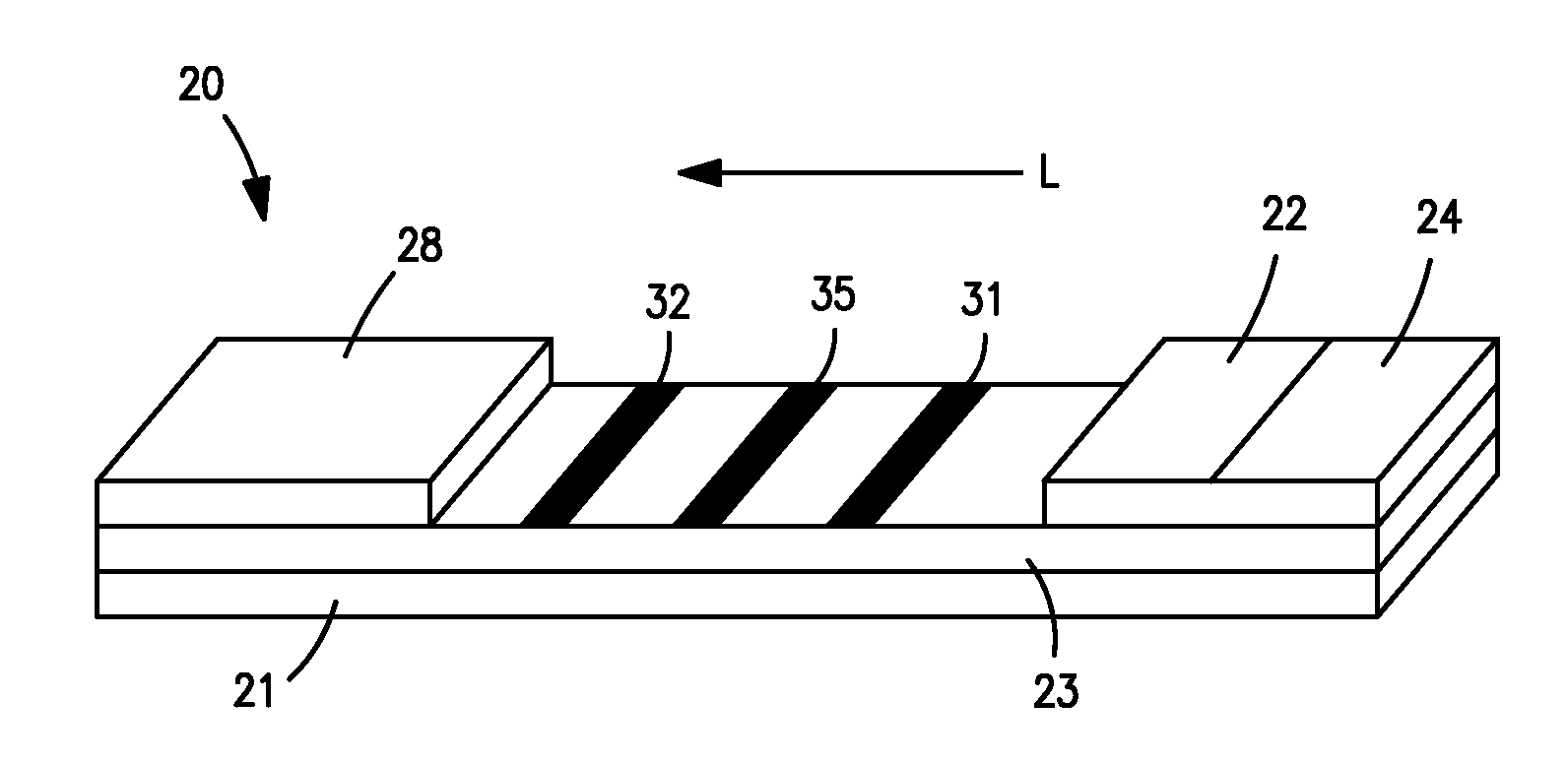
[0047] CRP monoclonal antibody (CRP MAb1 from Medimix (MedixMab), clone #6404, Lot #SP-179-2) and bovine serum albumin (BSA) buffer (10 mM phosphate buffered saline and 0.2% BSA, pH 7.3) were striped on clear-backed cards of laminated nitrocellulose (Millipore HF120). Specifically, CRP MAb1 (0.75 mg / ml) was striped at a dispense rate of 1.5 μl / cm. Two (2) BSA lines were then striped 1.25 mm on either side of the CRP MAb1 line at a dispense rate of 1.0 μl / cm. Finally, a single control line was striped downstream from the CRP MAb1 and BSA lines with 1.0 mg / ml CRP antigen (Scipac #P100-0, Lot #1049-20) at a dispense rate of 1.0 μl / cm. After striping, the cards were dried in an oven at 37° C. for 60 minutes. The cards were then assembled with an absorptive sink (Millipore CFSP203000) and a glass fiber conjugate pad (Millipore GFCP203000) pre-striped with three bands of gold particles conjugated with CRP antibody (MAb1). The conjugated particles had an optical density (“OD”) of 3.3. The ...

example 2

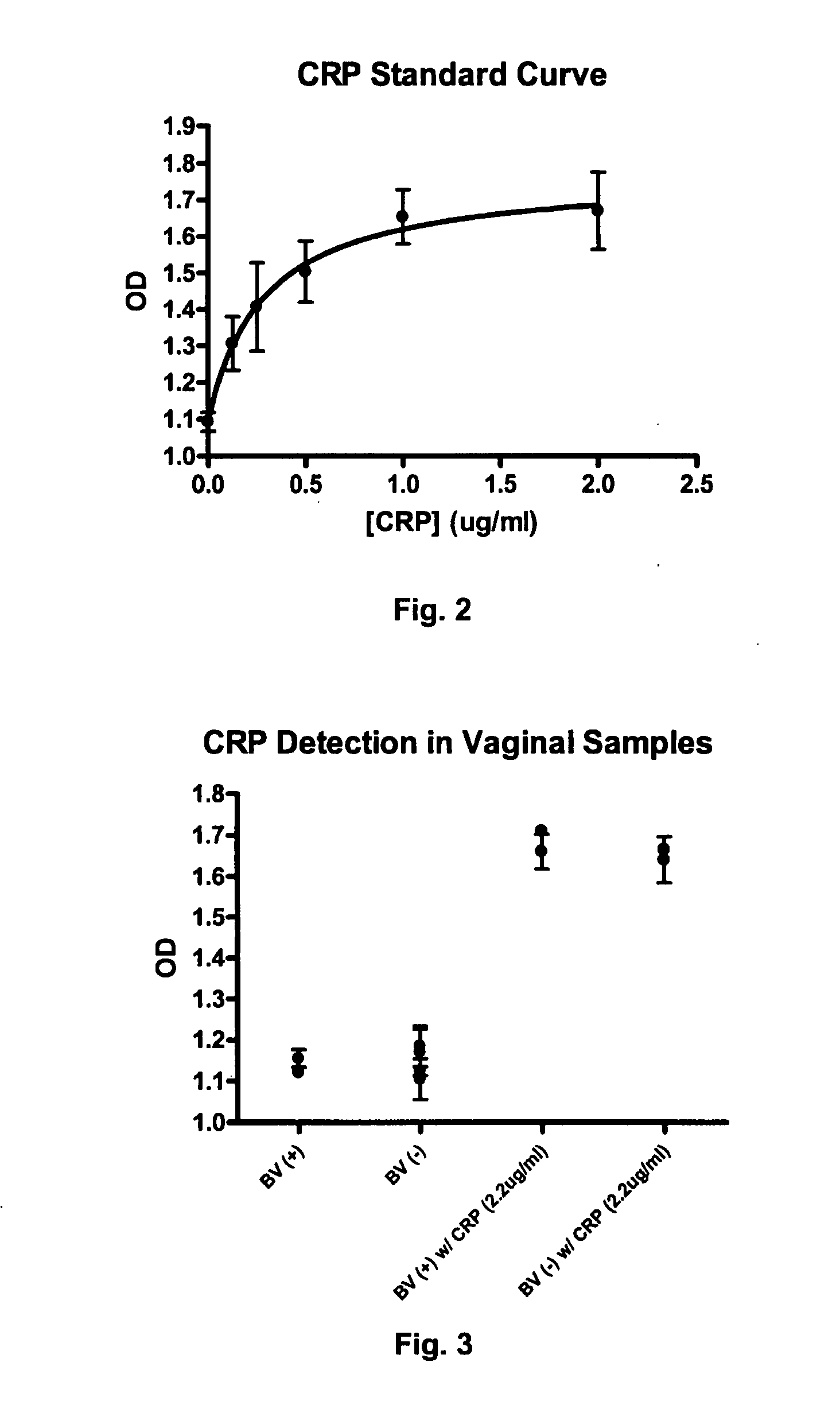
[0048] CRP levels were measured as described in Example 1 in vaginal samples that were known to be both positive and negative for bacterial vaginosis (“BV”) based on clinical trials conducted on non-pregnant women. Some of the vaginal samples were also spiked with CRP (2.0 μg / ml) for purposes of comparison. In addition to CRP, amine tests were also performed. More specifically, 50 μl of vaginal fluid or standards of putrescine hydrochloride solutions (5, 2.5, 1.25, 0.625, 0.312, 0.1506, 0.075, and 0.0 mg / ml) were placed in a microtiter plate wells. Thereafter, 100 μl of both a phenol-nitroprusside solution (10 mg of sodium nitroprusside and 2 ml of saturated phenol (from Sigma) in 18 ml of water) and a sodium hydroxide-hypochlorite solution (3 ml of Clorox™ bleach in 17 ml of ˜0.1N sodium hydroxide) were added to the well. The resultant solution was incubated for 10 minutes and read at 630 nanometers using microplate reader. Amine concentration of vaginal fluids was determined by a ...


PUM

PropertyMeasurementUnit
diameteraaaaaaaaaa
diameteraaaaaaaaaa
lengthaaaaaaaaaa

Abstract

A diagnostic test kit for simultaneously detecting the presence of amines (e.g., putrescine, cadaverine, tyramine, and trimethylamine) and C-reactive protein in a vaginal sample is provided. The test kit includes a lateral flow assay device containing a chromatographic medium (e.g., porous membrane, fluidic channel, etc.). The chromatographic medium defines a first detection zone within which is contained an amine-sensitive chromogen and a second detection zone within which is contained an immunoreactive receptive material that preferentially binds with C-reactive protein or a specific binding member thereof. The first and second detection zones may produce signals that are detectable, either visually or through the use of instrumentation. In this manner, the kit provides a complimentary system for both predicting and identifying the onset of premature membrane rupture. For example, the detection of amines in the vaginal sample may serve as a diagnosis of bacterial vaginosis, which may provide an early warning of the potential for premature membrane rupture. The complimentary detection of CRP may serve as an indicator of rupture and thus alert the patient to seek immediate medical care. Such a system may be equally effective in point of care (POC) and over-the-counter (OTC) applications.

Description

RELATED APPLICATIONS [0001] The present application is a continuation-in-part of U.S. patent application Ser. No. 10 / 790,617, filed on Mar. 1, 2004, the contents of which are incorporated herein by reference.BACKGROUND OF THE INVENTION [0002] Premature rupture of the amnichorionic membrane prior to the onset of labor is becoming an increasing problem for pregnant women. Upon premature rupture of the membrane, the fetus must be promptly delivered when the mother becomes clinically infected or the fetus shows signs of potential compromise. In either case, if left untreated, possible death to the fetus and the mother could result. The timing for the delivery of the baby becomes critical, as the risk of intrauterine infection increases significantly as more time passes following rupture. Accordingly, it becomes critical to provide a method of early detection of rupture. The problem is that leaking amniotic fluid is frequently confused by the mother with her own urine or vaginal discharg...

Claims


Application Information

Patent Timeline
Patent Type & Authority Applications(United States)
IPC IPC(8): G01N33/50G01N30/00G01N33/52
CPCG01N33/523
Inventor BOGA, RAMESHBABUTAKEUCHI, JAMESMACDONALD, J.
Owner KIMBERLY-CLARK WORLDWIDE INC
Why Eureka
Try Eureka