Labeled antimicrobial peptides and method of using the same to detect microorganisms of interest

Inactive Publication Date: 2007-10-04
ARCIDIACONO STEVEN MICHAEL +3
View PDF57 Cites 36 Cited by
  • Summary
  • Abstract
  • Description
  • Claims
  • Application Information

AI Technical Summary

Problems solved by technology

Although many such microorganisms are innocuous to humans, certain species of microorganisms are pathogenic and pose a serious health risk to people.
Unfortunately, the presence of pathogenic microorganisms in such media cannot typically be ascertained simply by visual or other sensory examination of the media, but rather, requires the use of specialized testing equipment and procedures.
Unfortunately, there are certain difficulties that are commonly encountered in using the above-described techniques to detect pathogens.
First,

Method used


Image

  • Labeled antimicrobial peptides and method of using the same to detect microorganisms of interest
  • Labeled antimicrobial peptides and method of using the same to detect microorganisms of interest
  • Labeled antimicrobial peptides and method of using the same to detect microorganisms of interest

Examples


example i

Preparation of Cy5-Labeled Antimicrobial Peptides

[0043] The antimicrobial peptides cecropin P1 (see Lee et al., “Antibacterial peptides from pig intestine: isolation of a mammalian cecropin,”Proc. Natl. Acad. Sci. U.S.A., 86:9159-62 (1989), which is incorporated herein by reference), PGQ (see Moore et al., “Antimicrobial peptides in the stomach of Xenopus laevis,” J. Biol. Chem., 266:19851-7 (1991), which is incorporated herein by reference), ceratotoxin A (see Marchini et al., “Purification and primary structure of ceratotoxin A and B, two antibacterial peptides from the female reproductive accessory glands of the medfly Ceratitis capitata (Insecta:Diptera),”Insect Biochem. Mol. Biol., 23:591-8 (1993), which is incorporated herein by reference), cecropin A (see Sun et al., “Peptide sequence of an antibiotic cecropin from the vector mosquito, Aedes albopictus,” Biochem. Biophys. Res. Commun., 249:410-5 (1998), which is incorporated herein by reference), CPF3 (see Maloy and Kari, “S...

example ii

Solution Binding Assay

[0047]E. coli O157:H7 (ATCC 43888) was grown in Luria broth to OD600 1 (approximately 108 CFU / ml) and washed 2× in equal volume PBST before being resuspended in PBST. 100 μl (107 CFU) cells were added to 900 μl PBST with 5 μg Cy5-CP1_c peptide for 30 minutes at ambient temperature, with rotary mixing (see reference numeral 41 of FIG. 4). Cells were harvested at 10,000×g for 3 minutes (see reference numeral 43 of FIG. 4), the supernatant removed with pipette tip, and washed three times with 1 ml PBST and spun as above (see reference numeral 45 of FIG. 4). Cells were then resuspended in 200 μl PBST and transferred to a black microplate (Nalge Nunc International, Rochester, N.Y.)(see reference numeral 47 of FIG. 4). 900 μl solution containing 5 μg peptide was added to 100 μl buffer without cells and assayed as a negative control. The microplate was imaged using the Storm 860 (Amersham Biosciences, Piscataway, N.J.) using red fluorescence at 1000 V PMT, 200 micron...

example iii

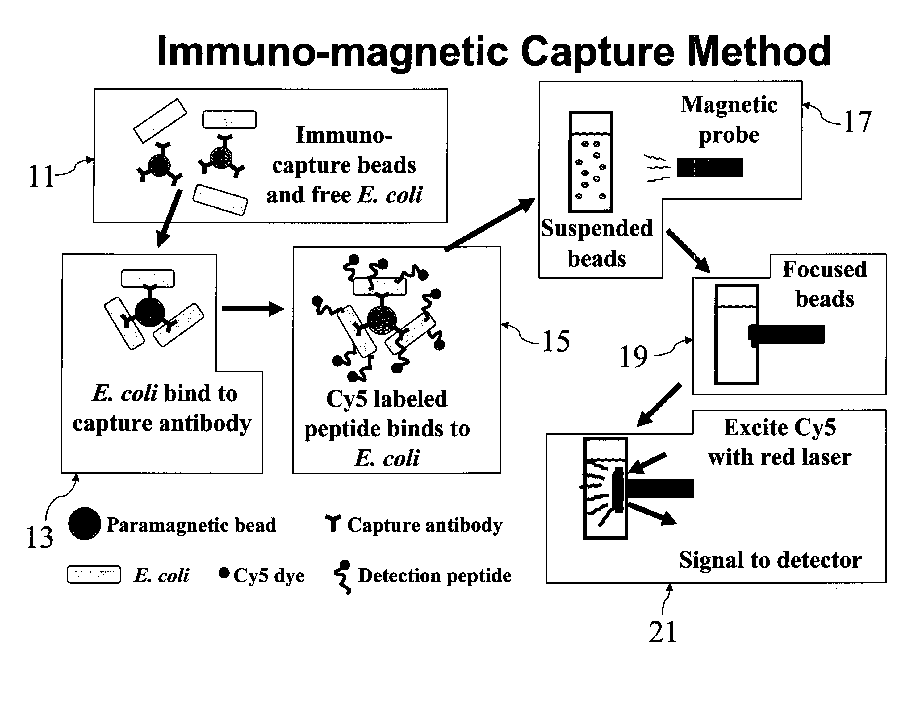
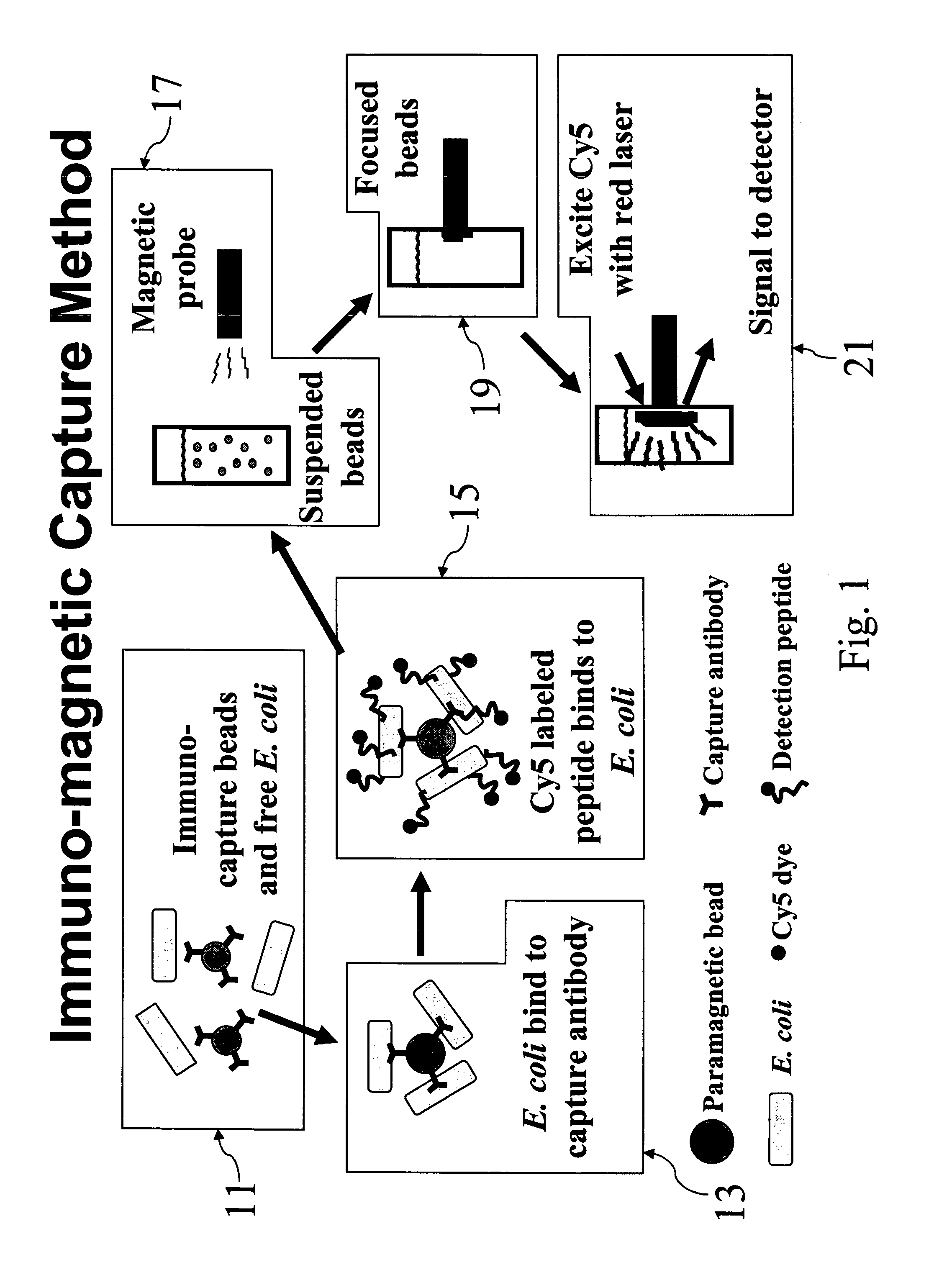
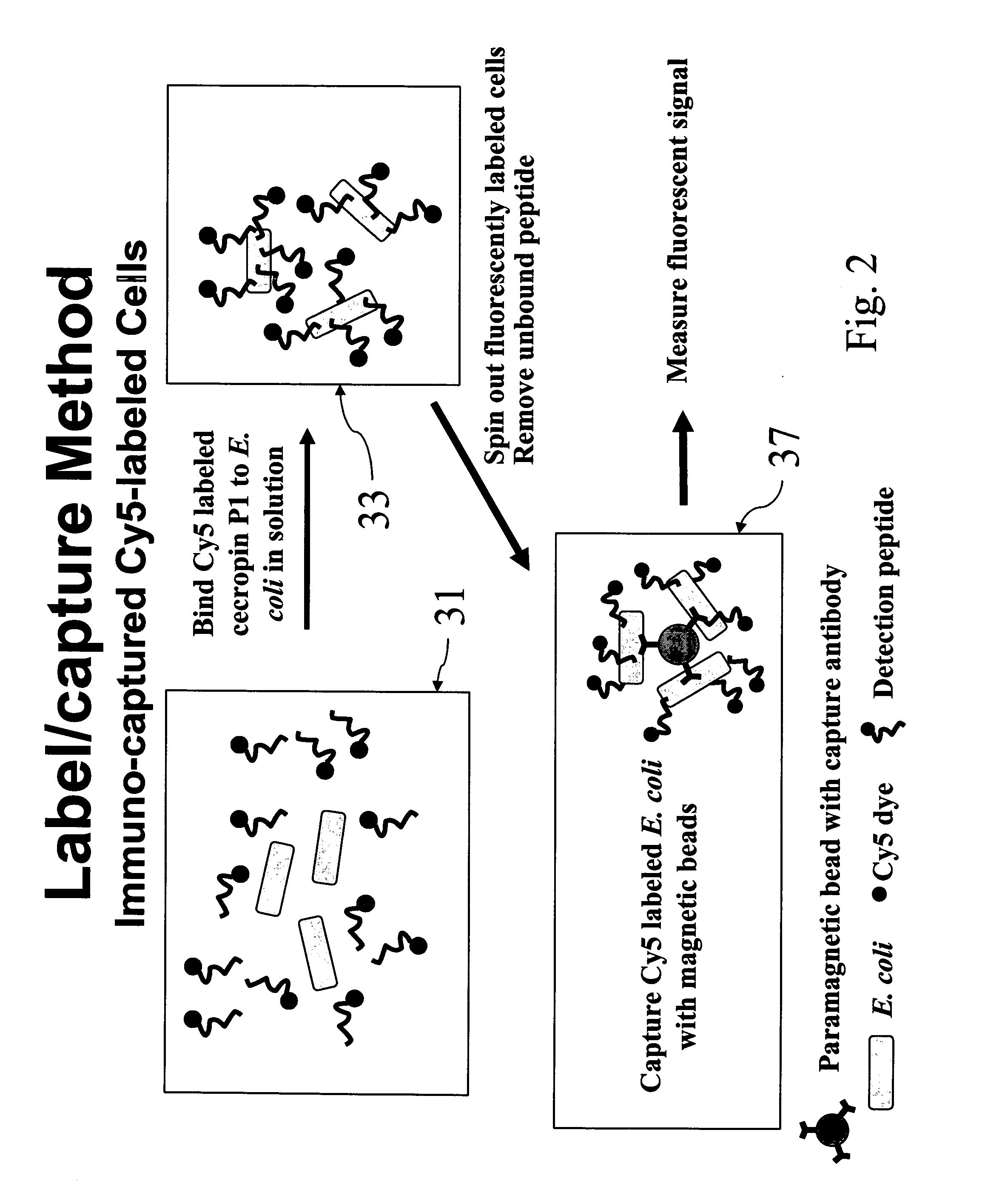
Immuno-Capture of Peptide Labeled Cells

[0049]E. coli O157:H7 cells were labeled with Cy5CP1_c in solution as described above. After removal of unbound excess peptide, 1 ml labeled cells was added to 20 μl anti-E. Coli O157:H7 Dyna-beads paramagnetic beads (Dynal Biotech, Browndeer, Wis.) and incubated 30 minutes by rotary mixing. Beads were collected after 2 minutes using a magnetic particle concentrator and washed three times with 1 ml PBST (0.05%). Samples were analyzed on Storm 860 and analyzed by TotaLab software to measure fluorescent signal.

[0050] Referring now to FIG. 6, it can be seen that, by following the above procedure, bacteria in concentrations as low as 103 CFU / ml, the lowest non-zero concentration tested, were capable of being detected.


PUM


Abstract

Labeled antimicrobial peptides and method of using the same to detect a microorganism of interest. In one embodiment, the method involves adding immuno-capture beads to a sample, the immuno-capture beads including capture antibodies coupled to a paramagnetic bead, the capture antibodies being specific for the type of microorganism of interest. After mixing, the target microorganism binds to the capture antibodies. Next, the beads are collected by positioning a magnet close to the sample, and the unbound material is removed from the sample. Then, a solution containing fluorescently-labeled antimicrobial peptide is added to the sample, the labeled peptide binding in great numbers to the immuno-captured microorganism. After removing unbound peptide, the beads are suspended in solution and a magnetic probe is used to collect the beads in a small volume. With the beads thus drawn together, the solution is excited with a laser. Such excitation causes the label to fluoresce, which fluorescence is then detected.

Description

STATEMENT OF FEDERALLY SPONSORED RESEARCH OR DEVELOPMENT [0001] The invention described herein may be manufactured and used by the U.S. Government for Governmental purposes without the payment of any royalty thereon.BACKGROUND OF THE INVENTION [0002] The present invention relates generally to techniques for detecting microorganisms and relates more particularly to a novel technique for detecting microorganisms. [0003] Microorganisms, such as bacteria, viruses, fungi and protozoa, are commonplace in the environment. Although many such microorganisms are innocuous to humans, certain species of microorganisms are pathogenic and pose a serious health risk to people. Exposure to such pathogenic microorganisms may be inadvertent, such as in the case of poorly handled or poorly prepared foods containing Salmonella, Listeria, E. coli O157:H7 or the like, or may be deliberate, such as in the case of biological weapons armed with spores of anthrax or the like. As can readily be appreciated, i...

Claims


Application Information

Patent Timeline
IPC IPC(8): G01N33/53
CPCG01N33/54326C12Q1/04
Inventor ARCIDIACONO, STEVEN MICHAELMELLO, CHARLENE MARIEPIVARNIK, PHILIP E.SENECAL, ANDRE
Owner ARCIDIACONO STEVEN MICHAEL
Why Eureka
Try Eureka