Overlay image contrast enhancement

a contrast enhancement and overlay technology, applied in image enhancement, instruments, applications, etc., can solve the problems of image dissimilarity and difficult comparison

Active Publication Date: 2007-10-11
FUJIFILM SONOSITE
View PDF18 Cites 92 Cited by
  • Summary
  • Abstract
  • Description
  • Claims
  • Application Information

AI Technical Summary

Benefits of technology

[0003] A method of creating an image difference overlay, or enhanced medical image, comprises identifying a set of reference images, or frames,, of a subject and identifying a set of data images, or frames, of the subject. A set of images, also referred to as a “loop” or “cine clip” can comprise one or more images acquired sequentially. The set of data images can be identified after an event, such as the administration of contrast agent to the subject, or after removal of a contrast agent already in the subject. A single reference image can be compared to one or more data set images and the reference set frame is associated with a data set image which closely resembles the reference set image. Each of the associated images can then be processed and used to create an enhanced medical image, also referred to throughout as an image difference overlay, or contrast overlay. In one non-limiting example, the contrast agent used is a microbubble contrast agent, and the image sets are acquired using ultrasound.

Problems solved by technology

For in vivo imaging, however, respiration, cardiac, and non-specific motion of patients and small animals causes post-contrast injection images to be dissimilar when compared to a pre-single injection image making the comparison difficult.

Method used


Image

  • Overlay image contrast enhancement
  • Overlay image contrast enhancement
  • Overlay image contrast enhancement

Examples


example 1

[0147] Embodiments of the invention comprise a method for the imaging of contrast agents for high-resolution, high-frequency ultrasound imaging of animal subjects including rodent models.

[0148] As described above, contrast agents can flow in the blood stream of the animal and can be deposited at any site that the blood flows. Contrast agents can also be prepared in such a way that they can attach themselves to specific markers in the animal. This “targeted” contrast agent technique uses ligands and antibodies to latch onto specific targets. Contrast agents can also be used for image enhancement to allow a user to easily distinguish small vessels in the organ. In one aspect, perfusion imaging is a way of injecting contrast agent into the animal, allowing the agent to circulate and then the agent can be destroyed using a destruction event. The time taken for the re-perfusion is meaningful to the researcher in quantifying perfusion. This image processing method is not specific to the ...

example 2

Materials and Methods

[0171] Inflammation in the mouse hindleg was induced by a one or three hour treatment with TNF-alpha injected subcutaneously into the hindpaw. Inflammation in the kidney was induced by ischemia-reperfusion injury. The left kidney was exposed and the renal artery was clamped for 32 minutes, followed by 2 hours of re-perfusion as described in Singbartl K, Green S A, Ley K. (2000) “Blocking P-selectin protects from ischemia / reperfusion induced acute renal failure”FASEB J. 14: 48-54. The wound was closed in layers and, covered with a saline-soaked gauze.

[0172] TargestarB microbubbles from Targeson (Charlottesville, Va.) were targeted to P-selectin by conjugating an anti-P-selectin monoclonal antibody to the surface of the MB per manufacturer's instructions. The MB were diluted to a concentration of 107 or 108 MB in 100 μL of phosphate-buffered saline, and injected as a bolus through a cannula placed in the left jugular vein.

[0173] Ultrasound imaging was performe...

example 3

Image Enhancement by Bolus Injection of Vascularized Tissue

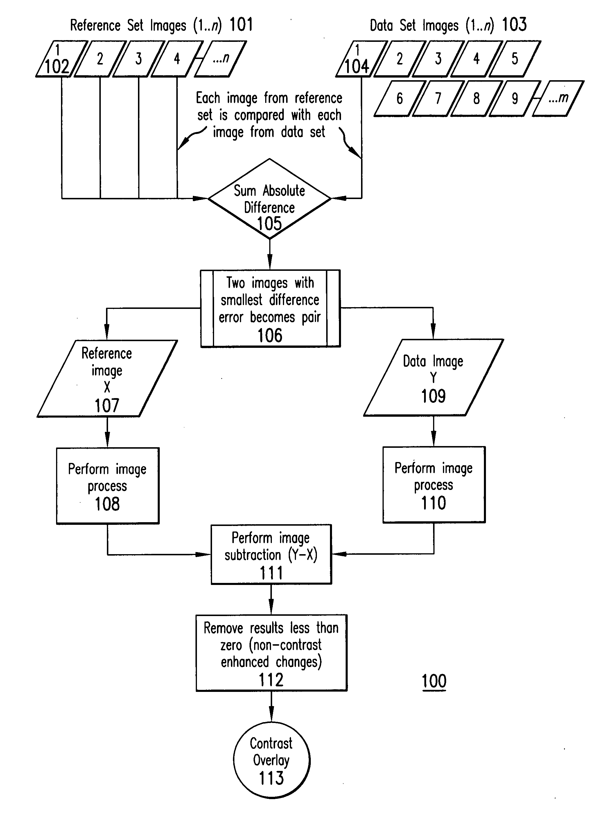
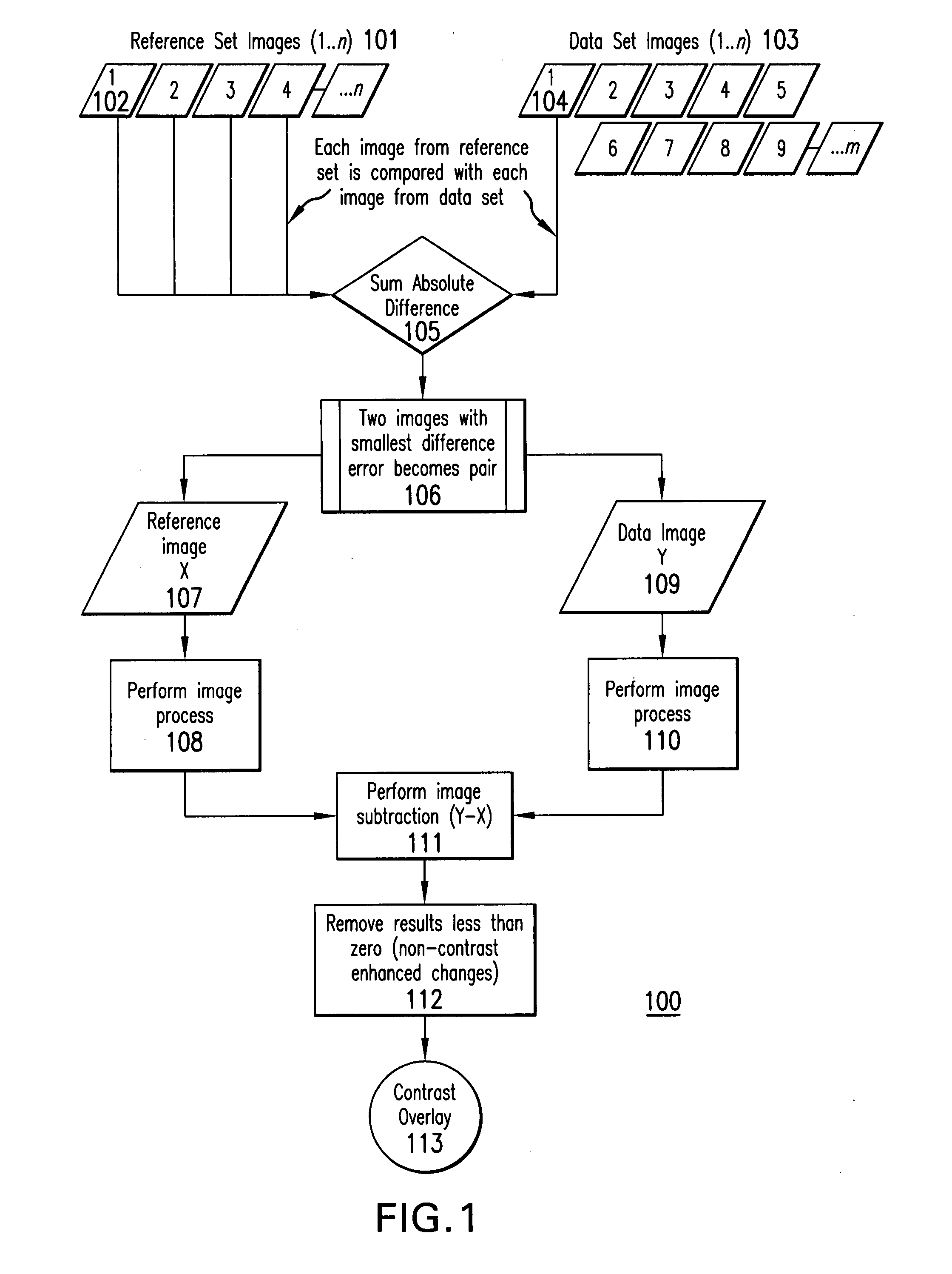
[0181] A Vevo 770® (VisualSonics Inc. Toronto, Calif.) high-resolution imaging system was used to acquire data set images and reference set images. The Vevo® system included software for comparing, matching and subtracting data set images and reference set images. For example, Vevo® Contrast Mode software was used.

[0182] The Default Cine Loop Size for Contrast Mode was set at 800, which defined the size of the Contrast Mode cine loop. A high frequency ultrasound imaging probe was connected to the to the Vevo® 770 imaging system. In this example, an RMV™-706 probe (VisualSonics Inc., Toronto, Calif.) was used.

[0183] The subject was positioned for scanning, and while scanning, the Field of View was adjusted to be 9×9 mm. Images were acquired at a frame rate between 10 and 20 Hz and the transmit power was set to 50%.

[0184] Contrast agent was prepared according to the instructions provided in VisualSonics, Inc. (Toronto, Ca...


PUM


Abstract

A method of creating an image difference overlay comprises identifying a loop of reference images of a subject and identifying a loop of data images of the subject. The loop of image data can be identified after an event, such as the administration of contrast agent to the subject. A reference loop image frame is compared to one or more data loop image frames and the reference loop frame is associated with a data loop image frame which closely resembles the data loop image frame. Each of the associated frames can then be processed and used to create an image difference overlay frame.

Description

CROSS REFERENCE TO RELATED APPLICATIONS [0001] This application claims the benefit of U.S. Provisional Patent Application No. 60 / 735,399 filed Nov. 11, 2005, which is fully incorporated herein and made apart hereof.BACKGROUND [0002] Sufficient contrast differences between tissues in acquired images of patients and small animals are important for medical diagnosis and biomedical research. To increase the signal intensity difference between tissues in such acquired images contrast agents specific for that imaging modality are often used. A comparison between a single post-contrast agent administrated image with a single pre-contrast agent image, as selected by the operator, can be made to identify specific tissue volumes that have undergone contrast enhancement. Identification of which tissues undergo contrast enhancement, and how much enhancement, is an important indicator for many biomedical research and diagnostic applications. For in vivo imaging, however, respiration, cardiac, an...

Claims


Application Information

Patent Timeline
Patent Type & Authority Applications(United States)
IPC IPC(8): A61B5/05
CPCA61B6/00A61B8/481G06T5/009G06T5/50G06T2207/10132A61B8/483G06T2207/30048G06T2207/30084A61B6/507A61B6/508G06T2207/30004
Inventor WHITE, CHRISTOPHER A.HIRSON, DESMONDPOON, STANLEYMEHI, JAMES
Owner FUJIFILM SONOSITE
Why Eureka
Try Eureka