Device and method for improving heart valve function

a heart valve and function technology, applied in the field of devices and methods for improving the function of heart valves, can solve the problems of heart valves that may not close properly, leak backwards, and serious impairment of heart valve function, and achieve the effect of improving the function of defective heart valves

Active Publication Date: 2007-11-22
EDWARDS LIFESCIENCES CORP +1
View PDF11 Cites 373 Cited by
  • Summary
  • Abstract
  • Description
  • Claims
  • Application Information

AI Technical Summary

Benefits of technology

[0009]Various embodiments of the present invention provide improved devices and methods for improving the function of a defective heart valve. Preferred embodiments are configured to be surgically implanted in a heart using a minimally invasive procedure wherein extracorporeal circulation is not required.
[0010]In one preferred embodiment, a blood flow controlling device is provided for improving valve function. The blood flow controlling device comprises a valve-blocking portion (such as an expandable valve portion) and an anchor portion. The valve-blocking portion is configured to be disposed between anterior and posterior leaflets of a mitral valve. In an embodiment of a valve-blocking portion, an expandable valve portion expands during ventricular systole to fill and conform to the gap between the mitral valve leaflets, thereby preventing regurgitation. In one variation, the expandable valve portion comprises a canopy or flap portion and a plurality of tethers. In one preferred embodiment, the blood flow controlling device is configured to be delivered into the heart in a two-stage procedure. More specifically, the anchor portion of the device is initially implanted within the heart and the valve portion is then coupled to the anchor portion after the anchor portion has become sufficiently embedded in the muscular wall of the heart.
[0011]In another preferred embodiment, a blood flow controlling device comprises an anchor portion and an expandable valve portion configured to be disposed between anterior and posterior leaflets of a mitral valve. In this embodiment, the expandable valve portion is provided with a plurality of apertures or other openings for allowing some blood to flow backward across the expandable portion. This embodiment advantageously reduces the formation of thrombi by eliminating the pooling of blood within the expandable member without substantially reducing the effectiveness of the device.
[0012]Preferred embodiments of the present invention include surgical devices and methods of treating heart valves wherein a blood flow controlling device is delivered into the heart through a small incision in the chest, such as, for example, in the sternum or between the ribs. Preferred embodiments of the surgical methods described herein do not require extracorporeal circulation. For example, in one preferred embodiment, a delivery catheter (or similar delivery device) is inserted through an incision in the chest wall and then through the cardiac tissue into a chamber of the patient's beating heart. The delivery catheter allows a blood flow controlling device to be delivered into the heart in a collapsed configuration and then expanded within the heart for treating a defective heart valve. Because the preferred delivery methods do not require extracorporeal circulation, complications are greatly reduced as compared with traditional open-heart surgery.

Problems solved by technology

The function of the heart may be seriously impaired if any of the heart valves are not functioning properly.
The heart valves may lose their ability to close properly due to e.g. dilation of an annulus around the valve or a leaflet being flaccid causing a prolapsing leaflet.
The inability of the heart valve to close properly can cause a leak backwards (i.e., from the outflow to the inflow side), commonly referred to as regurgitation, through the valve.
Heart valve regurgitation may seriously impair the function of the heart since more blood will have to be pumped through the regurgitating valve to maintain adequate circulation.
Heart valve regurgitation decreases the efficiency of the heart, reduces blood circulation, and adds stress to the heart.
In early stages, heart valve regurgitation leaves a person fatigued or short of breath.
If left unchecked, the problem can lead to congestive heart failure, arrhythmias or death.
Regurgitation through the mitral valve, sometimes referred to as mitral insufficiency or incompetence, is a particularly common problem that affects the health of millions of adults.
Changes in the geometric configurations of the left ventricle, papillary muscles and mitral annulus may adversely affect the function of the mitral valve and lead to regurgitation.
However, open-heart surgery is highly invasive and is therefore not an option for many patients.
However, due to variations in the type of mitral valve disease and the location of the coronary sinus relative to the mitral valve annulus, this approach may not be suitable for all patients.
Although this “edge-to-edge” approach has shown great promise, similar to the coronary sinus implant, it has been found that this approach may not be suitable for all patients.
However, there are conditions in which percutaneous procedures may not be appropriate.

Method used


Image

  • Device and method for improving heart valve function
  • Device and method for improving heart valve function
  • Device and method for improving heart valve function

Examples


Embodiment Construction

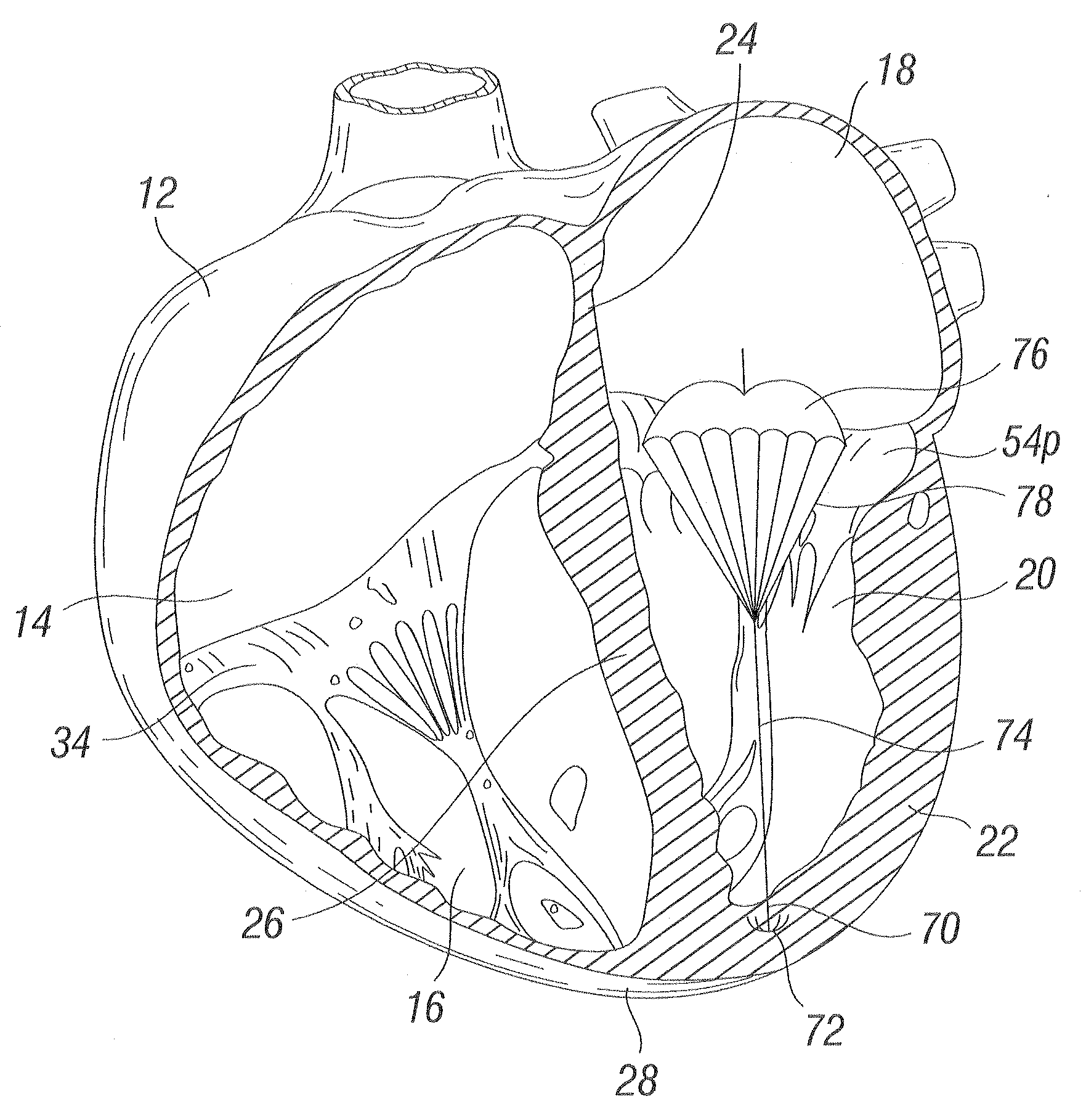
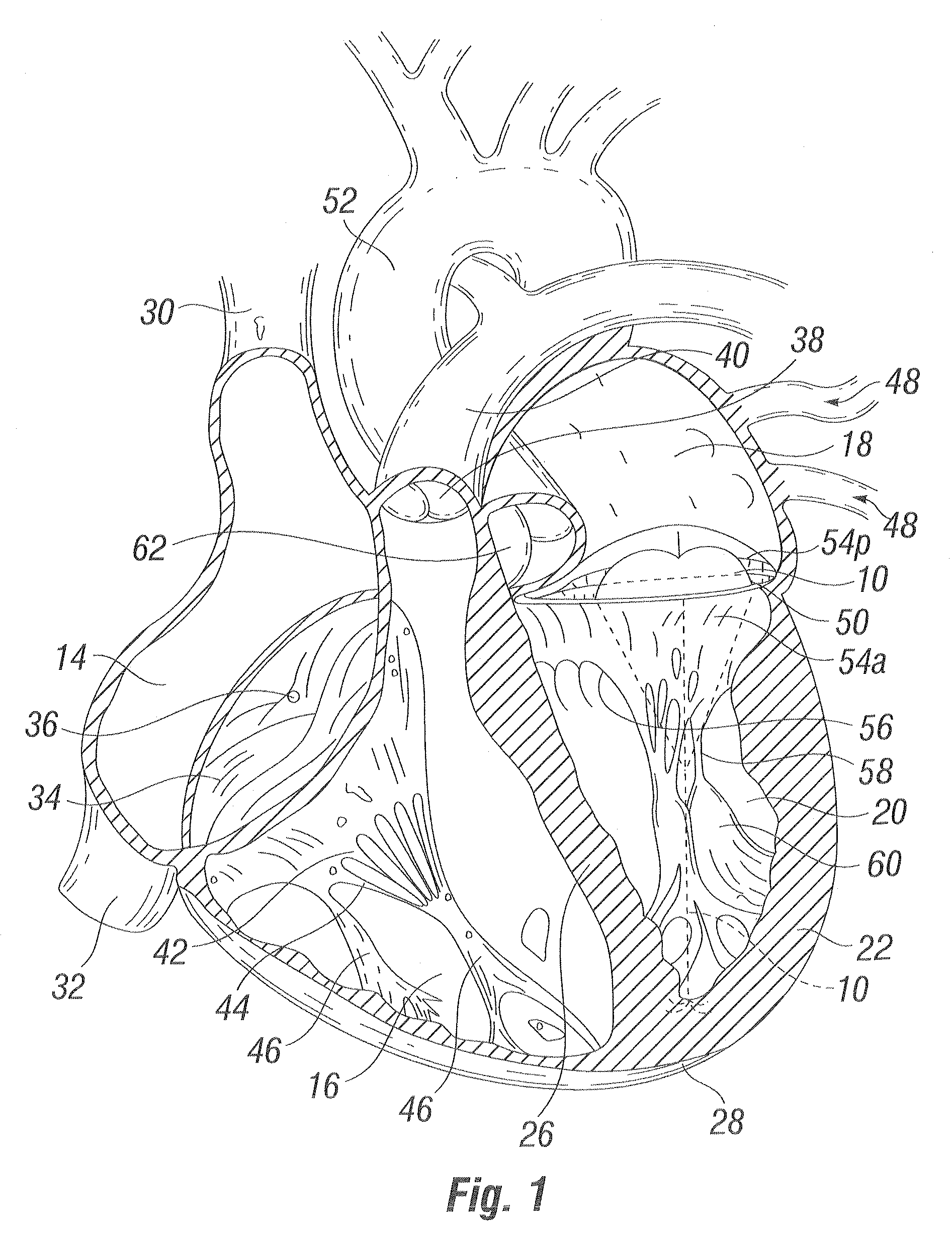
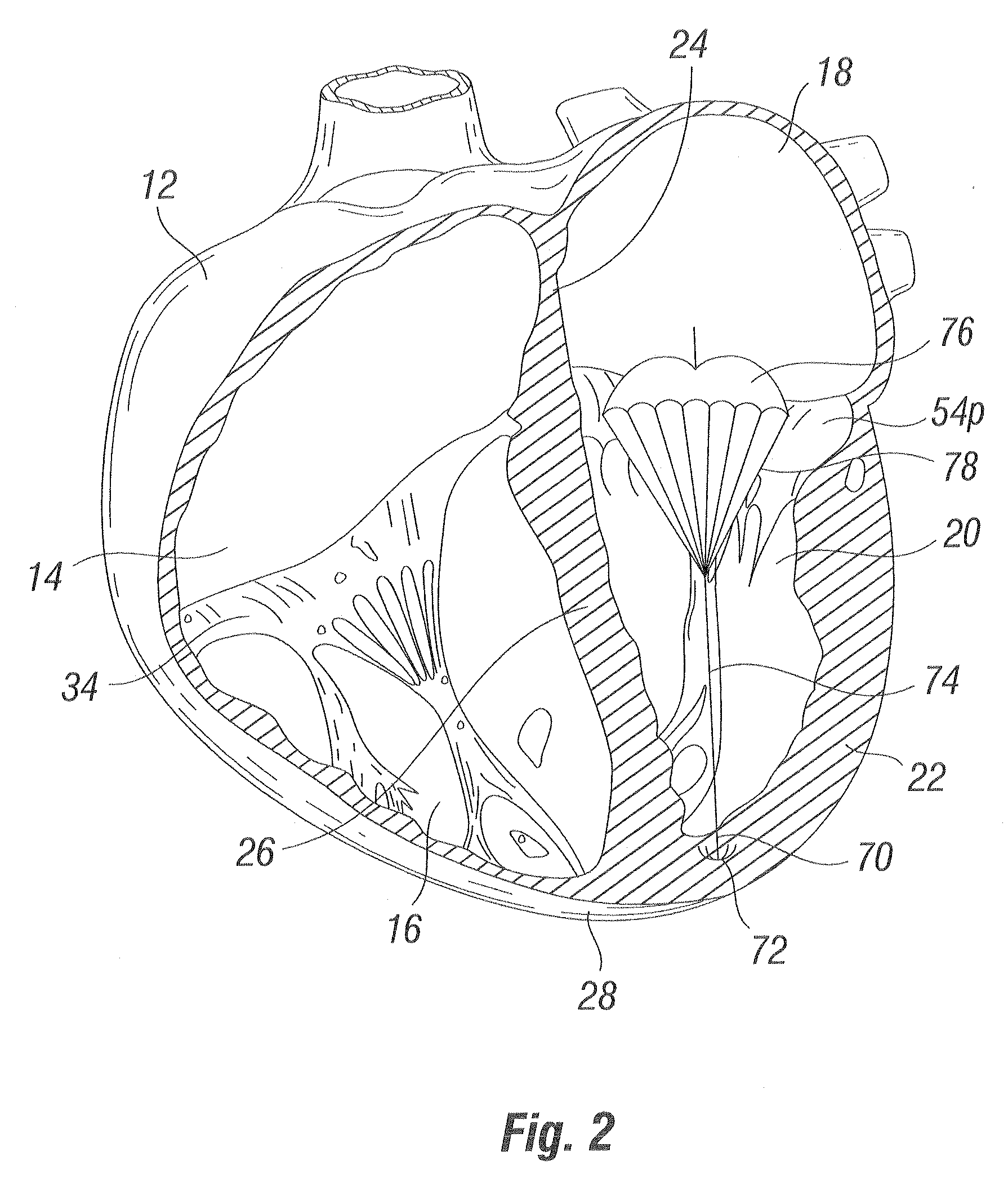
[0044]With reference to FIG. 1, a device 10 according to the invention is depicted in a heart 12. The heart 12 has four chambers, known as the right atrium 14, right ventricle 16, left atrium 18, and left ventricle 20. In the particular embodiment depicted, the device 10 is deployed in the left ventricle 20.

[0045]The general anatomy of the heart 12, which is depicted as viewed from the front of a patient, will be described for background purposes. The heart 12 has a muscular outer wall 22, with an interatrial septum 24 (not visible in FIG. 1, but visible in FIG. 3b, etc.) dividing the right atrium 14 and left atrium 18, and a muscular interventricular septum 26 dividing the right ventricle 16 and left ventricle 20. At the bottom end of the heart 12 is the apex 28.

[0046]Blood flows through the superior vena cava 30 and the inferior vena cava 32 into the right atrium 14 of the heart 12. The tricuspid valve 34, which has three leaflets 36, controls blood flow between the right atrium 1...


PUM


Abstract

The invention is device and method for reducing regurgitation through a mitral valve. The device and method is directed to an anchor portion for engagement with the heart wall and an expandable valve portion configured for deployment between the mitral valve leaflets. The valve portion is expandable for preventing regurgitation through the mitral valve while allowing blood to circulate through the heart. The expandable valve portion may include apertures for reducing the stagnation of blood. In a preferred configuration, the device is configured to be delivered in two-stages wherein an anchor portion is first delivered and the valve structure is then coupled to the anchor portion. In yet another embodiment, the present invention provides a method of forming an anchor portion wherein a disposable jig is used to mold the anchor portion into a three-dimensional shape for conforming to a heart chamber.

Description

RELATED APPLICATIONS[0001]The present application claims priority to Provisional Application No. 60 / 801,446 entitled “Device and Method for Improving Heart Valve Function,” filed May 18, 2006, and to Provisional Application No. 60 / 810,085, entitled “Prosthetic Insert for Improving Heart Valve Function,” filed Jun. 1, 2006, each of which is hereby incorporated by reference in its entirety.BACKGROUND OF THE INVENTION[0002]The present invention relates generally to devices and methods for improving the function of a defective heart valve. The devices and methods disclosed herein are particularly well adapted for implantation in a patient's heart for reducing regurgitation through a heart valve.[0003]The function of the heart may be seriously impaired if any of the heart valves are not functioning properly. The heart valves may lose their ability to close properly due to e.g. dilation of an annulus around the valve or a leaflet being flaccid causing a prolapsing leaflet. The leaflets ma...

Claims


Application Information

Patent Timeline
Patent Type & Authority Applications(United States)
IPC IPC(8): A61F2/24
CPCA61B17/0401A61B2017/00243A61B2017/0437A61F2240/002A61B2017/0453A61F2/246A61F2/2466A61B2017/0445A61F2220/0016A61F2230/0093
Inventor SOLEM, JAN OTTOALON, DAVID
Owner EDWARDS LIFESCIENCES CORP
Why Eureka
Try Eureka