Eureka AIR delivers breakthrough ideas for toughest innovation challenges, trusted by R&D personnel around the world.

Bioreactor for organ reconstruction and augmentation

a bioreactor and organ technology, applied in the field of tissue reconstruction, repair, augmentation and replacement, can solve the problems of deterioration of the urinary bladder in patients, deterioration of the bladder, and inability to achieve the same success in many other organ fields

Inactive Publication Date: 2007-11-29
TENGION
View PDF9 Cites 31 Cited by
  • Summary
  • Abstract
  • Description
  • Claims
  • Application Information

AI Technical Summary

Benefits of technology

[0017] Suitable antibiotics for use in the constructs and methods described herein include any antibiotic that does not inhibit or impede cell growth. For example, the antibiotic does not inhibit the cell growth of first cell population such as a smooth muscle cell population. Alternatively or in addition, the antibiotic does not inhibit the cell growth of a second cell population such as a urothelial cell population. Preferably, the antibiotic is selected from gentamicin and vancomycin, and more preferably, the antibiotic is gentamicin.

Problems solved by technology

Equal success has not been achieved in many other organ fields particularly in the field of bladder reconstruction.
The urinary bladder is subject to numerous maladies and injuries which cause deterioration of the urinary bladder in patients.
Further, bladder deterioration may also occur as a result of trauma such as, for example, car accidents and sports injury.
Lanza, R., Langer, R., and Chick, W, ACM Press, Colorado (1996) and references cited therein), many materials have proven to be unsatisfactory for use in bladder reconstruction.
Other attempts have usually failed due to either mechanical, structural, functional, or biocompatibility problems.
Permanent synthetic materials have been associated with mechanical failure and calculus formation.
De-epithelized bowel segments demonstrated an adequate urothelial covering for use in bladder reconstruction, but difficulties remain with either mucosal regrowth, segment fibrosis, or both.
Other problems have been reported with the use of certain gastrointestinal segments for bladder surgery including stone formation, increased mucus production, neoplasia, infection, metabolic disturbances, long term contracture and resorption.
These attempts with natural or synthetic materials have shown that bladder tissue, with its specific muscular elastic properties and urothelial impermeability functions, cannot be easily replaced.
However, auto-augmentation has been associated with disappointing long-term results and ureterocystoplasty is limited to cases in which a dilated ureter is already present.
A system of progressive dilation for ureters and bladders has been proposed, however, this has not yet been attempted clinically.
However, graft shrinkage and re-epithelialization of initially de-epithelialized bowel segments has been a recurring problem.
One significant limitation besetting bladder reconstruction is directly related to the availability of donor tissue.
The limited availability of bladder tissue prohibits the frequent routine reconstruction of bladder using normal bladder tissue.
For example, in a patient suffering from bladder cancer, the remaining bladder tissue may be contaminated with metastasis.

Method used


Image

  • Bioreactor for organ reconstruction and augmentation
  • Bioreactor for organ reconstruction and augmentation
  • Bioreactor for organ reconstruction and augmentation

Examples


example 1

Creation of Bladder-Shaped Polymers

[0088] The neo-organ constructs described herein are presented using neo-bladder constructs as an example. While reference is made here to neo-bladder constructs, it will be understood that the methods and materials described herein are useful for creating a variety of neo-organs and neo-vessel augmentation constructs, including, for example, neo-kidney augmentation constructs.

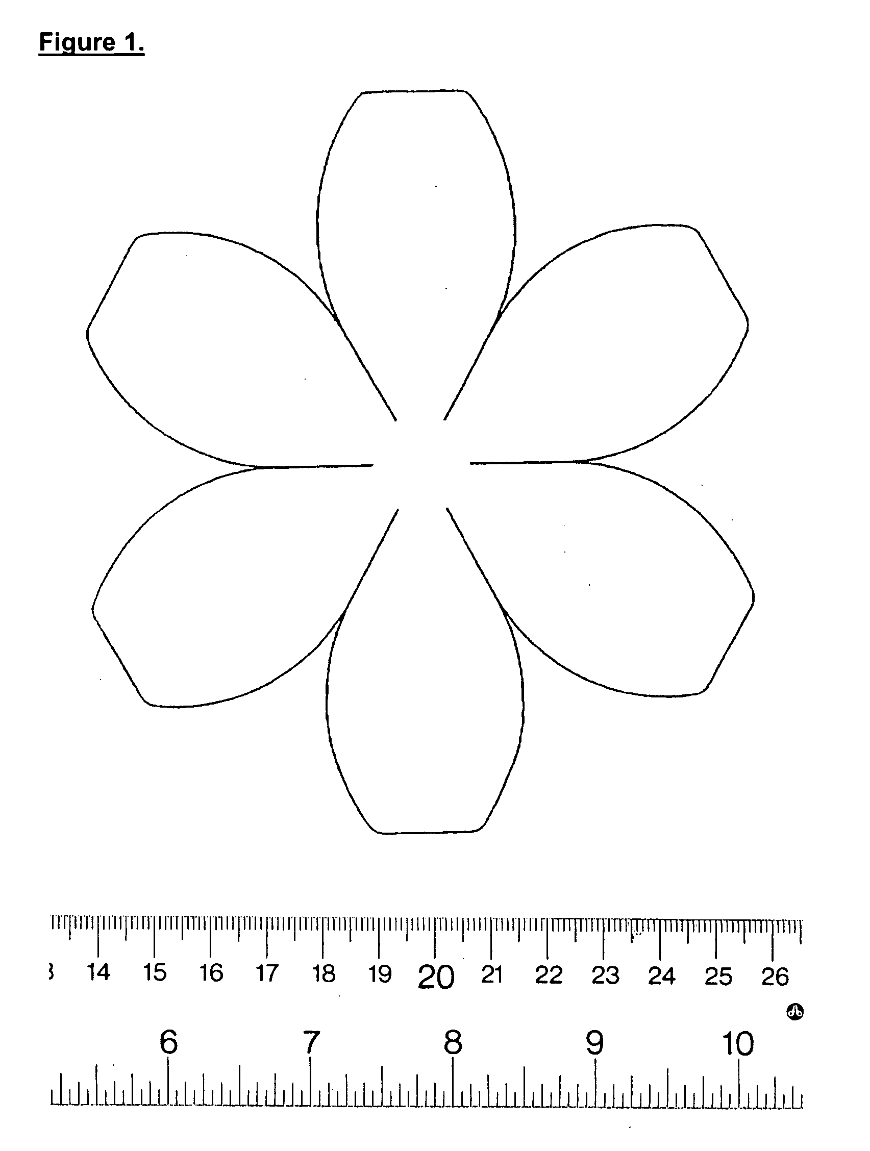
[0089] Manufacture of the neobladder matrix or scaffold. The neobladder matrices or scaffolds are constructed using polyglycolide-polyglycolic acid (PGA) non-woven felt (BMS or Concordia 2.5 mm thick, 58 mg / cc or 99 mg / ml). The PGA non-woven felt is cut using a neo-bladder pattern as a template. The neo-bladder pattern is for example, spherical, quasi-spherical, hemispherical, or quasi-hemispherical in shape, such that bladder repair, or augmentation procedures require one hemispherical or quasi-hemispherical neo-bladder construct, while total bladder reconstruction may req...

example 2

Cell Harvest and Culture

[0098] Biopsy procurement. In contrast to previous studies in which a 1×1 cm biopsy was taken from the side of the bladder using a scalpel to dissociate the tissue, the tissue samples used to create the neobladder constructs described in this Example were obtained by taking a 1×1 cm biopsy from the bladder apex, using a staple method. Previous biopsy procedures, such as the methods described in U.S. Pat. No. 6,576,019 by Atala et al., removed tissue from the vesical dome in general. In contrast, the biopsy procedures used herein remove tissue from a specific portion of the vesical dome, the bladder apex. Removing tissue from the bladder apex has been shown to provide a greater yield of useful cells. Useful cells refers to viable cells that are capable of expansion and seeding on the neobladder scaffolds described herein.

[0099] The staple method used herein involves making a loop in the apex of the bladder, stapling the base of the loop, and excising the loo...

example 3

Cell Seeding on Polymeric Matrix or Scaffold

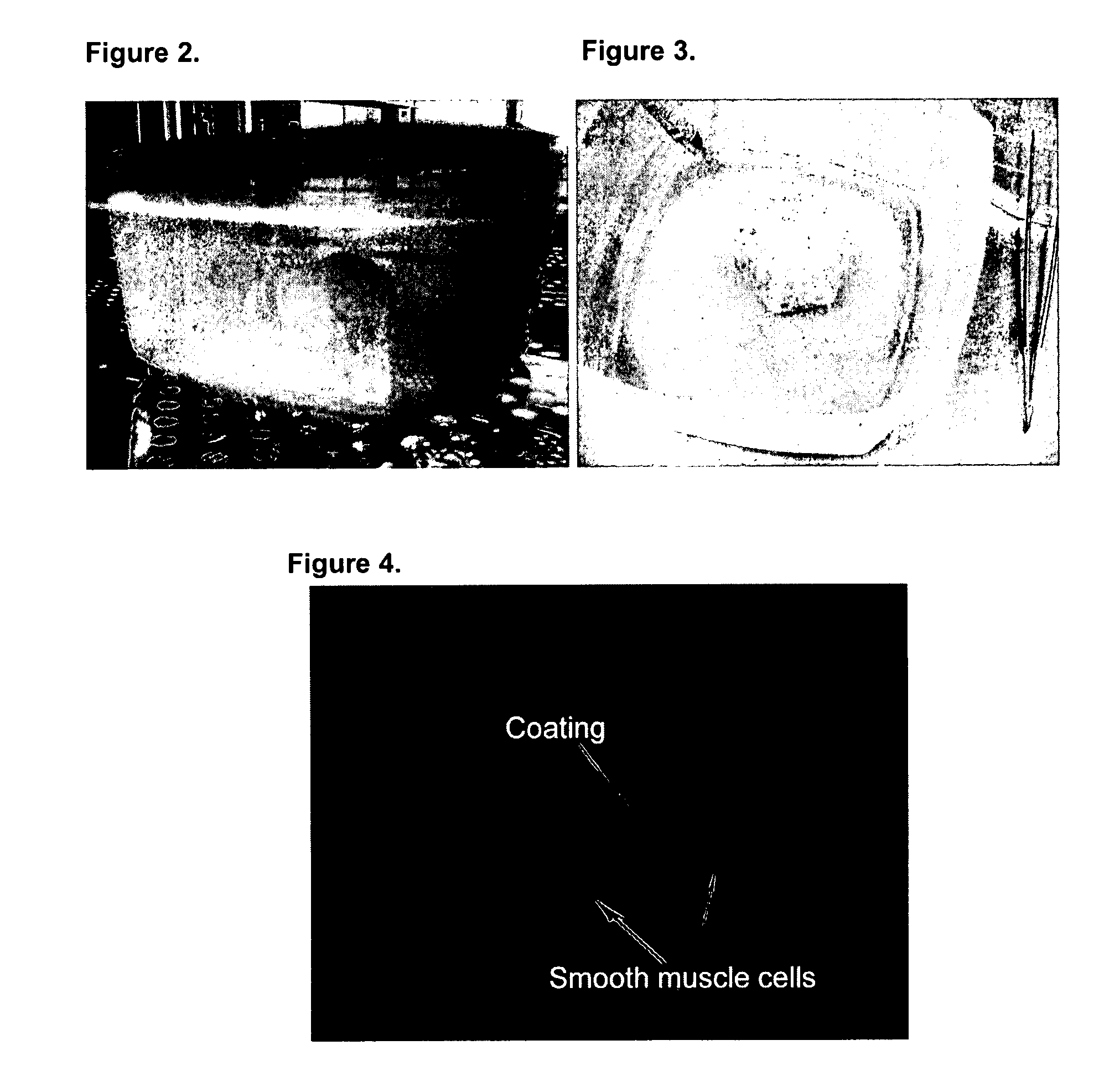
[0100] Neo-bladder matrix or scaffold seeding with SMC. After the smooth muscle cells (SMC) are harvested and expanded as described above in Example 2, the cell pellet is resuspended in 6 ml of SMC growth medium. The matrix or scaffold is removed from the pre-wetting container using forceps and is placed in an empty sterile cell-seeding container (see FIGS. 2 and 3, originally designed and manufactured by Tengion Inc.). The cells are distributed evenly on the outside surface of the scaffold.

[0101] Bright field microscopy (FIG. 4) confirmed that SMC do indeed take up residence within scaffolds seeded using the procedures described above.

[0102] Neobladder scaffold seeding with Urothelial Cells. After the urothelial cells (UC) are harvested and expanded, the cell pellet is resuspended in 6 ml of Construct Growth Medium 1:1 mixture of DMEM / 10% FBS:KSFM). The cells are distributed evenly on the inside surface of the scaffold (FIG. 5).


PUM

PropertyMeasurementUnit
pressuresaaaaaaaaaa
diameteraaaaaaaaaa
pore sizeaaaaaaaaaa

Abstract

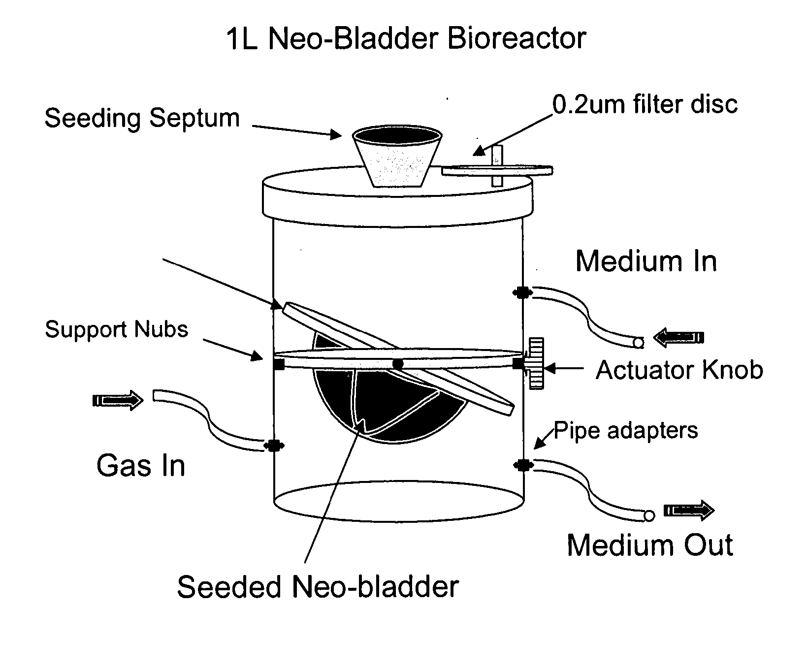
Bioreactors are used in neo-organ production to allow for an appropriate environment for the maintenance of healthy culturing conditions from pre-wetting to shipment of the neo-organ. The closed system “all-in-one bioreactor” is designed to allow for minimal exposure of the scaffold to the open air in order to maintain sterility. The design allows for the same container to be utilized for sterilization, pre-wetting, cell seeding, medium exchange, and shipment. The “all-in-one” bioreactor also remains completely closed after the urothelial cell seeding step to the implantation at the clinical site. This allows for sufficient time for release testing to occur so the neo-organ can be implanted into the patient.

Description

RELATED APPLICATIONS [0001] This application claims the benefit of U.S. Provisional Application No. 60 / 772,800, filed Feb. 10, 2006, the contents of which are hereby incorporated by reference in their entirety.FIELD OF THE INVENTION [0002] The invention is directed to methods and materials for tissue reconstruction, repair, augmentation and replacement, and particularly to use of such treatments in patients having a defect in urogenital tissues such as the bladder. The invention is also directed to a closed system bioreactor and methods and materials for using this closed system bioreactor for neo-organ sterilization, pre-wetting, seeding, medium exchange, and shipping. BACKGROUND OF THE INVENTION [0003] The medical community has directed considerable attention and effort to the substitution of defective organs with operationally effective replacements. The replacements have ranged from completely synthetic constructs such as artificial hearts to completely natural organs from anoth...

Claims


Application Information

Patent Timeline
Patent Type & Authority Applications(United States)
IPC IPC(8): A01N1/00C12M1/12
CPCA01N1/02A01N1/0231A01N1/0247C12M37/04C12M25/14C12M27/14C12M21/08
Inventor BERTRAM, TIMOTHY A.BRUCE, ANDREWJAIN, DEEPAKLUDLOW, JOHNMCCOY, DARRELLSANGHA, NAMRATA
Owner TENGION
Why Eureka
Eureka Blog
Learn More