Ocular Tissue Modification

a technology of ocular tissue and ocular artery, which is applied in the field of tissue modification, can solve the problems of tissue rejection, adversely affecting or eliminating vision, and damage to the monolayer during graft rejection, and achieves the effect of prolonging the survival of the graft and improving the healing of the ocular artery

Inactive Publication Date: 2007-11-29
THE FLINDERS UNIV OF SOUTH AUSTRALIA
View PDF1 Cites 2 Cited by
  • Summary
  • Abstract
  • Description
  • Claims
  • Application Information

AI Technical Summary

Benefits of technology

[0012] In a further embodiment of the invention there is provided a method of improving ocular graft healing and / or prolonging graft survival comprising exposing ocular tissue to an effective concentration for transfection of an expression vector comprising a nucleotide sequence encoding for an immunoglobulin or immunoglobulin fragment specific for an agent or agents associated with ocular graft healing and / or rejection, for a period sufficient to allow transfection such that cells of said ocular tissue will express the immunoglobulin or immunoglobulin fragment, harvesting said tissue from a donor and transplanting the ocular tissue to an eye of a recipient.

Problems solved by technology

There are numerous diseases and disorders that can affect corneal and other ocular tissue and which can, as a result, adversely affect or eliminate vision.
Although corneal transplant operations enjoy a high rate of success there are nonetheless some problems that can occur as rejection of the replacement cornea and ocular fibrosis or scarring.
Non-compliance by the patient with the prescribed dosing regime of immunomodulating agents may give rise to tissue rejection.
Human (but not rodent) corneal endothelium is essentially amitotic (8), so that damage to the monolayer during graft rejection cannot be repaired.

Method used


Image

  • Ocular Tissue Modification
  • Ocular Tissue Modification
  • Ocular Tissue Modification

Examples


example 1

Transplantation in the Outbred Sheep of Corneal Tissue Modified by Gene Therapy Using Various Expression Vectors to Produce the Immunomodulatory Cytokine IL-10

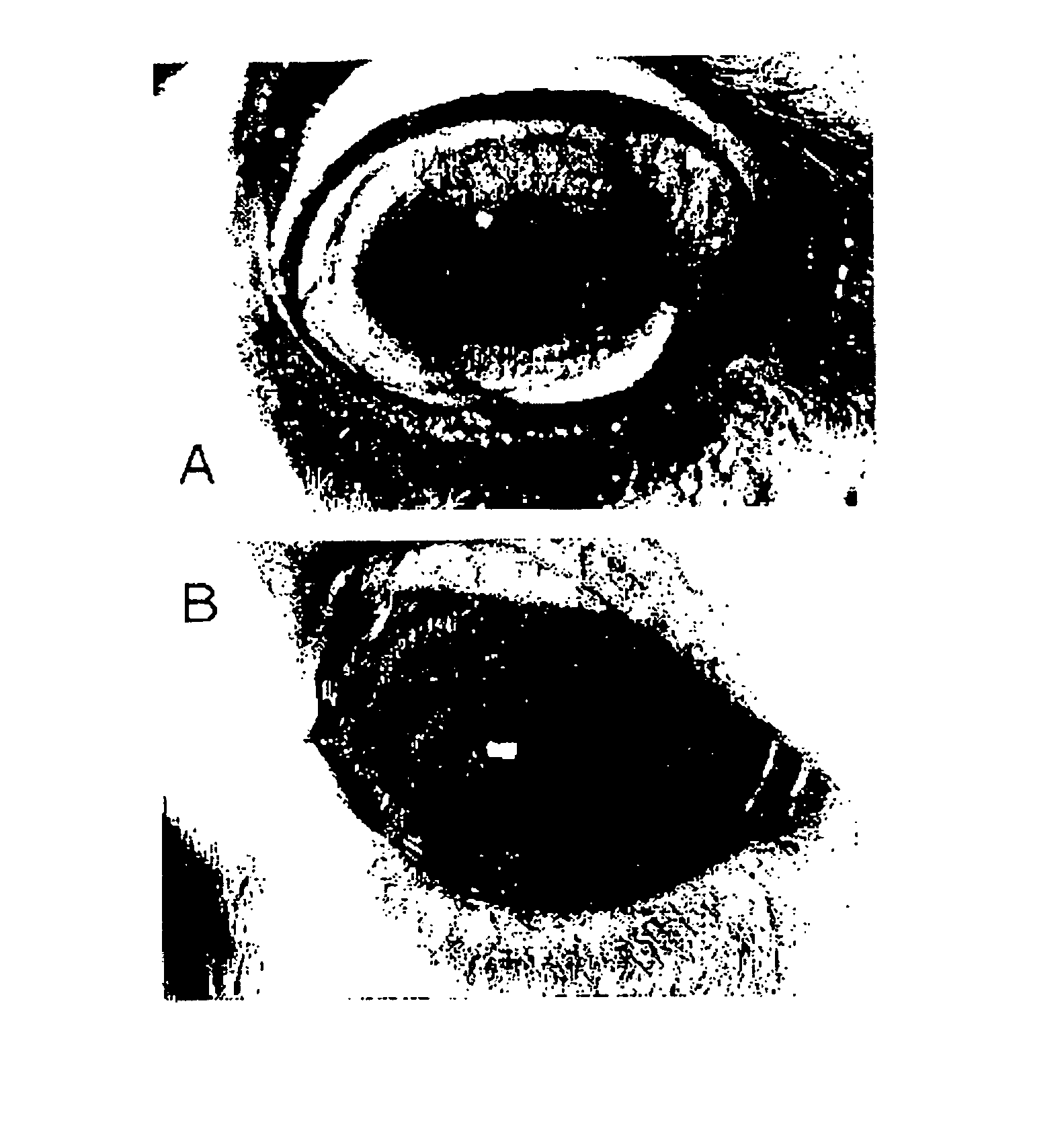
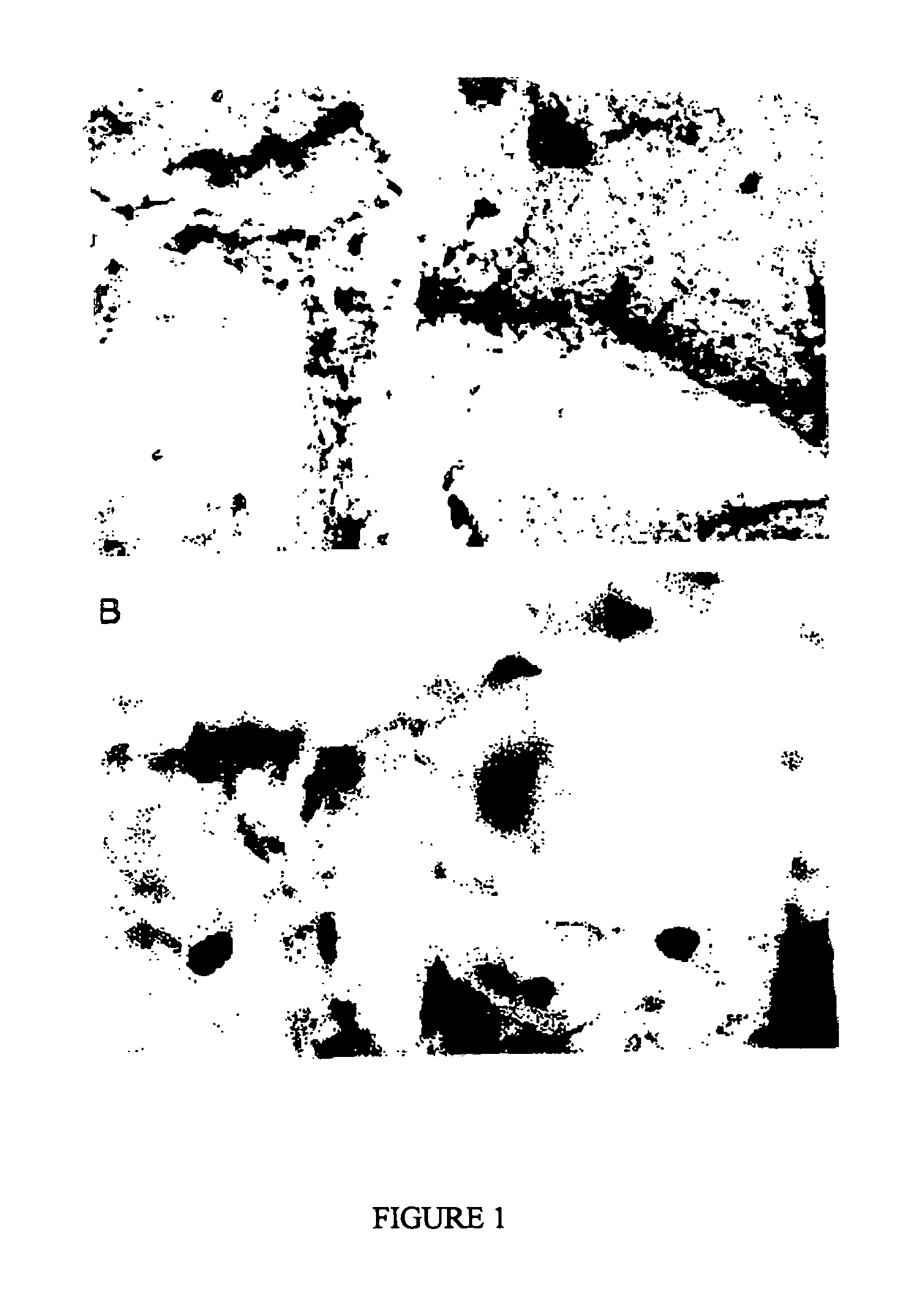
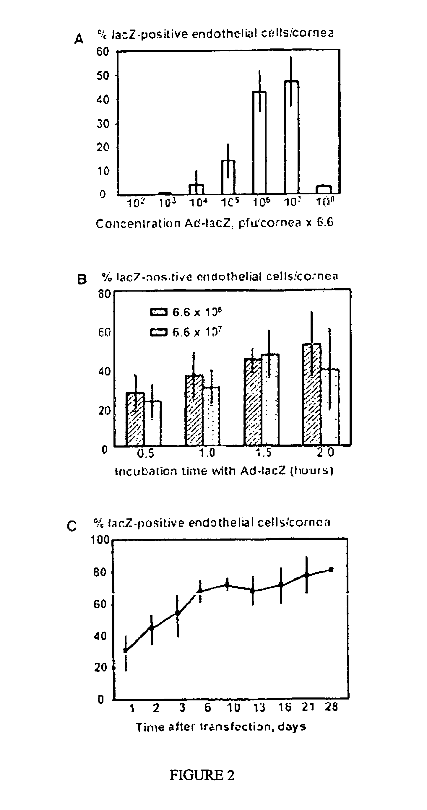
[0057] The inventors selected a model of orthotopic corneal transplantation in the outbred sheep, a relevant preclinical model in which unmodified corneal allografts undergo rejection at three weeks post-operatively in a manner that is very similar at a clinical level to human corneal graft rejection (9). Adenoviral vectors have already been shown to be capable of transferring reporter genes into corneal endothelium of various species (10-13) and the use of liposomal agents has also previously been explored (14, 15). Given that the mitotic potential of sheep corneal endothelium was unknown, the replicative capacity of this tissue was first examined, to allow an informed choice of the vector for gene therapy to be made. The immunomodulatory cytokine, IL-10, which down-regulates cell-mediated immune responses under some circums...

example 2

Prolongation of Rat Corneal Allograft Survival by Gene Transfer-Mediated Intraocular Expression of Anti-CD28

[0083] Corneal transplantation is a well-accepted treatment for sight-threatening corneal opacification, but some grafts fail. The main cause of corneal graft failure is irreversible rejection, a T cell-dependent process.

Aim

[0084] To determine whether gene transfer of cDNAs encoding engineered antibody fragments reactive with T cell costimulatory molecules to donor corneas will prolong rat corneal allograft survival.

Methods

[0085] Replication-deficient adenoviruses encoding the green fluorescent protein (eGFP) reporter gene and secretory single-chain variable-domain antibody fragments (scFv) against rat CD4 (Ad-CD4) or rat CD28 (Ad-CD28) were constructed and characterised. Adenovirus-infected corneas were examined by reverse transcription-PCR to detect expression of specific mRNAs and by fluorescence microscopy to detect expression of eGFP. Functional scFv proteins were ...


PUM

PropertyMeasurementUnit
total volumeaaaaaaaaaa
infection timeaaaaaaaaaa

Abstract

The present invention relates generally to the field of tissue modification, and in particular, but not exclusively, to methods of modifying cells of ocular and particularly corneal tissue to produce immunoglobulins or immunoglobulin fragments. The invention also relates to modified ocular cells and tissues, to expression vectors utilised in such methods, to methods of xeno- and allo-transplantion using the modified tissue and to methods of therapy of ocular diseases and disorders. In a preferred embodiment of the invention there is provided a method of modifying cells of ocular tissue to produce an immunoglobulin or immunoglobulin fragment of interest, which comprises exposing ocular tissue to an effective concentration for transfection of an expression vector comprising a nucleotide sequence encoding for the immunoglobulin or immunoglobulin fragment, for a period sufficient to allow transfection, such that cells of said ocular tissue produce the immunoglobulin or immunoglobulin fragment.

Description

FIELD OF THE INVENTION [0001] The present invention relates generally to the field of tissue modification, and in particular, but not exclusively, to methods of modifying cells of ocular and particularly corneal tissue to produce immunoglobulins or immunoglobulin fragments. The invention also relates to modified ocular cells and tissues, to expression vectors utilised in such methods, to methods of xeno- and allo-transplantion using the modified tissue and to methods of therapy of ocular diseases and disorders. BACKGROUND OF THE INVENTION [0002] There are numerous diseases and disorders that can affect corneal and other ocular tissue and which can, as a result, adversely affect or eliminate vision. For example, allergies, conjunctivitis, corneal infections, Fuchs' dystrophy (deterioration of corneal endothelial cells), varicella-zoster virus, iridocorneal endothelial syndrome, keratoconus, ocular herpes and a number of other conditions, as well as congenital ocular abnormalities, ca...

Claims


Application Information

Patent Timeline
Patent Type & Authority Applications(United States)
IPC IPC(8): C12P21/00A01K67/027A61K48/00C07K14/54C12N15/87
CPCA01K67/0271A01K2227/103A01K2267/03A61K38/208A61K48/005A61K35/30C07K14/5428C12N15/86C12N2710/10343A61K38/00A61K48/0075A61P27/02A61P43/00
Inventor JESSUP, CLAIRE F.BRERETON, HELEN M.COSTER, DOUGLAS J.WILLIAMS, KERYN A.
Owner THE FLINDERS UNIV OF SOUTH AUSTRALIA
Why Eureka
Try Eureka