
Method for determining the specific growth rate of distinct microbial populations in a non-homogeneous system

a technology of non-homogeneous system and specific growth rate, applied in the direction of microbiological testing/measurement, biochemistry apparatus and processes, etc., can solve the problems of limiting the acceptance of second wave tools, difficult method mastery, and ribosomes that cannot withstand major sequence changes or life ceases, etc., to achieve rapid measurement of specific growth rate or cell doubling time

Inactive Publication Date: 2008-01-10
UNIV OF SOUTH FLORIDA
View PDF6 Cites 7 Cited by
  • Summary
  • Abstract
  • Description
  • Claims
  • Application Information

AI Technical Summary

Benefits of technology

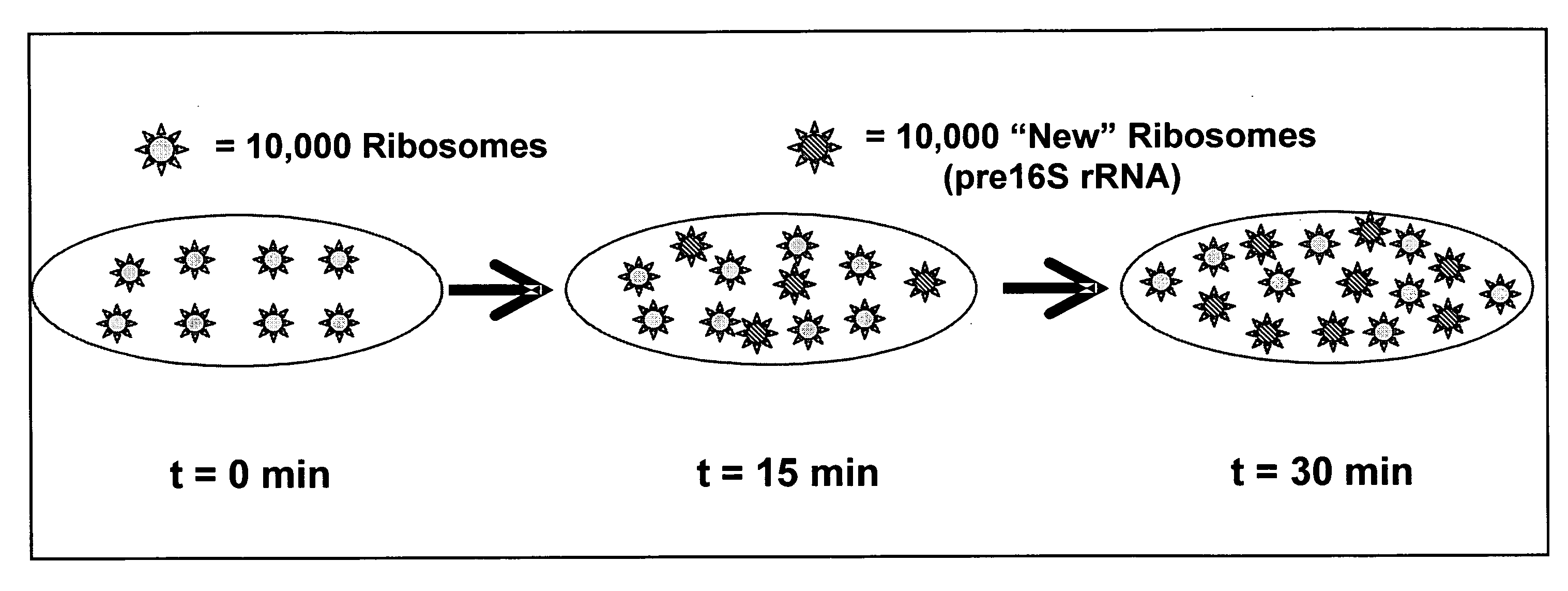
[0025] In a preferred embodiment, the method of the invention (also referred to herein as FISH-RiboSyn) is an in situ method that utilizes fluorescence in situ hybridization (FISH) with probes that target: (1) the 5′ or 3′ end of precursor 16S rRNA; or (2) the interior region of both precursor 16S rRNA and mature 16S rRNA. Images can be captured for a defined exposure time and the average fluorescent intensity for individual cells can be determined. The rate of increase of the whole cell fluorescent intensity is used to determine the specific rate of ribosome synthesis (or ribosome doubling time). The FISH-RiboSyn method can be attractive for rapidly measuring the specific growth rate (or cell doubling time) of distinct microbial populations within a mixed culture.

Problems solved by technology

Woese argued that since protein synthesis is an essential function for life, the ribosome could not withstand major sequence changes or life would cease.
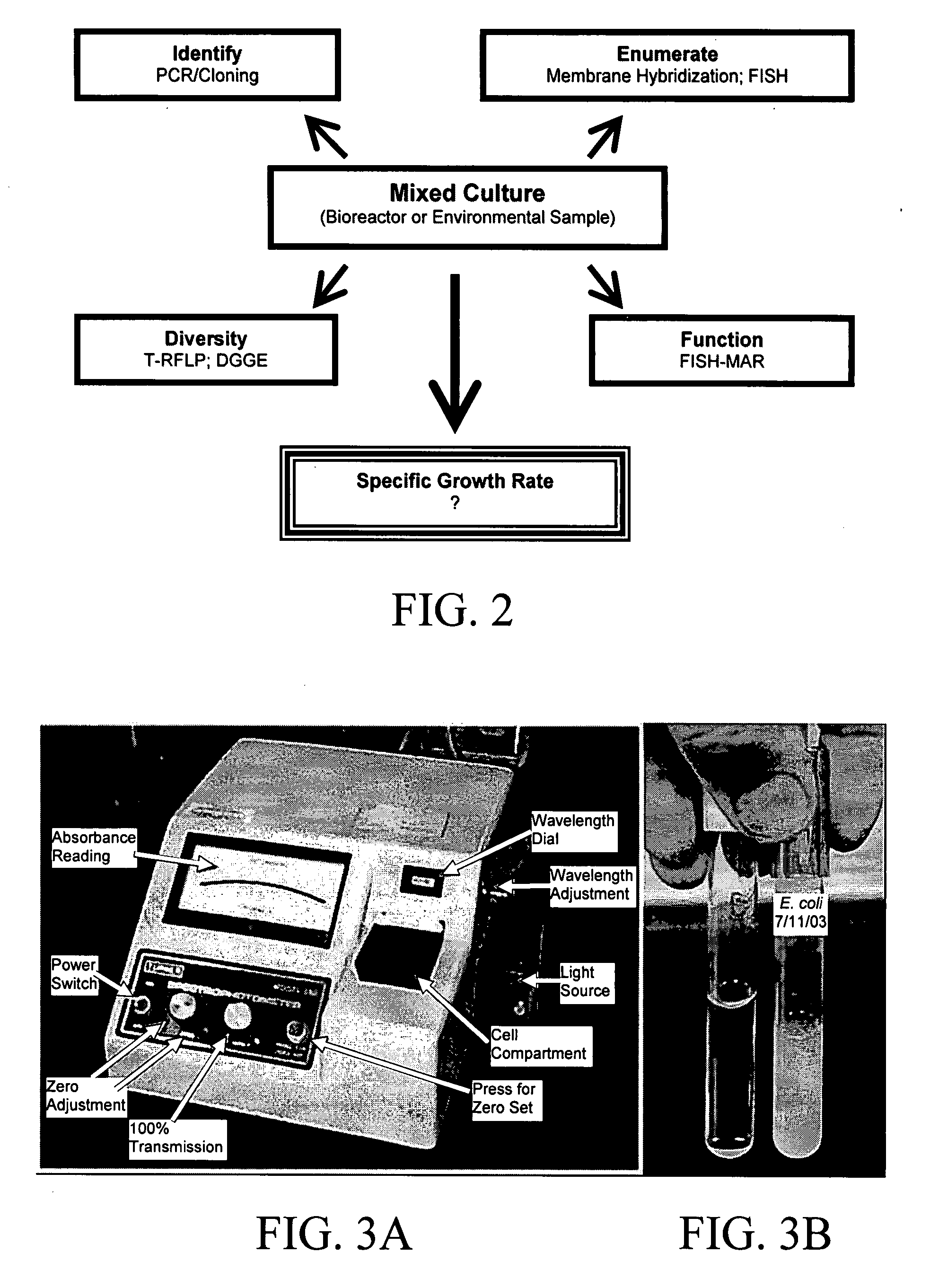
FISH-MAR is a difficult method to master, which limits its acceptance as a second wave tool.
The identification of the microbes containing these genes or mRNA is not always possible, since these biomolecules are not phylogenetic markers and are present at low cellular levels.
However, this approach was discarded as a method for measuring the specific growth rate (or cell doubling time), since cells maintain high levels of ribosomes during stationary phase which would be misinterpreted as rapidly growing cells.
embly. This secondary processing step is slower than the primary processing step, which results in an intracellular pool of precursor
Chloramphenicol disrupts ribosome synthesis.

Method used


Image

  • Method for determining the specific growth rate of distinct microbial populations in a non-homogeneous system
  • Method for determining the specific growth rate of distinct microbial populations in a non-homogeneous system
  • Method for determining the specific growth rate of distinct microbial populations in a non-homogeneous system

Examples


example 1

FISH Results Demonstrate a Strong Linear Relationship Between the dF / dtCm and Specific Growth Rate for A. calcoaceticus Cultured at Four Different Specific Growth Rates

[0164] The experimental results described below provide evidence that the FISH-RiboSyn method can be used to measure the dF / dtCm, which is directly related to the specific growth rate. In FIGS. 10A-10F, 11A-11C, 12A-12C, and 13A-13C, DAPI and FISH images are shown for the chloramphenicol-treated cultures of A. calcoaceticus. For the stationary phase culture (FIGS. 10A-10F), DAPI images (FIGS. 10A-C) reveal several bacteria cells, but the FISH images (FIGS. 10D-F) provide evidence that the chloramphenicol-treated cells did not accumulate pre16S rRNA because ribosome synthesis has ceased, which results in cells with low and constant fluorescent intensity. However, chloramphenicol-treated cells from the log growth cultures (FIGS. 11A-11C, 12A-12C, and 13A-13C) have active ribosome synthesis, which results in the accumul...


PUM

PropertyMeasurementUnit
melting temperatureaaaaaaaaaa
Tmaaaaaaaaaa
temperatureaaaaaaaaaa

Abstract

The present invention pertains to a molecular biology-based method and kit for measuring the specific growth rate (or cell doubling time) of distinct microbial populations. The method and kit can be used to analyze mixed culture samples that have been exposed to chloramphenicol or other protein synthesis inhibitors for defined times. In a preferred embodiment, the method of the invention (also referred to herein as FISH-RiboSyn) is an in situ method that utilizes fluorescence in situ hybridization (FISH) with probes that target: (1) the 5′ or 3′ end of precursor 16S rRNA; or (2) the interior region of both precursor 16S rRNA and mature 16S rRNA. Images can be captured for a defined exposure time and the average fluorescent intensity for individual cells can be determined. The rate of increase of the whole cell fluorescent intensity is used to determine the specific growth rate. The method of the invention can be attractive for rapidly measuring the specific growth rate (or cell doubling time) of distinct microbial populations within a mixed culture in industries such as environmental systems (water and wastewater treatment systems), bioremediation (optimization of conditions for microbial growth), public health (identification of rapidly growing infectious microbes), and homeland security (identification of rapidly growing bioterrorism agents).

Description

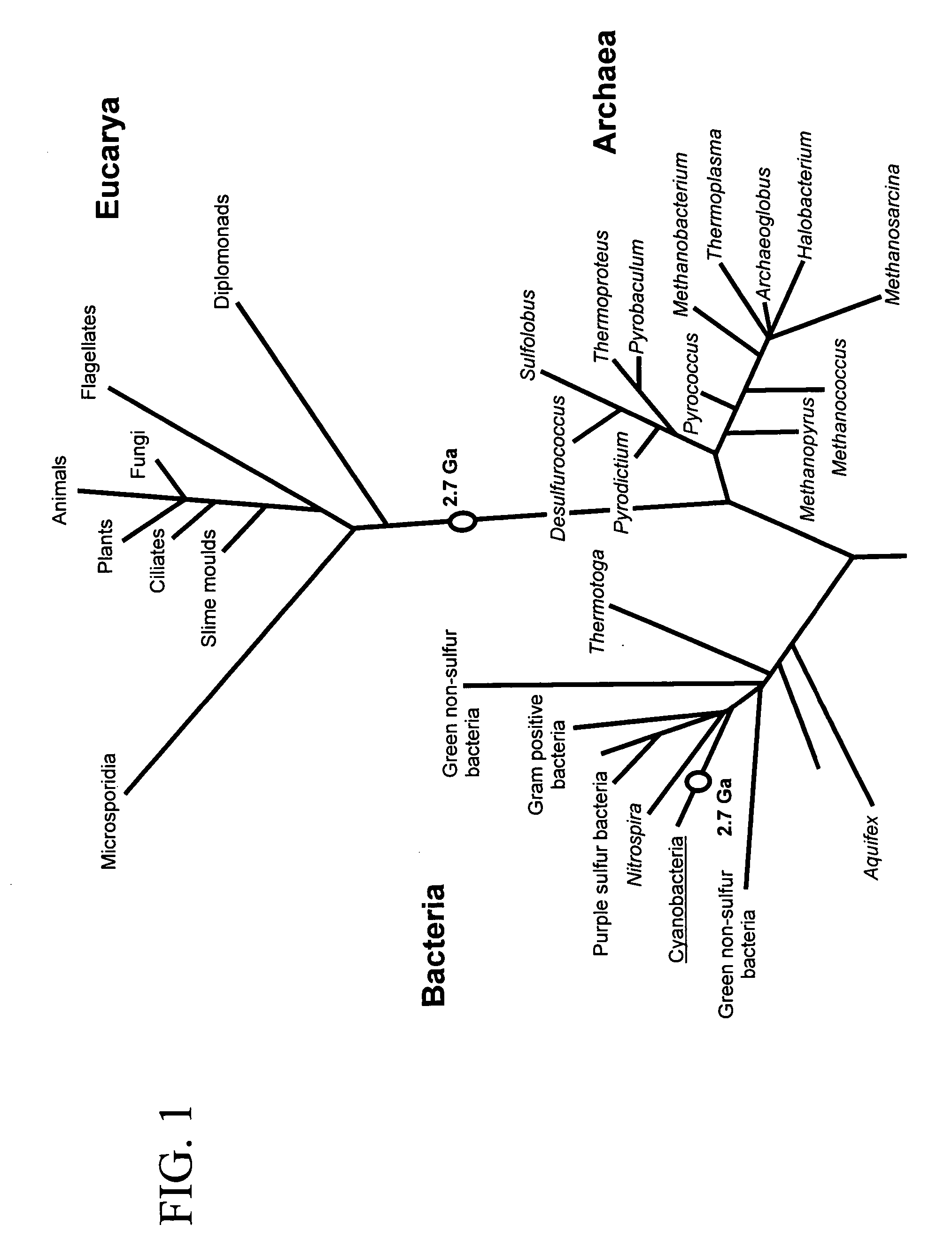
CROSS-REFERENCE TO RELATED APPLICATION [0001] The present application claims the benefit of U.S. Provisional Application Ser. No. 60 / 815,997, filed Jun. 23, 2006, which is hereby incorporated by reference herein in its entirety, including any figures, tables, nucleic acid sequences, amino acid sequences, and drawings.BACKGROUND OF THE INVENTION [0002] Before the 1970s, the phylogeny of the prokaryotes was based on crude comparisons of morphology and pattern of substrate utilization and was largely ignored due to the presumed simplicity of the organisms. Carl Woese used a different strategy to tackle prokaryotic phylogeny. He focused on sequence comparisons of the ribosome, a biomolecule found in all life forms. The ribosome is an essential macromolecule that is involved in the translation of messenger RNA into proteins. Woese argued that since protein synthesis is an essential function for life, the ribosome could not withstand major sequence changes or life would cease. He then tar...

Claims


Application Information

Patent Timeline
Patent Type & Authority Applications(United States)
IPC IPC(8): C12Q1/68
CPCC12Q1/6841C12Q2545/114
Inventor STROOT, PETER GEORGECUTTER, MATTHEW RAYMONDDUPONT, SAMUEL JAMES JR.
Owner UNIV OF SOUTH FLORIDA
Learn More