
Optical Imaging Contrast Agents for Imaging Lung Cancer

a technology of contrast agents and optical imaging, applied in the direction of in-vivo testing preparations, antineoplastic agents, drug compositions, etc., can solve the problems of prostate cancer, most other forms of cancer fatal, and urination

Inactive Publication Date: 2008-02-21
GE HEALTHCARE AS
View PDF9 Cites 4 Cited by
  • Summary
  • Abstract
  • Description
  • Claims
  • Application Information

AI Technical Summary

Benefits of technology

The present invention provides an optical imaging contrast agent that targets abnormal biological targets associated with prostate cancer. This agent can be used to detect and locate prostate cancer tissue and metastases in the body. The invention has the potential to improve the diagnosis and treatment of prostate cancer.

Problems solved by technology

The result is that the prostate gland increases in size and presses against the urinary system resulting in problems with urination.
Prostate cancer, on the other hand is as most other forms of cancer fatal if not treated.
The main symptoms of prostate cancer, however, can include: problems related to urination, such as pain, weak flow of urine and frequent urination.
Other symptoms include difficulty in obtaining erection, blood in urine or semen, painful ejaculation and lower back pain.
Staging of prostate cancer can be a difficult task.
There is today no reliable method for early diagnosis of prostate cancer.
PSA and other biomarkers are not selective for cancer, and existing in vivo imaging methods are not sensitive enough to diagnose prostate cancer at an early stage.
As pointed out prostate cancer is still a challenge to diagnose and treat.

Method used


Image

  • Optical Imaging Contrast Agents for Imaging Lung Cancer
  • Optical Imaging Contrast Agents for Imaging Lung Cancer
  • Optical Imaging Contrast Agents for Imaging Lung Cancer

Examples


example 1

Contrast Agent for Mapping of COX-2 Activity. Synthesis of COX-2 Ligand Coupled to Fluorescein

[0125]Step 1

[0126]2-Hydroxy-1-(4-methanesulfonylphenyl)ethanone is prepared from 2-bromo-1-(4-methanosulfonylphenyl)ethanone according to C. Puig et al in J. Med. Chem 2000,43, 214-223.

[0127]Step 2

[0128]A solution of 2-hydroxy-1-(4-methanosulfonylphenyl) ethanone (1.50 g, 7 mmol) and fluorescein isocyanate isomer I (2.72 g, 7 mmol) is heated in DMF at 120° C. for 5 hours.

[0129]The mixture is cooled, DMF evaporated off and acetic acid (25 ml) is added. The mixture is refluxed for 10 hours. The acetic acid is evaporated and the resulting mixture is purified on silica using chloroform / methanol as eluent.

example 2

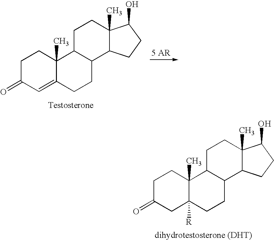
Contrast Agent for Mapping of Steroid 5 Alpha-Reductase Activity

[0130]Pentasilylated epigallocatechin is prepared according to J. J. Plattner et al in JACS (1972) 94 8613-5.

[0131]Step 1.

[0132]5(6)-Carboxyfluorescein (1 mmol) is dissolved in DMF (30 ml). Dicyclohexylcarbodiimide (1.1 mmol) is added. The mixture is stirred at room temperature for 12 hours. A solution of pentasilylated epigallocatechin (1 mmol) and dimethylaminopyridine (50 mg) in DMF (5 ml) is added and the mixture is stirred for 48 hours at room temperature. The mixture is evaporated. The ester product is isolated by flash chromatography (silica, hexane / chloroform).

[0133]Step 2.

[0134]The pentasilylated epigallocatechin-fluorescein conjugate (0.5 mmol) is dissolved in tetrahydrofuran (30 ml). Triethylamine trihydrofluoride (1 mmol) is added and the mixture is stirred at ambient temperature for 12 hours. The mixture is evaporated and the carboxyfluorescein epigallocatechin conjugate is isolated by flash chromatography ...

example 3

Contrast Agent for Mapping of EGFR / erB2 Tyrosine Kinase

[0135]Step 1. N-[4-((3-bromophenyl)amino)quinazolin-7-y-]acrylamide is prepared according to J. B. Smaill et al J. Med. Chem. (1999) 42 1803-1815.

[0136]Step 2. N-[4-((3-bromophenyl)amino)quinazolin-7-y-]acrylamide from step 1 (1 mmol) and ethylenediamine (10 mmol) are dissolved in DMF (25 ml). The mixture is stirred at 50° C. for 12 hours. The solvent is evaporated off and the conjugate compound is isolated by flash chromatography (silica, hexane, chloroform, methanol).

[0137]Step 3. Cy7-NHS ester (0.5 mmol), the conjugate compound from step 2 (0.5 mmol) and N-methylmorpholine (70 mg) are dissolved in DMF (30 ml). The mixture is stirred at 40° C. for 3 days. The Cy7 amide conjugate is isolated by flash chromatography (silica, hexane, ethyl acetate, methanol).


PUM

PropertyMeasurementUnit
molecular weightaaaaaaaaaa

Abstract

The invention provides contrast agents for optical imaging of prostate cancer in patients. The contrast agents may be used in diagnosis of prostate cancer, for follow up of progress in disease development, for follow up of treatment of prostate cancer and for surgical guidance. Further, the invention provides methods for optical imaging of prostate cancer in patients.

Description

FIELD OF THE INVENTION [0001]The present invention provides contrast agents for optical imaging of prostate cancer in patients. The contrast agents may be used in diagnosis of prostate cancer, for follow up of progress in disease development, and for follow up of treatment of prostate cancer.[0002]The present invention also provides new methods of optical imaging of prostate cancer in patients, for diagnosis and for follow up of disease development and treatment of prostate cancer.DESCRIPTION OF RELATED ART[0003]Prostate cancer is the most common cancer type for men in the western world. More than 25% of all men with a cancer diagnosis have prostate cancer.[0004]The prostate is a gland with the size of a walnut located between the rectum and the bladder. The gland stores a fluid that is released to form semen. The function of the prostate gland is dependent on testosterone.[0005]Tumours in the prostate gland can be benign or malignant. Benign prostatic hyperplasia (BPH) is not a can...

Claims


Application Information

Patent Timeline
Patent Type & Authority Applications(United States)
IPC IPC(8): A61K49/00A61P35/00
CPCA61K49/0032A61K49/0056A61K49/0052A61K49/0043A61P35/00
Inventor KLAVENESS, JOJOHANNESEN, EDVINTOLLESHAUG, HELGE
Owner GE HEALTHCARE AS
Learn More