Devices and Methods for In-Vivo Pathology Diagnosis

a pathology and in-vivo technology, applied in the field of endoscopy, can solve the problems of inability the inability of the human eye to distinguish the various cellular outlines and internal cell structures of in-vivo tissue, and the inability of the contact endoscope to penetrate into the deeper tissues. the effect of tissue streamlined or cutting

Inactive Publication Date: 2008-02-21
FRITSCH MICHAEL H +2
View PDF99 Cites 47 Cited by
  • Summary
  • Abstract
  • Description
  • Claims
  • Application Information

AI Technical Summary

Benefits of technology

[0027]According to one aspect of the present invention, materials may be placed at a destination location within tissue. For example, staining fluids may be placed for precisely staining certain target tissue in preparation for pathologic diagnosis. By using multiple endoscope working channels one or more irrigation-suction streams may be created.
[0028]According to another aspect of...

Problems solved by technology

Such a contact endoscope cannot access or penetrate into deeper tissues because it has a slim, fragile optical rod and a flat non-streamlined lens tip.
Such a conventional contact endoscope entering into the body and pushing to deeper target tissues would not be not practical because such would cause damage to the endoscope and to the body tissues.
Without staining, the human eye, even with microscopic magnification, cannot distinguish the various cellular outlines and internal cell structures of in vivo tissue.
Such conventional endoscope tips, spe...

Method used


Image

  • Devices and Methods for In-Vivo Pathology Diagnosis
  • Devices and Methods for In-Vivo Pathology Diagnosis
  • Devices and Methods for In-Vivo Pathology Diagnosis

Examples


Embodiment Construction

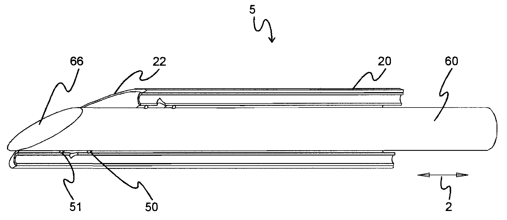
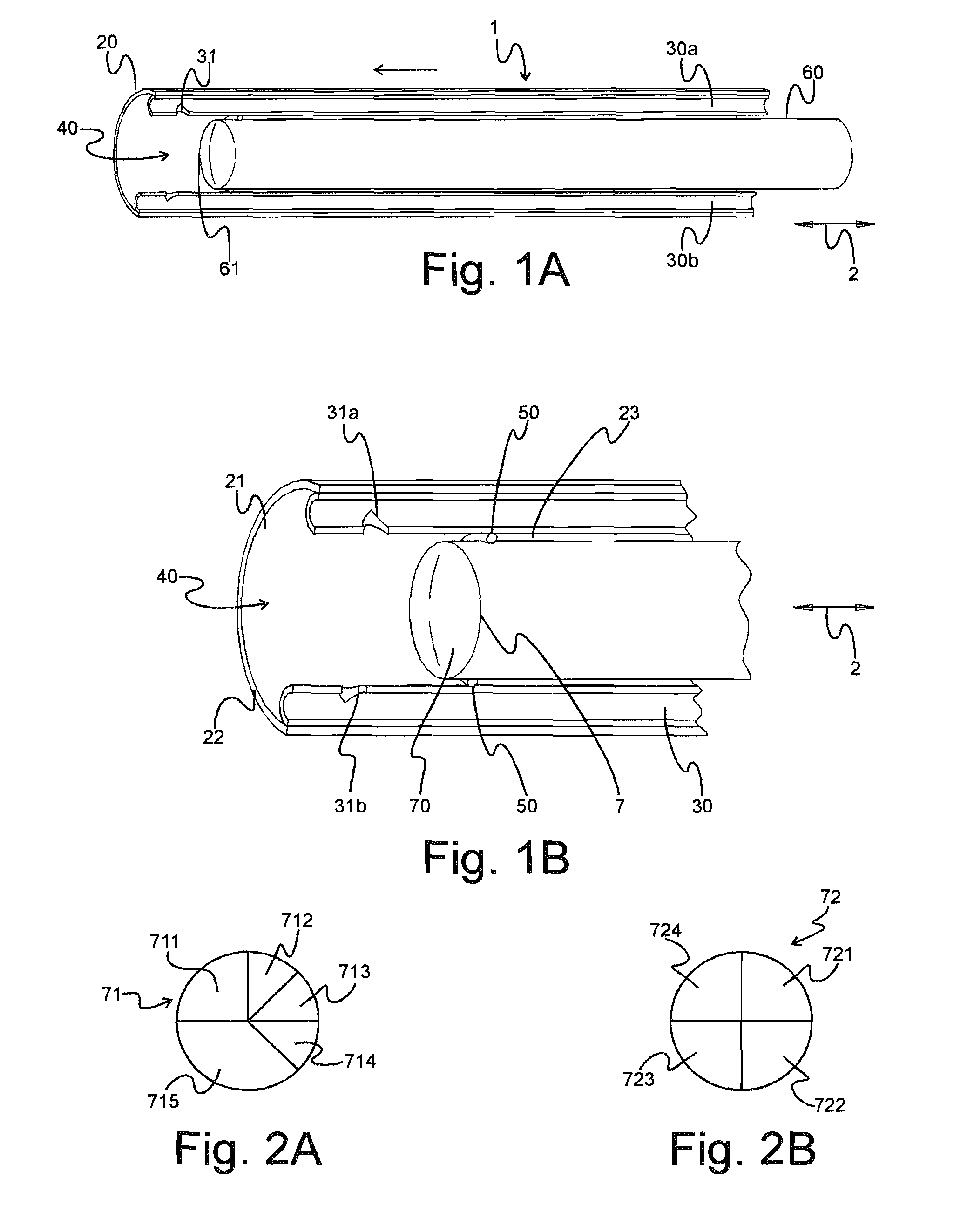
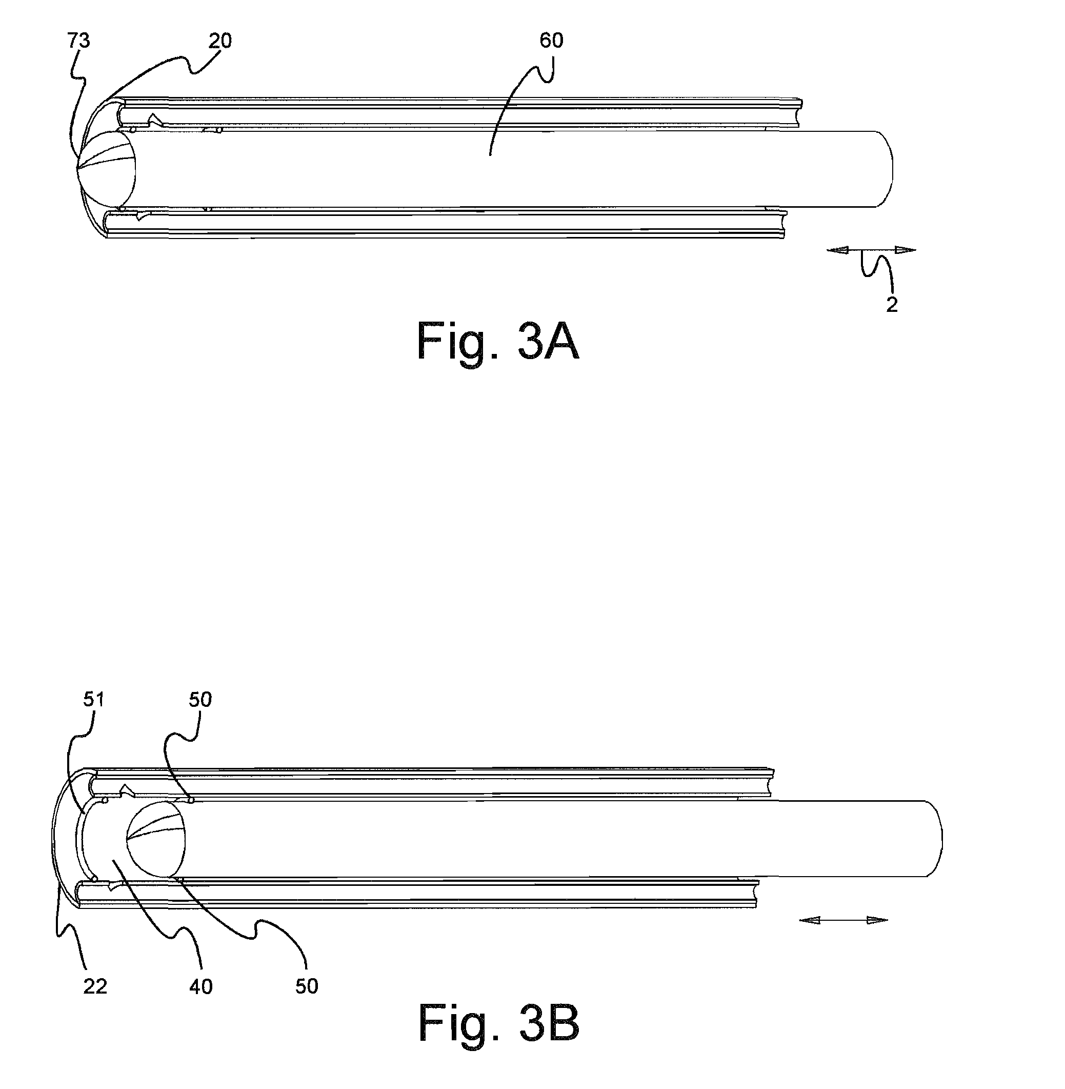
[0052]FIGS. 1 and 1B schematically show an endoscope system 1 having a sheath 20 (illustrated in cross section) adapted for protecting a contact endoscope 60 within a cylindrical inner space 21. Contact endoscope 60 is longitudinally movable within sheath 20, which effects a protective sleeve. Sheath 20 contains one or more irrigation channels 30 that are preferably in a same longitudinal direction 2 of movement of contact endoscope 60. Channels 30 are used to flush staining fluids into and across body tissues of interest in order to stain the tissue cells (not shown) in preparation for pathological diagnosis. A single channel may be used to first flush and then remove the stains. In a preferred embodiment, at least two or more channels 30 are used. With such a use of two or more channels, a continuous irrigation-suction stream of one or more stains can be used. Stains may thereby flow over the tissues sequentially or simultaneously, by entering an inflow port (not shown) of sheath ...


PUM


Abstract

An in vivo pathology diagnosis system includes a penetrating device that is at least one of an endoscope sheath and an endoscope, the penetrating device having irrigation and working channels with openings at a distal end chamber of the device. The device is structured for penetrating tissue until the distal end is proximate a target tissue area. A method includes supplying stain to the chamber via the irrigation channels, thereby staining the target tissue area. Pathology of the stained target tissue is performed by viewing such stained tissue through the penetrating device. The viewing may be performed through a selected lens segment of a tissue contact type endoscope or endoscope sheath. A suction channel may be provided for removing material from the chamber, and cauterizing electrodes may be provided as part of the device. Control of in vivo staining may be performed by a computer program.

Description

RELATED APPLICATIONS[0001]This application claims benefit of priority from Fritsch et al., U.S. Provisional Patent Application Ser. No. 60 / 838,614 filed on 19 Aug. 2006, which is incorporated herein by reference in its entirety.I. FIELD OF THE INVENTION[0002]The invention relates generally to devices and methods of endoscopy and, more particularly, to devices and methods for accessing and diagnosing pathologic histologic tissue including deep tissue.II. BACKGROUND OF THE INVENTION[0003]Physicians who practice pathology conventionally diagnose and characterize disease in living patients by examining biopsies and other specimens. Such examination of tissues and cells has generally involved gross and microscopic visual examination of tissues, with special stains and immunohistochemistry, and other procedures, being employed to visualize specific proteins and other substances in and around cells.[0004]When certain surgeries are performed, a corresponding typical pathology histology diag...

Claims


Application Information

Patent Timeline
IPC IPC(8): A61B10/04A61B1/00
CPCA61B10/0045A61B17/3417A61B19/5212A61B2017/320044A61B1/015A61B2018/00982A61B2019/545A61B1/00135A61B18/148A61B2018/00595A61B2019/5433A61B90/361A61B2090/3933A61B2090/395
Inventor FRITSCH, MICHAEL H.FRITSCH, JOHN H.FRITSCH, JOSEPHINE M.
Owner FRITSCH MICHAEL H
Why Eureka
Try Eureka