

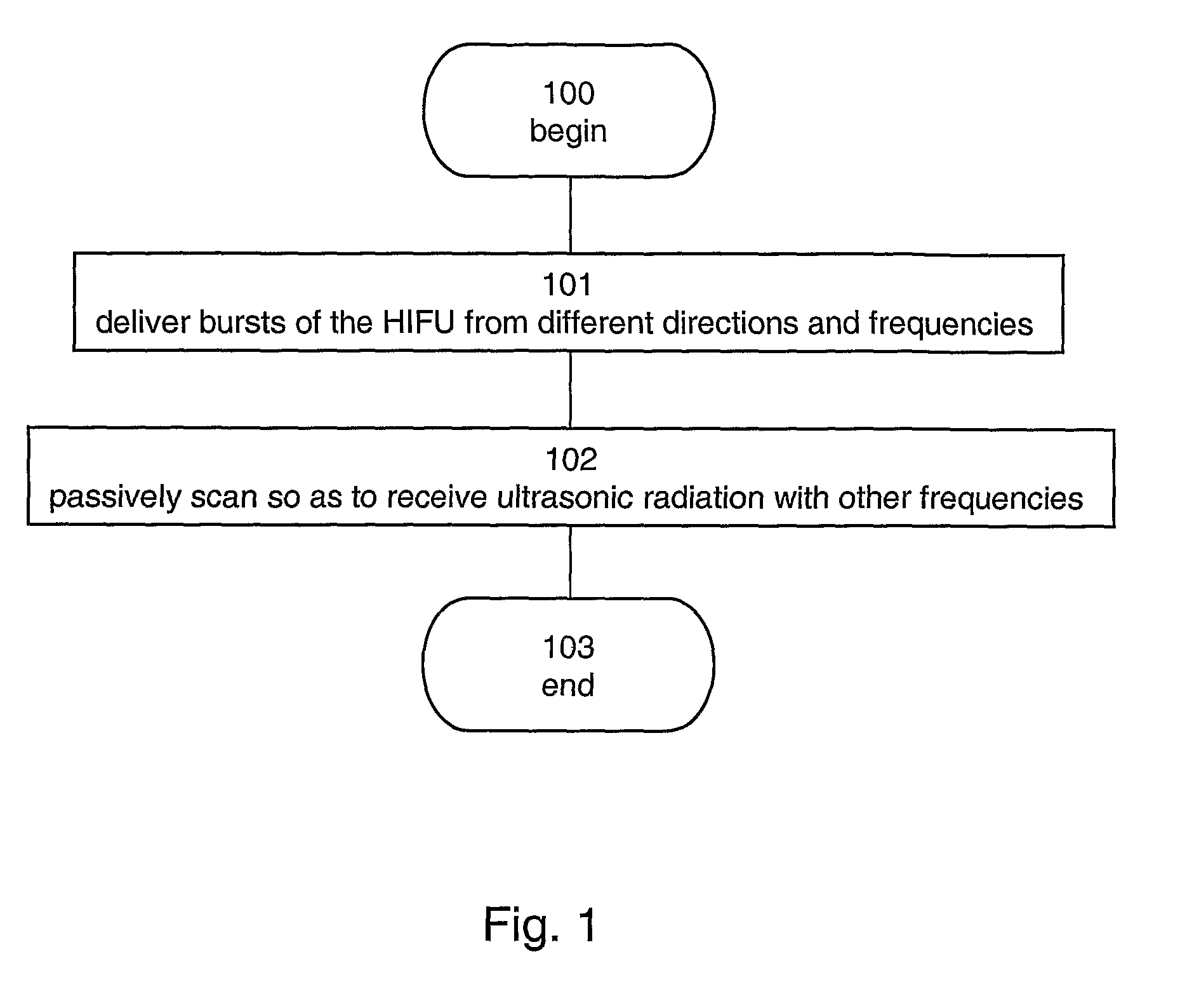
Ultrasonic Image-Guided Tissue-Damaging Procedure
a tissue damage and ultrasonic imaging technology, applied in the field of ultrasonic image-guided tissue damage procedures, can solve the problems of limited access to patients within the mri scanner, cosmetically undesirable, and high cost of most of these systems
- Summary
- Abstract
- Description
- Claims
- Application Information
AI Technical Summary
Benefits of technology
Problems solved by technology
Method used
Image

Examples
example 1
TUCT Image
[0180]FIG. 8 illustrates a three-dimensional phantom reconstruction of breast 14 based on attenuation-coefficient imaging, as obtained by the spiral transmission ultrasound computerized tomography (SUCT) and reported in a paper by Azhari H. and Sazbon D., entitled “Volumetric imaging using spiral ultrasonic computed tomography”, published in Radiology, 1999, 212(1):270-275. A three-dimensional computerized reconstruction section 14A was virtually cut at about 10 mm from the base of breast 14, to depict target masses 15 simulating abnormal tissue. Related work in the field includes that of Greenleaf, et al. [Greenleaf James F., and Bahn, Robert C., “CLINICAL IMAGING WITH TRANSMISSIVE ULTRASONIC COMPUTERIZED TOMOGRAPHY,” IEEE Trans, Biomed Eng, v BME-28, n 2, February 1981, p 177-185], which describes transmission ultrasound computer-assisted tomography for detection and diagnosis of cancer in the breast, and Jago, et al. [Jago, J. R., Whittingham, T. A., “Practical system f...
example 2
TUCT System
[0181]FIGS. 9A-C schematically illustrate configurations 30A and 30B, for producing a temperature image (thermal map) using a TUCT system 32, according to various exemplary embodiments of the invention.
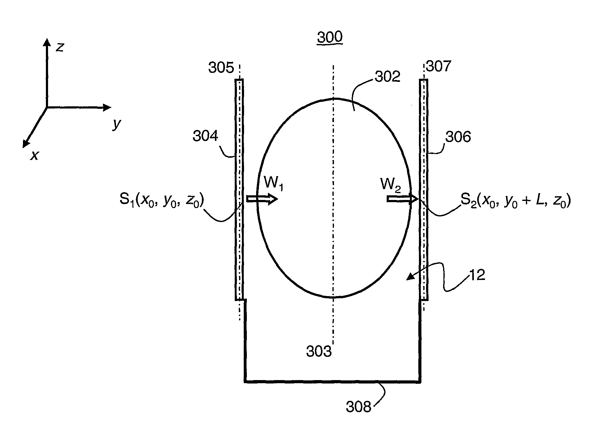
[0182]As shown in FIG. 9A, configuration 30A comprises a TUCT system 32, which comprises ultrasonic transducers or transducer arrays 16 and 18. Transducers 16 and 18 are in communication with an imaging unit 24. Data processor 22 is further operative to perform data analysis and display, by graphical means, by printout, or by other means. It will be appreciated that data processor 22 may be integrated with imaging unit 24, so as to form a single unit. Preferably, transducers 16 and 18 and organ 14 (shown in FIGS. 9A-B as a breast) are immersed in or applied with tissue coupling medium 12.
[0183]Additionally, a surgical-procedure unit, which is operative to heat a portion of the tissue, for example, a HIFU system 34A, is preferably also in communication with data processor 22...
example 3
Configuration for HIFU Treatment of a Breast
[0196]FIGS. 10A-C schematically illustrate configuration 30A of Example 2 (see FIG. 9A), designed for image guided HIFU treatment of a woman's breast, in accordance with various exemplary embodiments of the invention.
[0197]As shown in FIGS. 10A and 4C, a woman 50 lies prone on bed 40, with her breast 14 inserted through hole 42 and sleeve 46 (FIG. 10C) into water tank 44, where both ultrasound imaging and ultrasound ablation are performed, under water.
[0198]As seen in FIGS. 10A and 4C, configuration 30A includes a special bed 40, which defines a hole 42, into which breast 14 (FIGS. 9A-B) is to fit. Hole 42 is in communication with a water tank 44. Preferably a removable, washable or disposable sleeve 46 is employed for hygienic purposes.
[0199]FIGS. 10A and 10B further illustrate HIFU system 34A, comprising HIFU operating unit 26A and an HIFU transducer or transducer array 20A, and TUCT system 32, comprising transducers 16 and 18, imaging u...
PUM
 Abstract
Description
Claims
Application Information