
Biosensors for Detecting Bond Rupture

a biosensor and bonding technology, applied in the field of biosensors, can solve the problems of non-specific adsorption of all affinity binding methods, affecting the accuracy of the detection, and requiring a minimum of several hours to complete the analysis,

Inactive Publication Date: 2008-09-11
IND RES LTD
View PDF12 Cites 1 Cited by
  • Summary
  • Abstract
  • Description
  • Claims
  • Application Information

AI Technical Summary

Benefits of technology

[0009]It is the object of the present invention to provide a biosensor with a separate oscillator and rupture detector, or to at least provide the public with a useful choice.

Problems solved by technology

Such assays, while quite sensitive, take a minimum of several hours but often considerably longer.
Even greater delays may be involved if transport of a sample to a city laboratory from a remote area is required.
All affinity binding methods suffer from non-specific adsorption, the process by which proteins other than those sought attach themselves to a surface, on sites other than the prepared “capture” molecules.
This is a limiting factor with regard to detection threshold.

Method used


Image

  • Biosensors for Detecting Bond Rupture
  • Biosensors for Detecting Bond Rupture
  • Biosensors for Detecting Bond Rupture

Examples


example

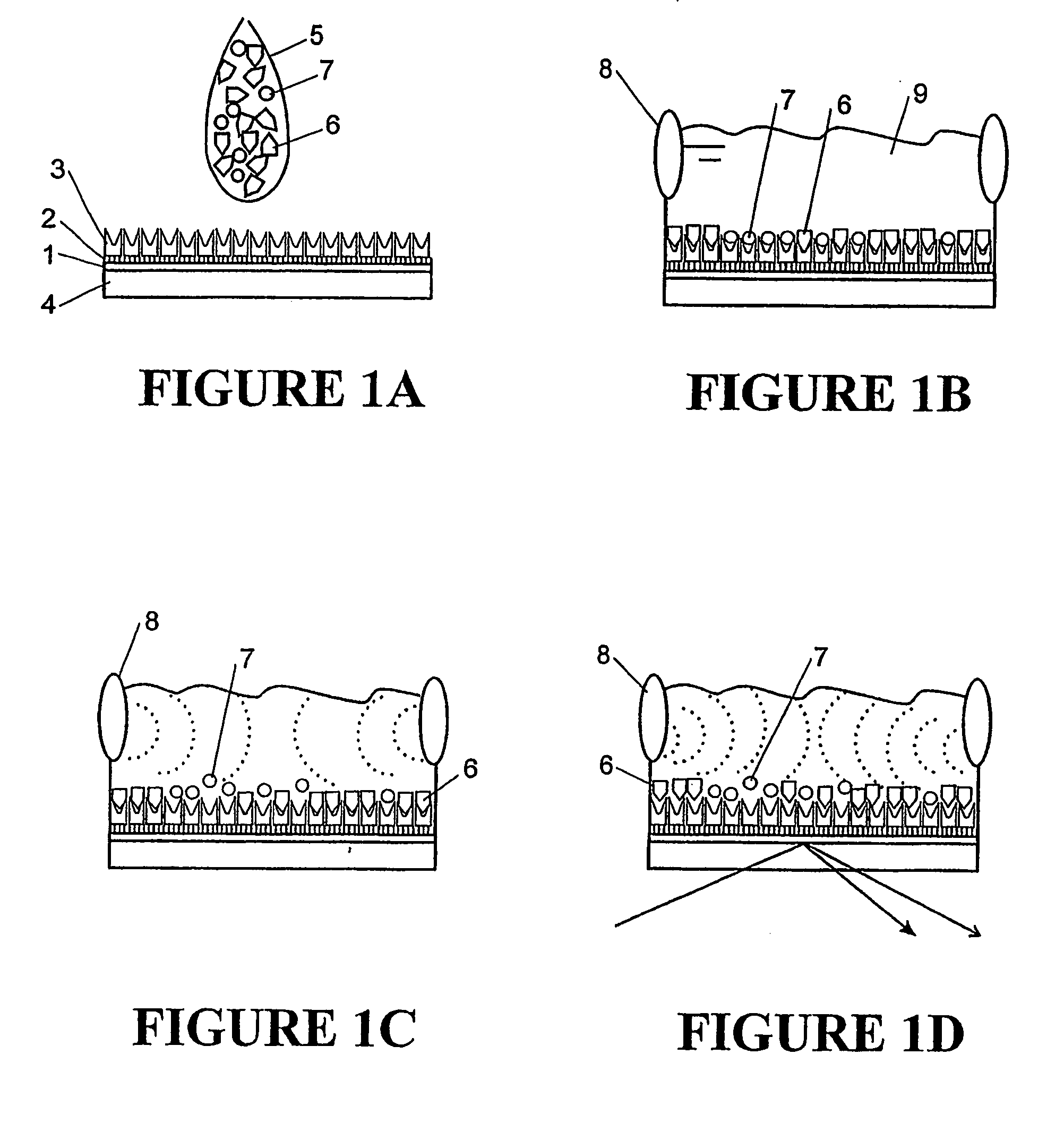
[0060]FIG. 5 shows an example of one embodiment of biosensor that uses an SPR device to detect bond rupture. The biosensor includes an oscillation source 50, delay line 51, fluid channel 52, surface 53, reflective layer 54, incident light beam 55, and reflected light beam 56. A light detector is also included (not shown) that detects changes on the angle of the reflected light beam 56. In this example oscillator 50 is a 10 MHz transducer that can be connected to a wave form generator.

[0061]This example shows both surface immobilisation and bond rupture scanning. These were monitored in situ by integration of both SPR detection and acoustic waveform induction into a thin layer flow cell.

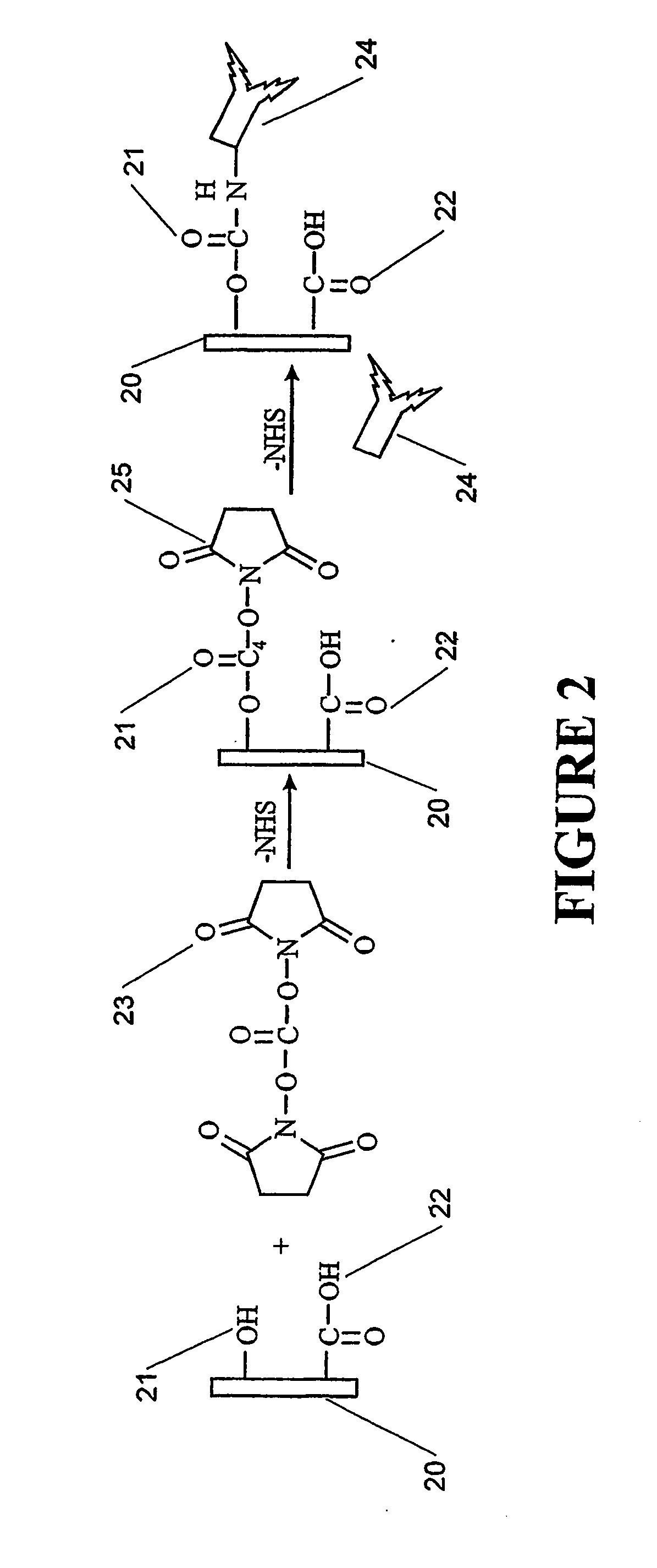
[0062]Surface 53, in this example a gold surface, provides a surface on which a self assembled monolayer may be formed. In this example 5 mg of biotin-PEO3-amine was dissolved in 250 mL of 0.1 M phosphate buffered saline (PBS) solution (pH 7.4). The amine and PBS solution was flushed through the bare ...


PUM

PropertyMeasurementUnit
pHaaaaaaaaaa
flow rateaaaaaaaaaa
reflectanceaaaaaaaaaa

Abstract

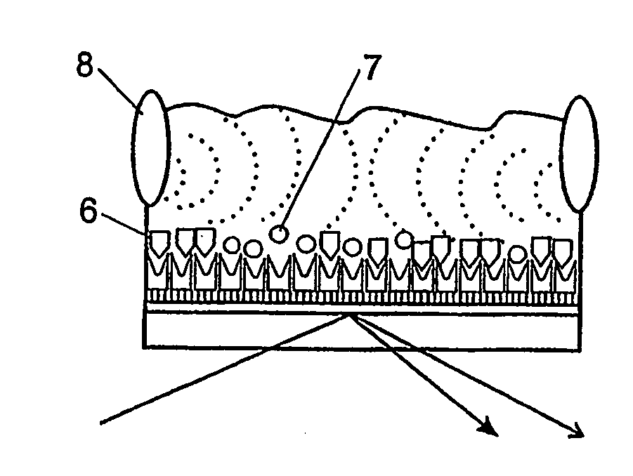
A biosensor comprises a surface onto which bio-macromolecules are bound, the surface and bio-macromolecules being immersed in liquid, a bond rupture detector associated with the surface and arranged to detect the rupture of bonds between the bio-macromolecules and a target substance (6), and an oscillator (8) associated with the liquid and spaced from the surface and arranged to produce oscillations in the liquid to cause bonds between the bio-macromolecules and the target substance (6) to rupture.

Description

FIELD OF INVENTION[0001]The invention relates to biosensors for detecting a variety of chemical and biological agents and in particular to biosensors for detecting a variety of chemical and biological agents with a separate detector and oscillator.BACKGROUND[0002]In current New Zealand medical practices when a patient is ill a swab is taken and analysed in a laboratory. For example to identify a strain of influenza in a patient an aliquot of appropriate fluid is incubated with the purified antibody to a characteristic molecule or marker on the surface of the influenza virus. By means of amplifying agents and secondary antibodies, which are tagged with enzymes or radioisotopes, the concentration of the virus can be determined. Such assays, while quite sensitive, take a minimum of several hours but often considerably longer. Thus it may be several hours or even a day before an accurate diagnosis is available for the patient. Even greater delays may be involved if transport of a sample...

Claims


Application Information

Patent Timeline
Patent Type & Authority Applications(United States)
IPC IPC(8): G01N29/00G01N21/01C12Q1/00C12M1/34C12Q3/00G01N21/55G01N29/02G01N29/14G01N29/22G01N29/24G01N33/48G01N33/53G01N33/543
CPCB82Y15/00B82Y30/00G01N21/553G01N29/14G01N2291/0426G01N29/24G01N33/54373G01N2291/0251G01N2291/0423G01N29/222
Inventor YUAN, YONGANDREWS, MICHAELARNOLD, W MICHAELMARLOW, BARRY
Owner IND RES LTD
Learn More