Multi-well micropatterning by ablation

a multi-well, ablation technology, applied in the field of multi-well micropatterning by ablation, can solve the problem of limiting the practicality of integration with high throughput formats, and achieve the effect of not increasing experimental complexity

Inactive Publication Date: 2008-09-11
MASSACHUSETTS INST OF TECH
View PDF8 Cites 35 Cited by
  • Summary
  • Abstract
  • Description
  • Claims
  • Application Information

AI Technical Summary

Benefits of technology

[0006]Micropatterning biomolecules and cells allows precise experimentation investigating the role of the microenvironment on cellular fate and function; however, the fabrication complexity of many micropatterning techniques limits the practicality of integration with high throughput formats. Successful large scale biological investigations require simple procedures and standard materials in order to integrate seamlessly into the process flow of the biological research lab. By merging micropatterning with standard lab materials, large scale experimentation can exploit the benefits of micropatterning without increasing experimental complexity.

Problems solved by technology

Micropatterning biomolecules and cells allows precise experimentation investigating the role of the microenvironment on cellular fate and function; however, the fabrication complexity of many micropatterning techniques limits the practicality of integration with high throughput formats.

Method used


Image

  • Multi-well micropatterning by ablation
  • Multi-well micropatterning by ablation
  • Multi-well micropatterning by ablation

Examples


example 1

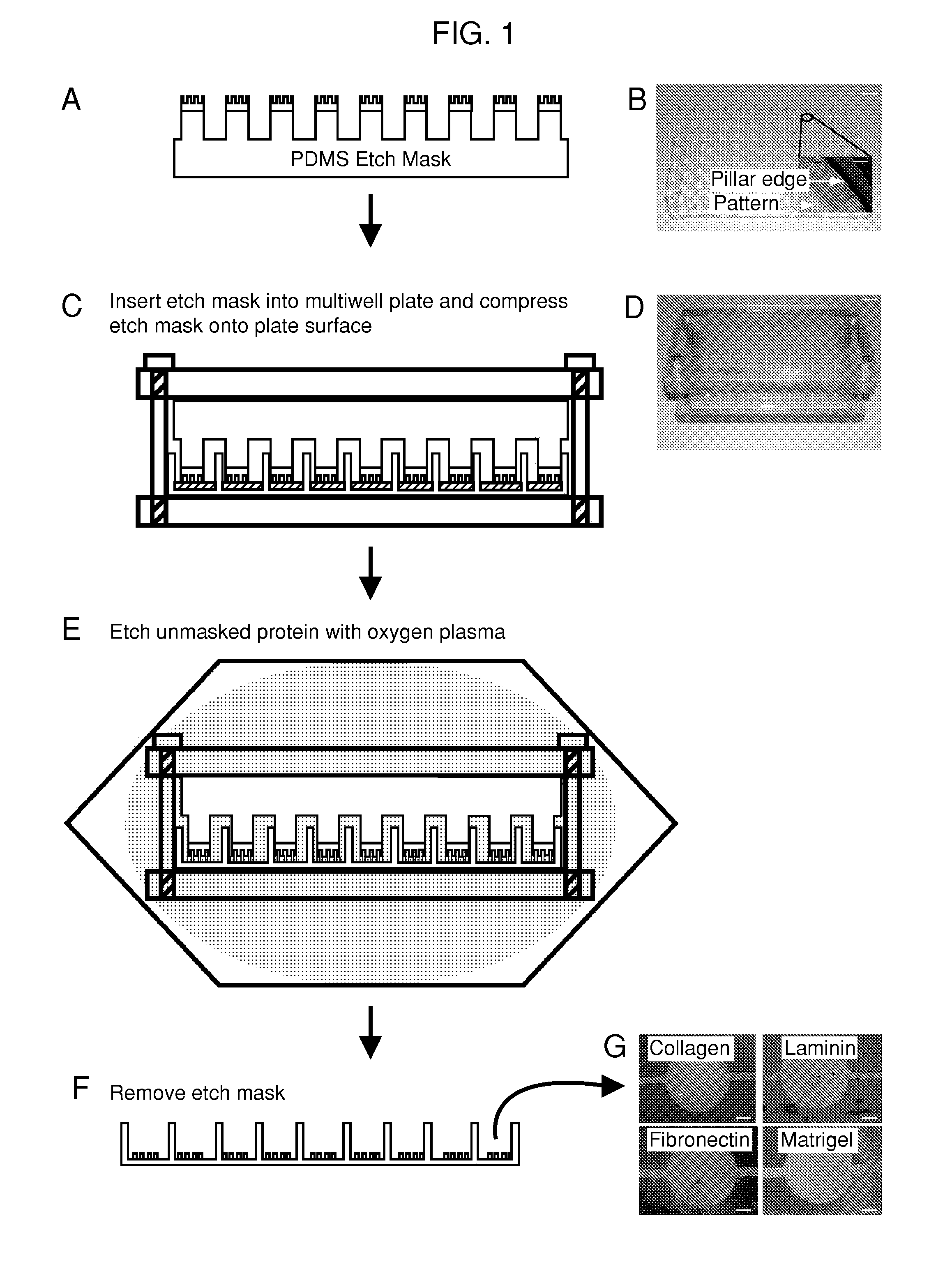
Fabrication of the 96-Well Etch Mask

[0128]A mold was machined with the same center to center spacing as a standard 96-well plate and was used to generate the support structure for the 96-well etch mask. This support structure consisted of an array of 96 pillars spaced evenly to allow nesting in a standard 96-well plate. The support structure is molded out of poly(dimethylsiloxane) (PDMS) (sylgard 184, Dow Corning, Midland, Mich.) and prepared using standard techniques. Duffy, D. C.; McDonald, J. C.; Schueller, O. J. A.; Whitesides, G. M. Analytical Chemistry 1998, 70, 4974. In a separate step, mold masters for the micropatterns were fabricated with SU8 photoresist (Microchem, Newton, Mass.) using a high resolution transparency photomask. The molds were fabricated to have 50 μm thick features. These mold masters were coated with a layer of PDMS 2 mm thick The PDMS was peeled from the mold and circles were punched out with a cork borer and glued to the pillars of the PDMS support stru...

example 2

Patterning Protein and Cells in 96-Well Plates

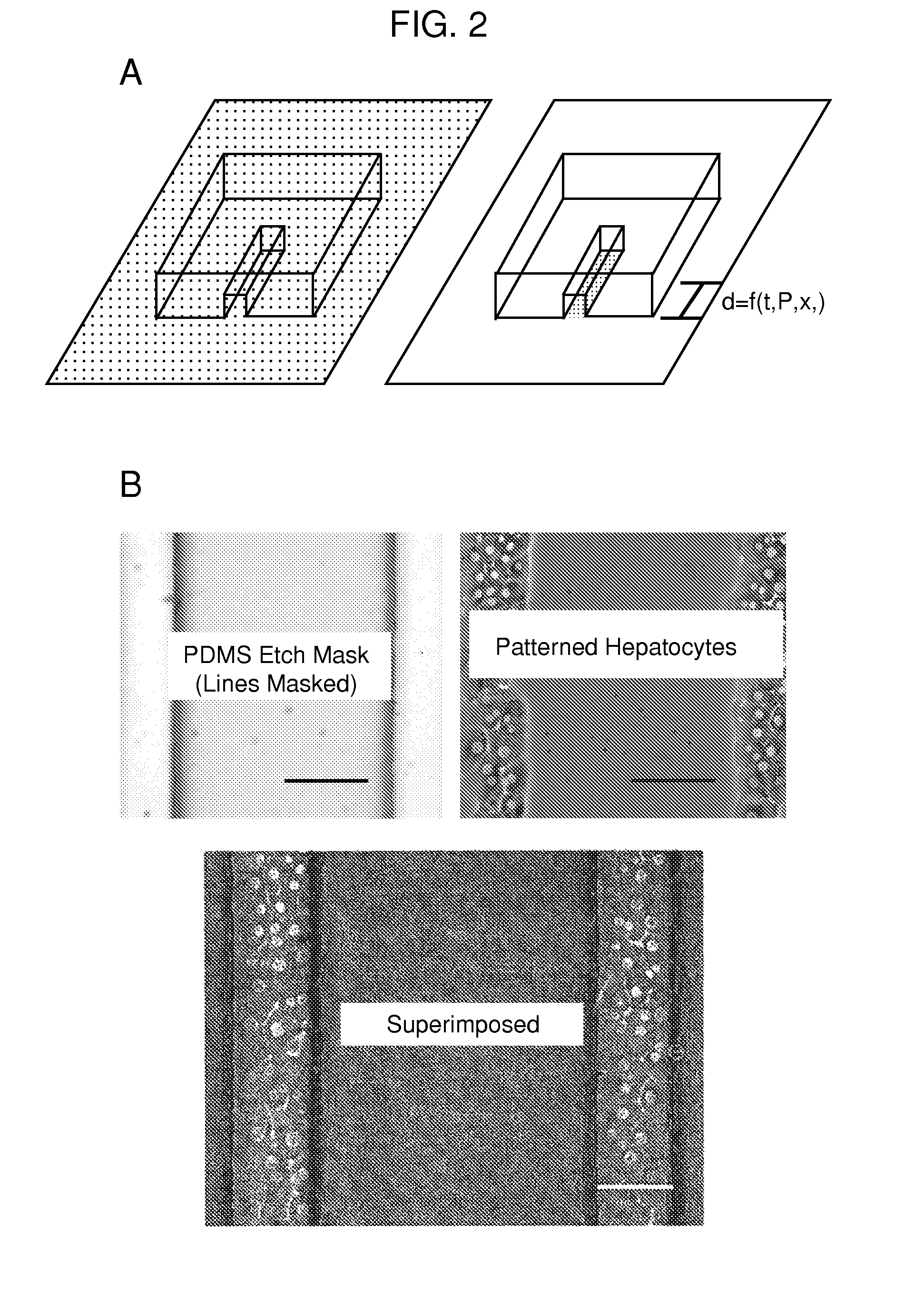
[0129]Biomolecules were physisorbed to each well of a standard multi-well plate (solutions of type-I collagen, fibronectin, Matrigel and laminin at 50 μg / mL in water. Note that 1×PBS may be used instead of water. The multi-well plates were incubated for 1 hour at 37° C. followed by rinsing with water and allowed to air dry. This results in an adsorbed thickness of approximately 150 nm. Gurdak, E.; Dupont-Gillain, C. C.; Booth, J.; Roberts, C. J.; Rouxhet, P. G. Langmuir 2005, 21, 10684-10692. In some cases, micropatterned proteins were fluorescently labeled via incubation (1 hour at room temperature) with Alexa Fluor® 488 carboxylic acid, succinimidyl ester (Invitrogen, Carlsbad, Calif.) dissolved in phosphate buffered saline (PBS) at 20 μg / mL. A single etch mask was inserted into the multi-well plate and compressed in a custom clamp consisting of two blocks of polycarbonate flanking the masked multi-well plate and compression was applie...

example 3

Ablation Quantification

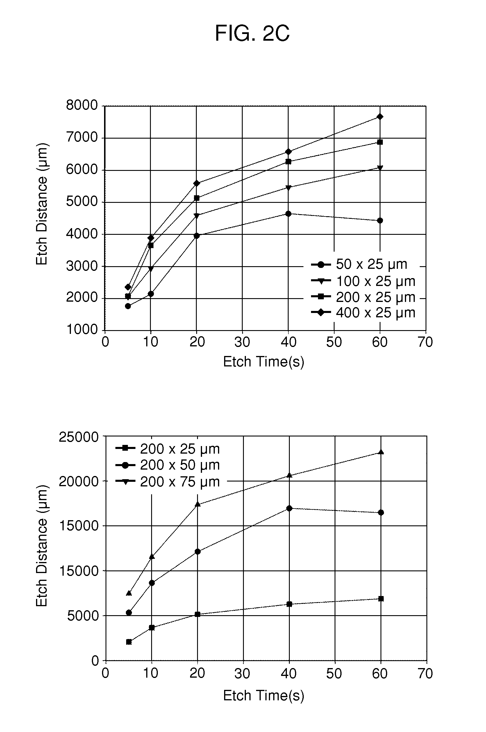
[0131]A PDMS etch mask containing dead-end microchannels with channel widths of 50, 100, 150, and 200 μm repeated for three channel heights of 25, 50, and 75 μm (12 channels total) was used to quantify the etching rate. The mask was placed onto p60 petri dishes physisorbed with type-I collagen and exposed to oxygen plasma for several time points (5, 10, 20, 40, and 60 seconds) with each time point on a separate dish. Primary rat hepatocytes were then seeded onto the patterned dishes and cultured for 24 hours. The hepatocytes specifically attached to regions with type-I collagen, hence plasma ablated distances could be directly correlated to hepatocyte attachment as hepatocytes will not attach to adsorbed collagen substrates from solutions less than 0.5 μ / mL. The distances were measured using Metamorph (Universal Imaging, Sunnyvale, Calif.) and plotted as a function of time.


PUM

PropertyMeasurementUnit
thickaaaaaaaaaa
feature sizeaaaaaaaaaa

Abstract

The present invention is drawn to the generation of micropatterns of biomolecules and cells on standard laboratory materials through selective ablation of a physisorbed biomolecule with oxygen plasma. In certain embodiments, oxygen plasma is able to ablate selectively physisorbed layers of biomolecules (e.g., type-I collagen, fibronectin, laminin, and Matrigel) along complex non-linear paths which are difficult or impossible to pattern using alternative methods. In addition, certain embodiments of the present invention relate to the micropatterning of multiple cell types on curved surfaces, multiwell plates, and flat bottom flasks. The invention also features kits for use with the subject methods.

Description

CROSS-REFERENCE[0001]This application claims the benefit of U.S. Provisional Application No. 60 / 851,101, filed Oct. 12, 2006, which application is incorporated herein by reference in its entirety.GOVERNMENT SUPPORT[0002]The U.S. government may have certain rights in this invention, pursuant to the Ruth L. Kirschstein National Research Service Award (NRSA), grant no. # 1F32DK072601.BACKGROUND OF THE INVENTION[0003]The growth and function of anchorage dependent cells is closely tied to local microenvironmental cues surrounding the cell including the nature of the underlying substrate, the degree of cell-cell contact (both homotypic and heterotypic), paracrine signaling, and physical forces. Mooney, D.; Hansen, L.; Vacanti, J.; Langer, R; Farmer, S.; Ingber, D. J Cell Physiol 1992, 151, 497-505; Reid, L. M.; Fiorino, A. S.; Sigal, S. H.; Brill, S.; Holst, P. A. Hepatology 1992, 15, 1198-1203; Nelson, C. M.; Chen, C. S. Febs Letters 2002, 514, 238-242; Bhatia, S, N.; Balis, U. J.; Yarmu...

Claims


Application Information

Patent Timeline
Patent Type & Authority Applications(United States)
IPC IPC(8): C12M1/22B44C1/22
CPCB01L3/5025G01N33/5067B01L2200/12B01L2300/0636G01N33/5008G01N33/54353G01N33/5436C12M23/12C12M23/20C12M25/04C12N5/0671B01J2219/0898B01J2219/00587B01J2219/00317B01J19/0046C12N2535/10C12N2502/14B01L2200/0647
Inventor EDDINGTON, DAVID T.BHATIA, SANGEETA N.
Owner MASSACHUSETTS INST OF TECH
Why Eureka
Try Eureka