Imaging system with a single color image sensor for simultaneous fluorescence and color video endoscopy

a color video and endoscopy technology, applied in the field of imaging systems with a single color image sensor for simultaneous fluorescence and color video endoscopy, can solve the problems of additional time and difficulty in switching between imaging modes with an automated device, and achieve the effect of improving the attainable spatial resolution

Active Publication Date: 2008-10-02
STRYKER EUROPEAN OPERATIONS LIMITED
View PDF30 Cites 213 Cited by
  • Summary
  • Abstract
  • Description
  • Claims
  • Application Information

AI Technical Summary

Benefits of technology

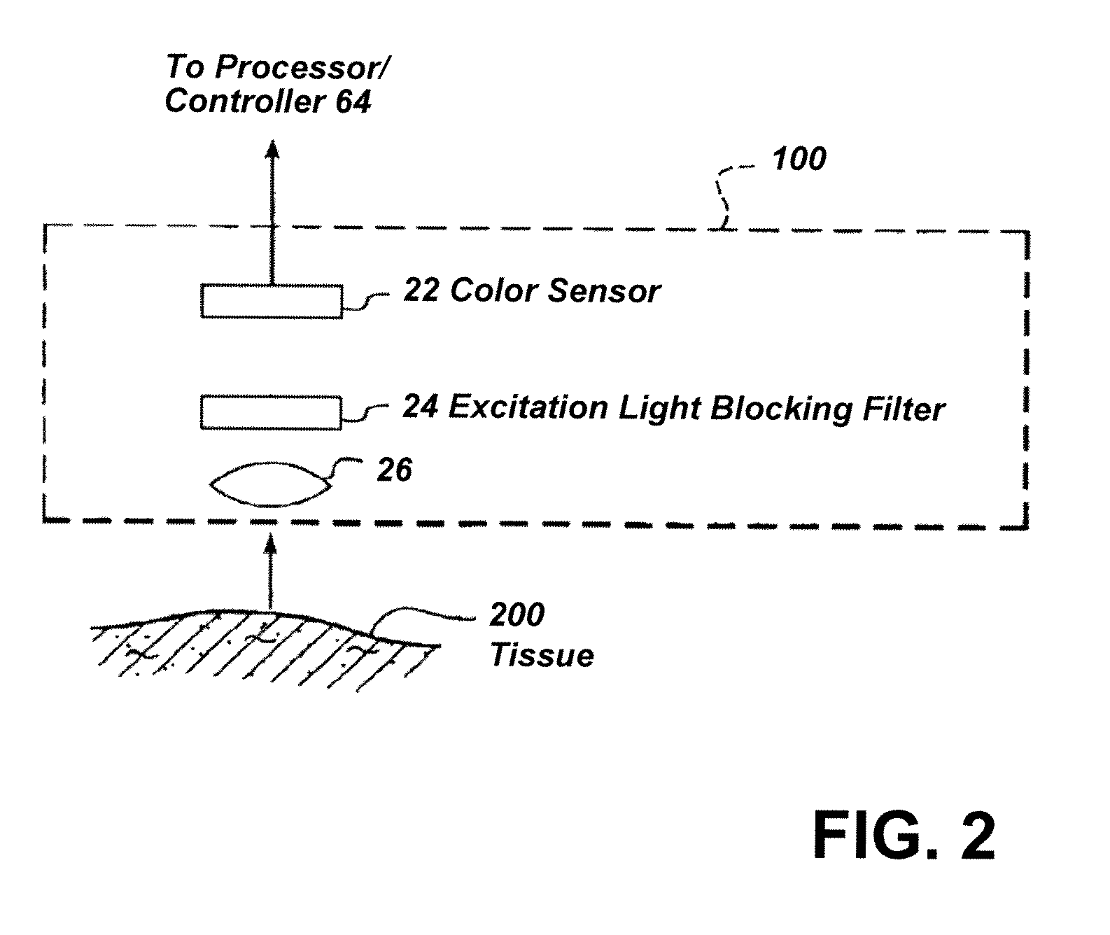
[0014]In one embodiment, a high-resolution video image may be generated by computing a luma image of the combined full-frame fluorescence and white-light image signals and colorizing the luma image based on a ratio of red reflectance to fluorescence signals to produce a superimposed fluorescence / color image for display. Processing an image based on the luma data enhanced the attainable spatial resolution. A change in tissue pathology, as indicated by a change in the fluorescence signal from that tissue, can be represented as a change in color in the video image.

Problems solved by technology

Although switching between imaging modes with an automated device is not difficult, additional time may be required to complete the endoscopic procedure because areas of interest are examined sequentially in each mode.

Method used


Image

  • Imaging system with a single color image sensor for simultaneous fluorescence and color video endoscopy
  • Imaging system with a single color image sensor for simultaneous fluorescence and color video endoscopy
  • Imaging system with a single color image sensor for simultaneous fluorescence and color video endoscopy

Examples


first embodiment

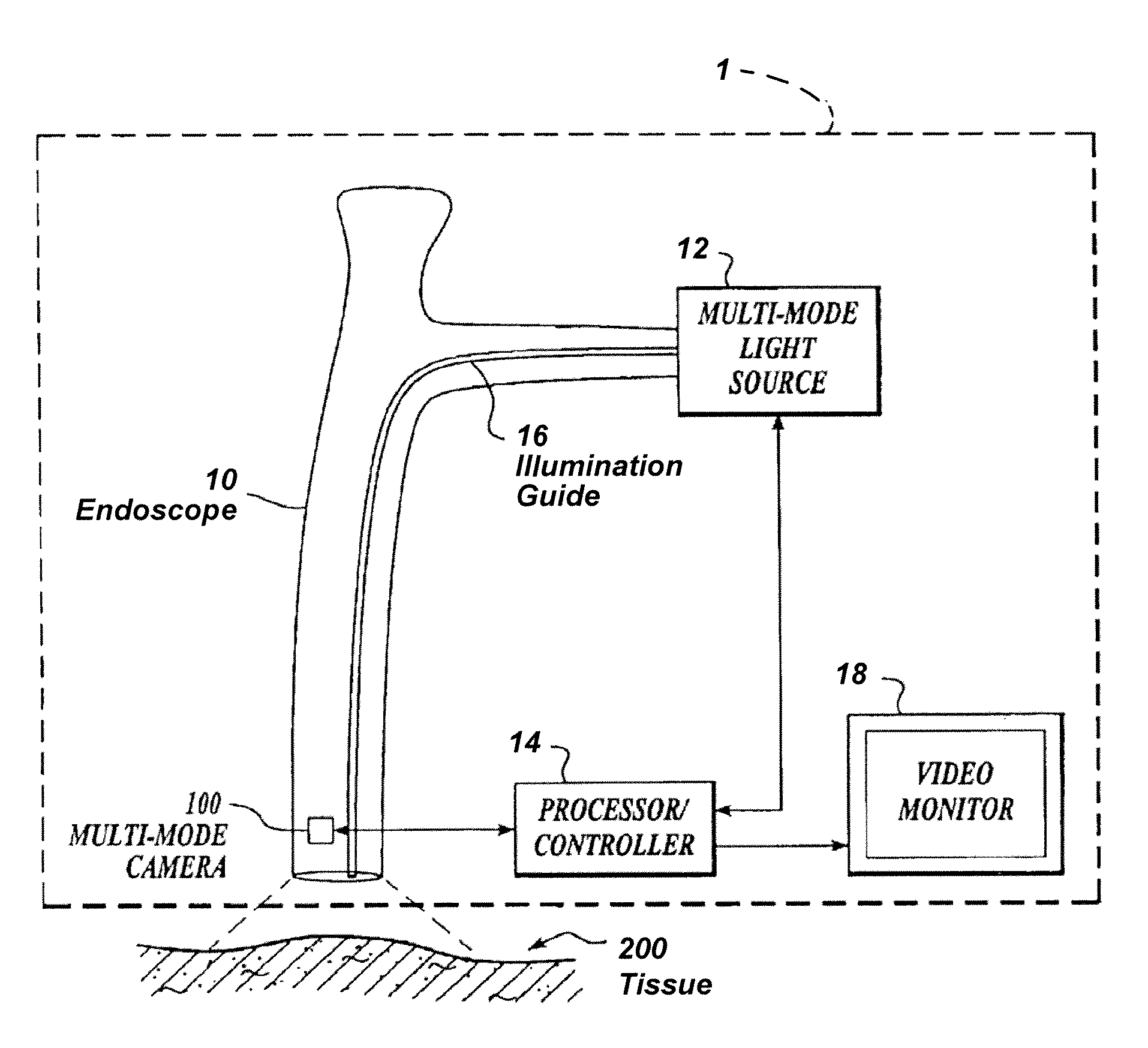
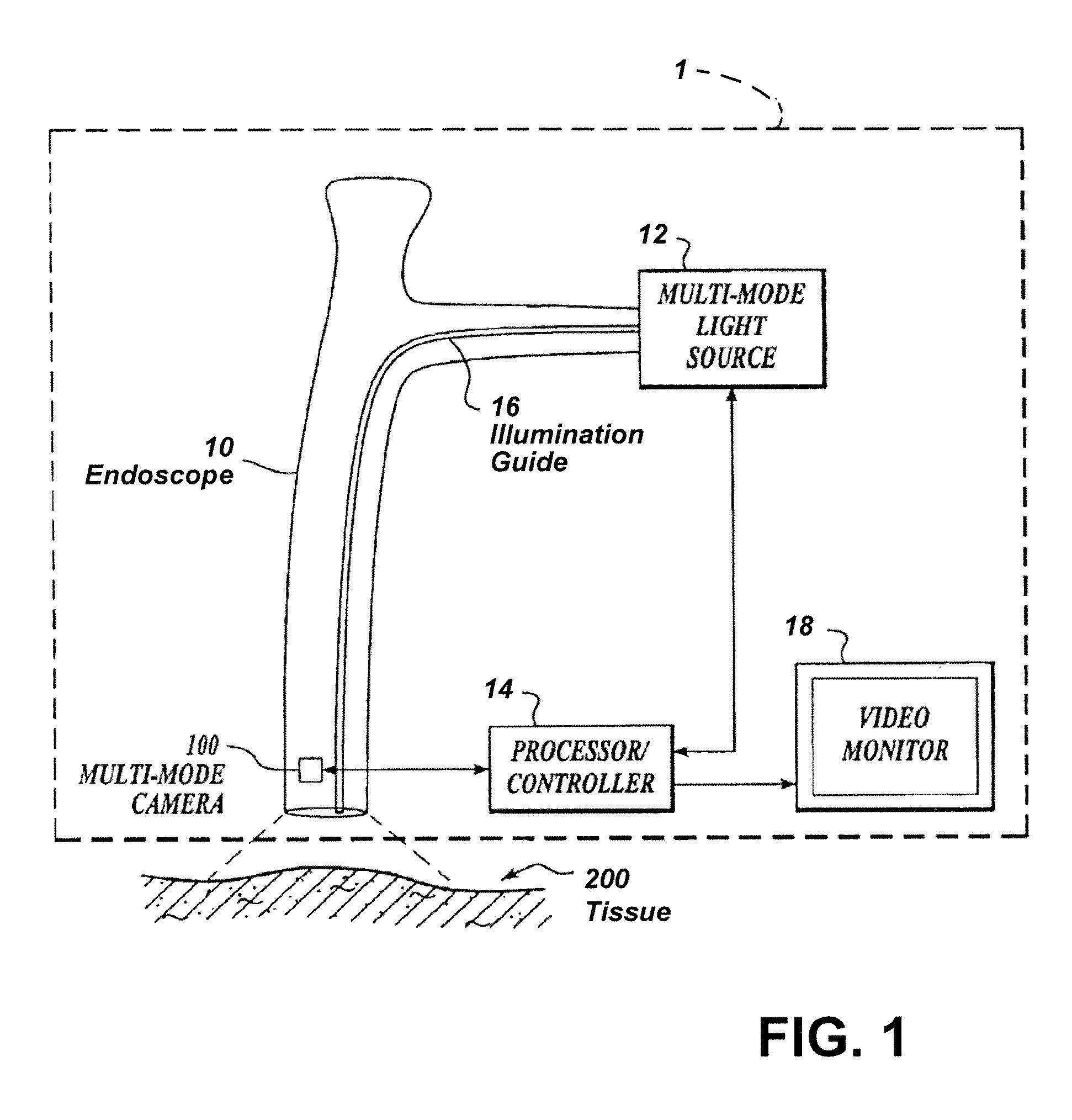
[0034]FIG. 3 shows in more detail a multi-mode light source 30 for simultaneously illuminating a tissue sample 200 with continuous fluorescence excitation light and switched illumination light. Light source 30 includes a first light source 31, for example, an arc lamp, and a collimating lens 33 for producing a high intensity, preferably collimated spectral output Ssource which includes an excitation wavelength range. A bandpass filter 34 filters out spectral components outside the excitation wavelength range Sexcitation and allows only spectral components within the excitation wavelength range Sexcitation to pass. Light source 30 further includes a second light source 32, for example, a halogen lamp, for producing a preferably collimated spectral output Sillumination with a high intensity in an imaging wavelength range covering, for example, the visible spectral range. Light source 32 may be switched with timing signals produced by processor / controller 14, for example, by placing a ...

second embodiment

[0035]FIG. 4 shows a multi-mode light source 40 for simultaneously illuminating a tissue sample 200 with continuous fluorescence excitation light and switched illumination light. Light source 40 includes an excitation / illumination light source 31, for example, an arc lamp, and a collimating lens 33 for producing a high intensity, preferably collimated spectral output Ssource which includes an excitation wavelength range Sexcitation. A dichroic mirror 41 reflects the spectral illumination component Sillumination and passes the excitation wavelength range Sexcitation which may be additionally narrow-band filtered by bandpass filter 34. The light component reflected off a first dichroic mirror 41 is then reflected by mirror 42, passes through a shutter 45 (mechanical or electronic) and is then further reflected by mirror 43 and reflected at second dichroic mirror 44 to become collinear with the excitation light passing through filter 34. As before, the combined collimated excitation / im...


PUM


Abstract

An endoscopic video system and method using a camera with a single color image sensor, for example a CCD color image sensor, for fluorescence and color imaging and for simultaneously displaying the images acquired in these imaging modes at video rates in real time is disclosed. The tissue under investigation is illuminated continuously with fluorescence excitation light and is further illuminated periodically using visible light outside of the fluorescence excitation wavelength range. The illumination sources may be conventional lamps using filters and shutters, or may include light-emitting diodes mounted at the distal tip of the endoscope.

Description

RELATED APPLICATIONS[0001]This application claims the benefit of U.S. Provisional Application Ser. Nos. 60 / 876,597, filed Dec. 22, 2006, and 60 / 908,373, filed Mar. 27, 2007, the disclosures of all of which are incorporated herein by reference in their entirety as if fully set forth herein.BACKGROUND OF THE INVENTION[0002]The invention is directed to methods and systems for simultaneous real-time fluorescence and color video endoscopy at close to video frame rates. The invention is also directed to high-efficiency illumination sources and to methods and systems for controlling temporal and spectral output of these light sources.[0003]Medical endoscopy is increasingly employing specialized optical imaging techniques, such as fluorescence (i.e. autofluorescence and photodynamic) endoscopy, narrow band imaging and other techniques, for improved visualization and for the detection and diagnosis of diseases. Endoscopic imaging systems that provide specialized imaging modes typically also ...

Claims


Application Information

Patent Timeline
Patent Type & Authority Applications(United States)
IPC IPC(8): H04N7/18
CPCA61B1/045A61B1/0638A61B1/0676A61B1/0684H04N5/2256H04N9/045A61B1/043A61B1/0646A61B1/0669A61B1/128H04N7/183H04N23/56H04N23/84H04N25/136H04N23/843A61B3/18A61B1/041H04N23/10H04N23/555A61B1/051
Inventor WESTWICK, PAULPOTKINS, DAVIDFENGLER, JOHN
Owner STRYKER EUROPEAN OPERATIONS LIMITED
Why Eureka
Try Eureka