
Immunoglobulin Fusion Proteins

a technology of immunoglobulin and fusion protein, which is applied in the direction of fusion polypeptide, peptide/protein ingredients, dna/rna fragmentation, etc., can solve the problems of low in vivo stability of the protein, decrease of the bioactivity of the protein, and ineffective method to develop effective pharmaceutical formulations containing the active protein, etc., to increase the expression level of the polypeptide and increase the serum half-life of the biologically active molecul

Active Publication Date: 2008-12-04
POSTECH ACADEMY IND FOUND OF POHANG UNIV OF SCI & TECH POSTECH +1
View PDF0 Cites 66 Cited by
  • Summary
  • Abstract
  • Description
  • Claims
  • Application Information

AI Technical Summary

Benefits of technology

[0013]The present invention provides a hybrid Fc, which is derived from combinations of human IgG subclasses or combinations of human IgD and IgG. The hybrid Fc is effective, when joined to a biologically active molecule, to increase serum half-life of the biologically active molecule as well as increase expression level of the polypeptide when a nucleotide coding for the Fc-polypeptide fusion protein is expressed.
[0040]In another embodiment, there is provided a method of (i) reducing the symptoms of, preventing or treating an autoimmune disease, (ii) inhibiting rejection of a graft, or (iii) treating or preventing endotoxin-induced shock, including administering a therapeutically effective amount of the polypeptide described above, wherein the polypeptide is fused to a biologically active molecule.

Problems solved by technology

However, they may have disadvantages as a therapeutic agent because their in vivo stability is low.
However, since the coupling of an active protein to human albumin only slightly increases its residence time, it was not an effective method to develop an effective pharmaceutical formulation containing the active protein which is coupled to human albumin.
But, the increase in glycosylation of the proteins also leads to a decrease of bioactivity of the proteins.
However, it was reported that PEG conjugation to cytokines or hormones results in a reduced receptor binding affinity due to steric hindrance caused by the conjugation.
However, unlike the fusion with the extracellular domains of cell surface receptors, the fusion with soluble proteins to IgGs leads to reduced biological activities, compared to the non-fused cytokine or growth factors.
The other limitation of the Fc fusion technology is the presence of undesirable immune responses.

Method used


Image

  • Immunoglobulin Fusion Proteins
  • Immunoglobulin Fusion Proteins
  • Immunoglobulin Fusion Proteins

Examples


example 1

Preparation of Expression Vectors for hFc-1, hFc-2, hFc-3, hFc-4, hFc-5, and hFc-6 Fusion Proteins

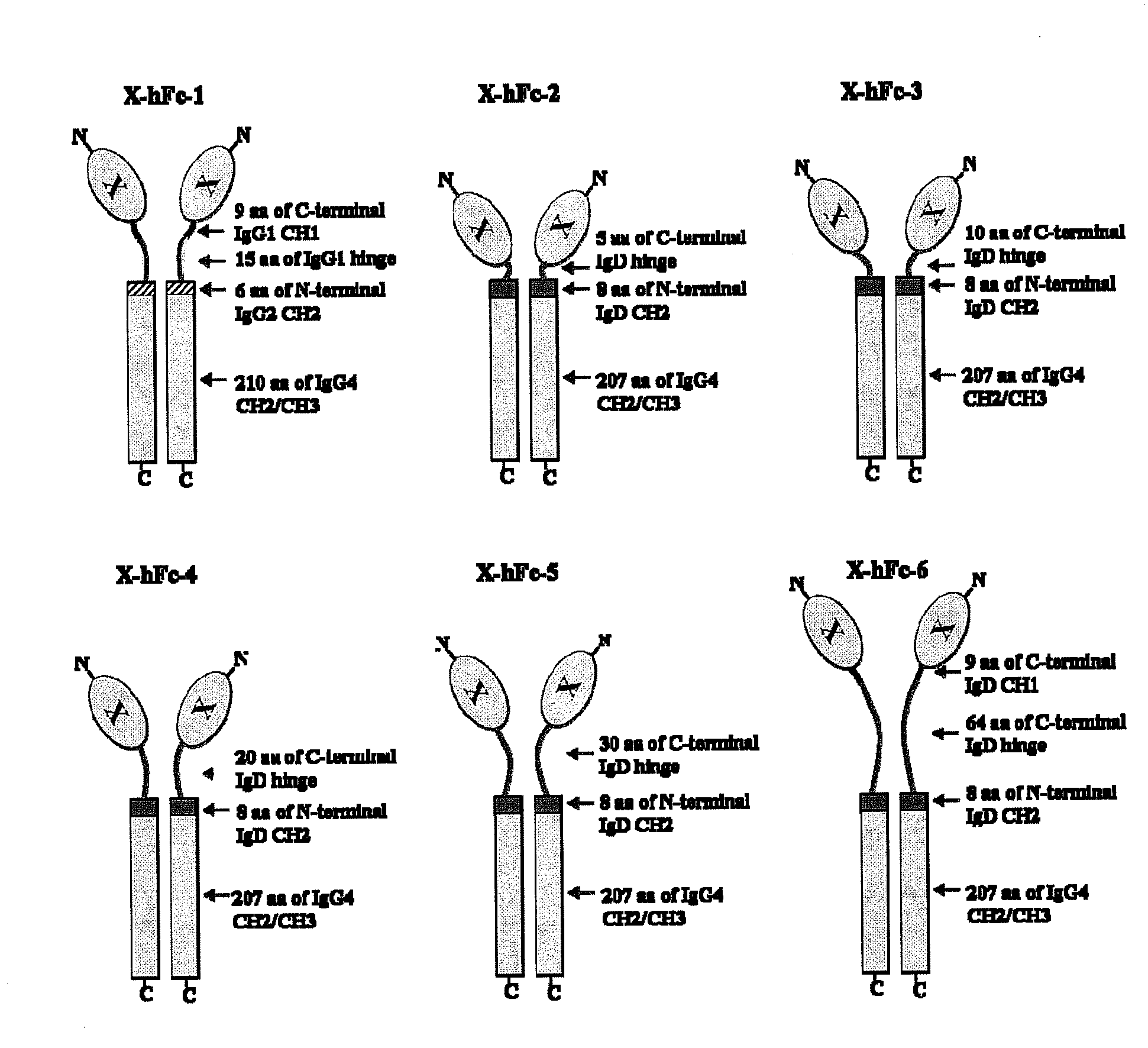
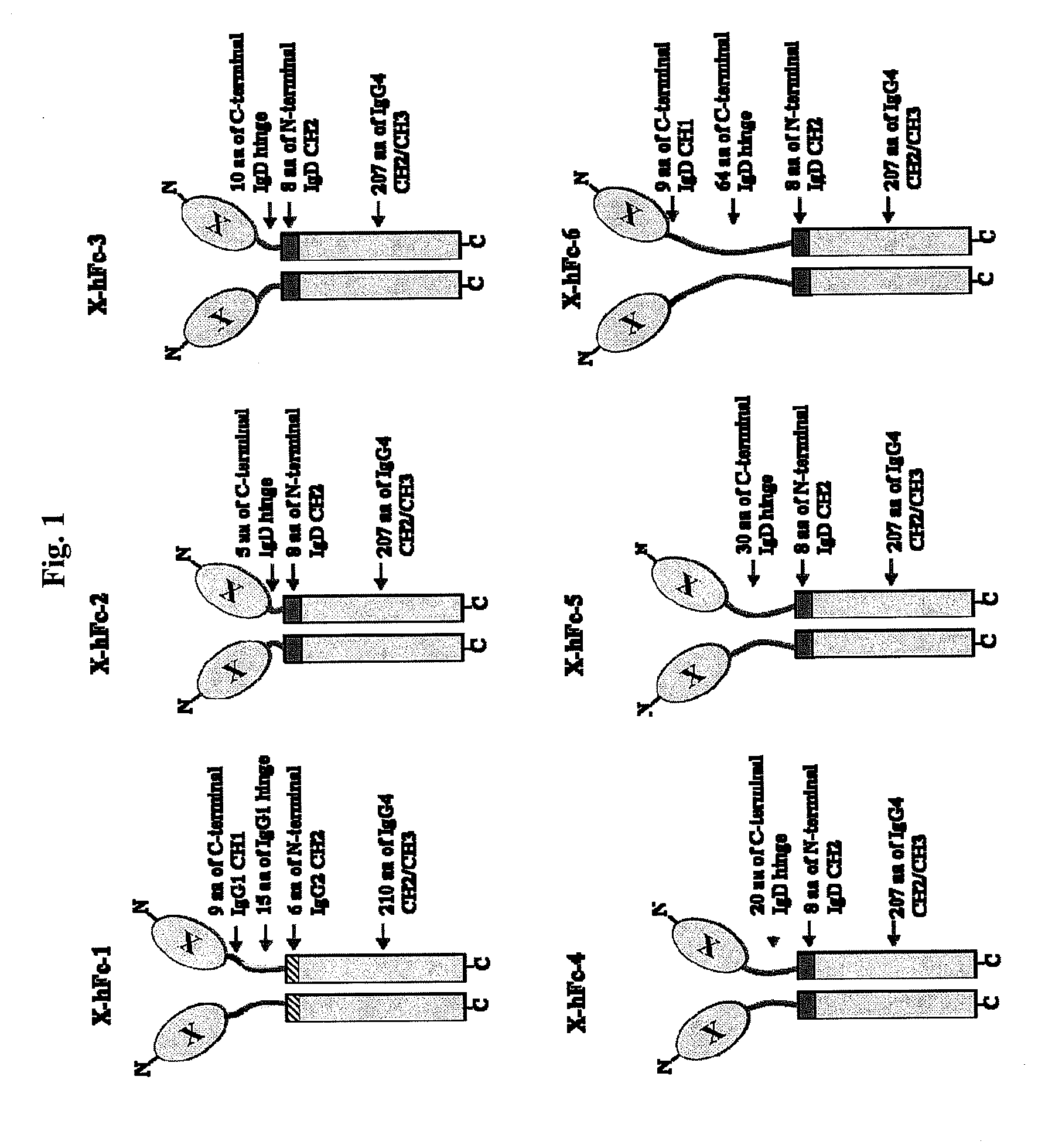
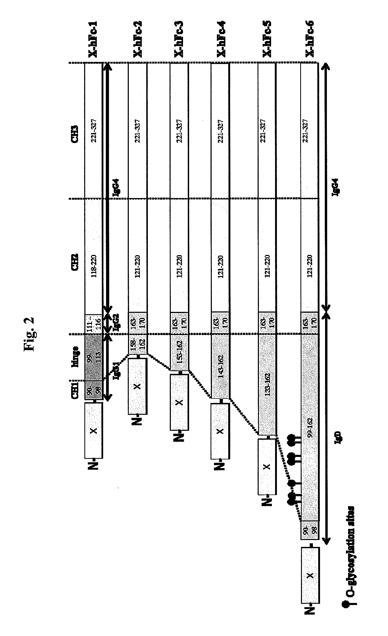
[0143]The hFc-1 includes 9 amino acids (90-98) of C-terminal IgG1 CH1 region, hinge region (99-113) of IgG1, 6 amino acids (111-116) of N-terminal IgG2 CH2 region, 103 amino acids (118-220) of IgG4 CH2 region, and 107 amino acids (221-327) of IgG4 CH3 region (FIGS. 1 and 2). An amino acid sequence of hFc-1 is shown in SEQ ID NO: 18. To obtain codon-optimized nucleotides each coding for hFc-1 (SEQ ID NO: 1), human EPO (SEQ ID NO: 7), human G-CSF (SEQ ID NO: 8) and human p40N303Q (a mutant derived from substitution of Asn with Gln at the 303rd amino acid of human p40 subunit) (a nucleotide sequence of p40N303Q is shown as SEQ ID NO: 9, and an amino acid sequence of human p40 is shown as SEQ ID NO: 17), respectively, these nucleotide molecules were synthesized by custom service of TOP Gene Technologies (Quebec, Canada) (www.topgenetech.com). To increase the level of protein expression, it ...

example 2

Preparation of Expression Vectors for thFc-1 and thFc-2 Coupled to IFN-β

[0145]The thFc-1 includes 23 amino acids (MDAMLRGLCCVLLLCGAVFVSPS) of signal sequence of human tissue plasminogen activator (tPA), 15 amino acids (99-113) of IgG1 hinge region, 6 amino acids (111-116) of N-terminal IgG2 CH2 region, 103 amino acids (118-220) of IgG4 CH2 region, and 107 amino acids (221-327) of IgG4 CH3 region (FIG. 3). An amino acid sequence of thFc-1 is shown in SEQ ID NO: 28. The thFc-2 includes 23 amino acids (MDAMLRGLCCVLLLCGAVFVSPS) of tPA signal sequence, 15 amino acids (148-162) of IgD hinge region, 8 amino acids (163-170) of N-terminal IgD CH2 region, 100 amino acids (121-220) of IgG4 CH2 region, and 107 amino acids (221-327) of IgG4 CH3 region (FIG. 3). An amino acid sequence of thFc-2 is shown in SEQ ID NO: 29. To obtain codon-optimized nucleotides coding for thFc-1 (SEQ ID NO: 26) or thFc-2 (SEQ ID NO: 27) coupled to the N-terminus of human IFN-beta deleted its signal sequence, these n...

example 3

Expression of Human EPO-hFcs, Human G-CSF-hFcs, Human p40N303Q-hFcs, Human TNFR-hFc-5 and thFcs-IFN-beta Proteins

[0147]COS-7 cells were used for expression test and cultured with DMEM media (Invitrogen, Carlsbad) supplemented with 10% fetal bovine serum (Hyclone, South Logan) and antibiotics (Invitrogen, Carlsbad). The vectors encoding EPO-hFcs, G-CSF-hFcs, p40N303Q-hFcs, TNFR-hFc-5, thFcs-IFN-beta were transfected to 5×10⁶ COS-7 cells using conventional electroporation methods. At 48 h after transfection, supernatants and cells were harvested. To check the expression of fusion protein from each vector, all the samples were used for ELISA assay with several kits (R&D system, Minneapolis, #DEP00 for EPO; Biosource, Camarillo, #KHC2032, for G-CSF; R&D system, Minneapolis, #DY1240 for p40N303Q; R&D system, Minneapolis, #DRT200 for TNFR, PBL Biomedical Laboratories, #41410-1A for IFN-beta) and western blot analysis with anti-human IgG antibodies (Santa Cruz Biotechnology, Santa Cruz). ...


PUM

PropertyMeasurementUnit
acidaaaaaaaaaa
solubleaaaaaaaaaa
nucleic acidaaaaaaaaaa

Abstract

Disclosed are fusion proteins comprising a biologically active molecule and an immunoglobulin (Ig) Fc domain which is linked to the biologically active molecule. The Fc domain is a hybrid human Fc domain of (i) IgG1, IgG2 or IgG4 or (ii) IgG4 and IgD. The hybrid Fc is useful as a carrier of biologically active molecules.

Description

[0001]This is a non-provisional application under 35 U.S.C. §111(a) which claims benefit from U.S. provisional application 60 / 940,753 filed on May 30, 2007, which is incorporated herein by reference.TECHNICAL FIELD[0002]The present invention relates to a hybrid human Fc and an immunoglobulin fusion protein in which the hybrid human Fc is joined to a biologically active molecule. In particular, it relates to a hybrid human Fc, which is derived from combinations of human immunoglobulin G (IgG) subclasses or combinations of human IgD and IgG, and a fusion protein in which such an Fc is coupled to a biologically active molecule via a covalent bond.RELEVANT ART[0003]Biologically active molecules may be of great interest therapeutically. However, they may have disadvantages as a therapeutic agent because their in vivo stability is low. Their circulating half-life or serum half-life is short because they are digested by various enzymes in living body. Therefore, it has been desired to impr...

Claims


Application Information

Patent Timeline
Patent Type & Authority Applications(United States)
IPC IPC(8): A61K38/16C07K14/00C07K14/54C07K14/555C07K14/575C12N15/63A61K31/7052A61P1/00A61P19/02C12N5/00C07H21/00C07K14/745C07K16/00C12P21/00
CPCA61K38/00C07K2317/52C12N15/62C07K2319/30C07K2317/53C07K2317/60A61P1/00A61P1/04A61P17/06A61P19/02A61P19/08A61P19/10A61P25/00A61P29/00A61P31/00A61P35/02A61P37/02A61P37/06A61P37/08A61P43/00A61P5/06A61P7/00C07K19/00C07K16/00
Inventor YANG, SEHWANSUNG, YOUNG CHUL
Owner POSTECH ACADEMY IND FOUND OF POHANG UNIV OF SCI & TECH POSTECH
Learn More