
Tissue Ablation by Shear Force for Sampling Biological Fluids and Delivering Active Agents

a technology of biological fluids and tissue ablation, which is applied in the field of tissue ablation by shear force for sampling biological fluids and delivering active agents, can solve the problems of painful lancing, diabetic patients, and painful lancing, and achieve the effect of enhancing sufficient but not excessive membrane ablation

Inactive Publication Date: 2009-01-01
KOLLIAS NIKIFOROS +2
View PDF0 Cites 118 Cited by
  • Summary
  • Abstract
  • Description
  • Claims
  • Application Information

AI Technical Summary

Benefits of technology

The present invention is a method for transporting molecules through a mammalian barrier membrane, such as the skin, buccal, vaginal, and rectal membranes. The method involves ablating the membrane with a shear device containing a shear sheet and a shear member, and then using a driving force to move the molecule through the perforated membrane. The driving force can be selected from a variety of methods, such as iontophoresis, electro-osmosis, reverse iontophoresis, electroporation, phonophoresis, pressure gradients, and concentration gradients. The device can also include a sensor to modify the driving force, such as a pressure sensor, conductivity sensor, or pH sensor. The technical effect of this invention is the ability to transport molecules through mammalian barrier membranes with greater efficiency and precision.

Problems solved by technology

This lancing procedure, however, can be quite painful due to the highly sensitive skin at the fingers.
Unfortunately, diabetic patients have to endure this painful lancing procedures several times a day, in addition to insulin injections, in order to have a tight control of their blood glucose levels, as the treatment requires.
Repeated lancing in limited surface areas (such as on the fingertip) may result in the formation of calluses.
This leads to increased difficulty in drawing blood and increased pain.

Method used


Image

  • Tissue Ablation by Shear Force for Sampling Biological Fluids and Delivering Active Agents
  • Tissue Ablation by Shear Force for Sampling Biological Fluids and Delivering Active Agents
  • Tissue Ablation by Shear Force for Sampling Biological Fluids and Delivering Active Agents

Examples


example 1

Extraction of Interstitial Fluid (ISF) from Human Skin Following Shear Perforation

[0060]Shear perforation of human skin was performed on the ventral leg (calf) of one male volunteer using a commercially available shaver for facial hairs (National Battery-operated Shaver, ES815B, Mastushita Electric Works, Ltd, Japan). The screen of the shaver was covered with a metallic tape (Ideal Tape Co., Lowell, Mass., USA), so that only 1 cm2 square area at the center of the screen was exposed. The shaver was pressed firmly on the skin for about 5 seconds. Neither pain nor discomfort was experienced by the volunteer. The perforation skin site (0.7 cm2) showed only slight erythema after the skin perforation procedure. A hand-held suction device (MityVac II® vacuum pump, Prism Enterprises, San Antonio, Tex., USA) was applied to the perforation skin site with about 70 cm Hg of suction for a certain time duration. Interstitial fluid (a clear, slightly yellowish liquid) extracted out the pores from ...

example 2

Subcutaneous Injection of Insulin as a Control Experiment for Transdermal Insulin Delivery

[0064]The subcutaneous administration of insulin provides a dose response to which a comparative measure of transdermally delivered insulin can be made. An insulin dose of 5 IU insulin (Regular Iletin® II, Insulin Injection, USP, Purified Pork, Eli Lilly and Company, Indianapolis, Ind., USA) was subcutaneously injected in the rear mid-ventral area of each of four female Yorkshire pigs (weight: 22-26 kg) with a 1 cc Sub-Q Precision Glide® Needle (Becton-Dickinson and Company, Franklin Lakes, N.J., USA). FIG. 5a shows that subcutaneous insulin administration resulted in the expected rapid reduction in blood glucose concentration.

[0065]FIG. 5b shows that a 5 IU insulin subcutaneous injection resulted in a peak insulin concentrations of 31-55 μIU / ml. As the insulin concentrations went back to its basal concentration (FIG. 5b), the blood glucose concentrations also were gradually returned toward its...

example 3

Transdermal Delivery of Insulin into Swine Following Shear Perforation of the Skin

[0066]The stratum corneum is recognized as the primary barrier to percutaneous absorption of drug applied to the skin surface. The densely structured outmost skin layer consisting of flattened and keratinized dead skin cells provides a formidable permeation barrier to most small and virtually all large compounds. It is well known in the field of transdermal drug delivery that a protein drug such as insulin cannot penetrate into the intact skin of human and swine. Our own experiment confirmed this (result not shown). This example investigates in vivo transdermal delivery of insulin into swine following application of shear perforation to remove the stratum corneum barrier at the test site.

[0067]Two female Yorkshire pigs of the same body weight range as those in Example 2 were immobilized with anesthetics. The test site on the side of the chest was prepared by clipping lightly off the hair and cleaning w...


PUM

PropertyMeasurementUnit
areaaaaaaaaaaa
current densityaaaaaaaaaa
frequenciesaaaaaaaaaa

Abstract

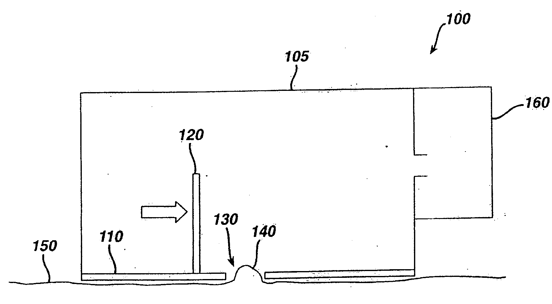
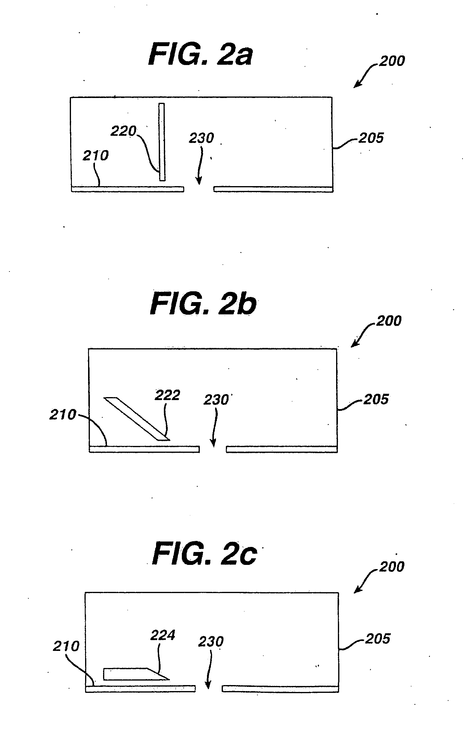
A shear device for use in transporting a molecule through a mammal's mammalian barrier membrane of at least one layer of cells includes a sheet containing an opening(s) and a shear member(s). The sheet is configured for contacting the mammalian barrier membrane (e.g., human skin) and the shear member is configured for ablating a portion of the mammalian barrier membrane forced through the opening(s). This ablating is accomplished by movement of the shear member(s) over the sheet and the opening(s). The ablation of the portion of the mammalian barrier membrane is such that a driving force can be employed to transport the molecule therethrough.

Description

FIELD OF THE INVENTION[0001]The present invention relates to methods and devices for the ablation of barrier membranes using a shear device in order to enable sampling of biological fluids for diagnostic purposes and to enable delivering of active compounds for therapeutic purposes.BACKGROUND OF THE INVENTION[0002]Many medical procedures in use today require a relatively small sample of blood (e.g., 5-50 micro-liters) This blood is often obtained by lancing or piercing the skin (e.g., of the finger) with a needle of a spring-loaded lancet to enable the collection of 1 or 2 drops of blood. See, e.g., U.S. Pat. Nos. 4,503,856 and 4,517,978. This lancing procedure, however, can be quite painful due to the highly sensitive skin at the fingers. Furthermore, to facilitate the blood collection, some forms of pressure gradients, such as either applying a positive pressure (e.g., by squeezing) or a negative pressure (i.e., a suction), is often required to be applied to the cut. See, e.g., U....

Claims


Application Information

Patent Timeline
Patent Type & Authority Applications(United States)
IPC IPC(8): A61B5/00A61N1/30G01N33/66A61B5/05A61B5/145A61B5/15A61B5/157A61B17/20
CPCA61N1/303
Inventor KOLLIAS, NIKIFOROSSUN, YINGANTHONY F., COSTON
Owner KOLLIAS NIKIFOROS
Learn More