Oct-ivus catheter for concurrent luminal imaging

a luminal imaging and octave catheter technology, applied in the field of in vivo imaging apparatus, can solve the problems of myocardial infarction, prone to rupture, no methods are currently available to the cardiologist,

Active Publication Date: 2009-02-12
VOLCANO CORP
View PDF116 Cites 235 Cited by
  • Summary
  • Abstract
  • Description
  • Claims
  • Application Information

AI Technical Summary

Problems solved by technology

Unfortunately, no methods are currently available to the cardiologist that may be applied to specify which coronary plaques are vulnerable and thus prone to rupture.
Although treadmill testing has been used for decades to identify patients at greater cardiovascular risk, this approach does not have the specificity to differentiate between stable and vulnerable plaques that are prone to rupture and frequently result in myocardial infarction.
While OCT imaging provides high resolution (2-20 μm) tomographic visualization of coronary arteries, OCT, however, lacks penetration with a maximum penetration depth of only 2-3 mm into the tissue.
While IVUS uses frequencies from 20 to 40 MHz and provides good depth penetration, it lacks sufficient resolution (˜120 μm) to study thin-cap thrombus or atheroma lesions and other fine details with the vasculature.
Conversely, while OCT provides high resolution (2-20 μm) tomographic visualization of coronary arteries, OCT, however, lacks penetration with a maximum penetration depth of only 2-3 mm.
The current high resolution capabilities of OCT are well suited for imaging vulnerable plaques but poor depth penetration hamper full characterization of coronary lesions and plaque burden.

Method used


Image

  • Oct-ivus catheter for concurrent luminal imaging
  • Oct-ivus catheter for concurrent luminal imaging
  • Oct-ivus catheter for concurrent luminal imaging

Examples


Embodiment Construction

[0025]In the accompanying figures, like elements are identified by like reference numerals among the several preferred embodiments of the present invention.

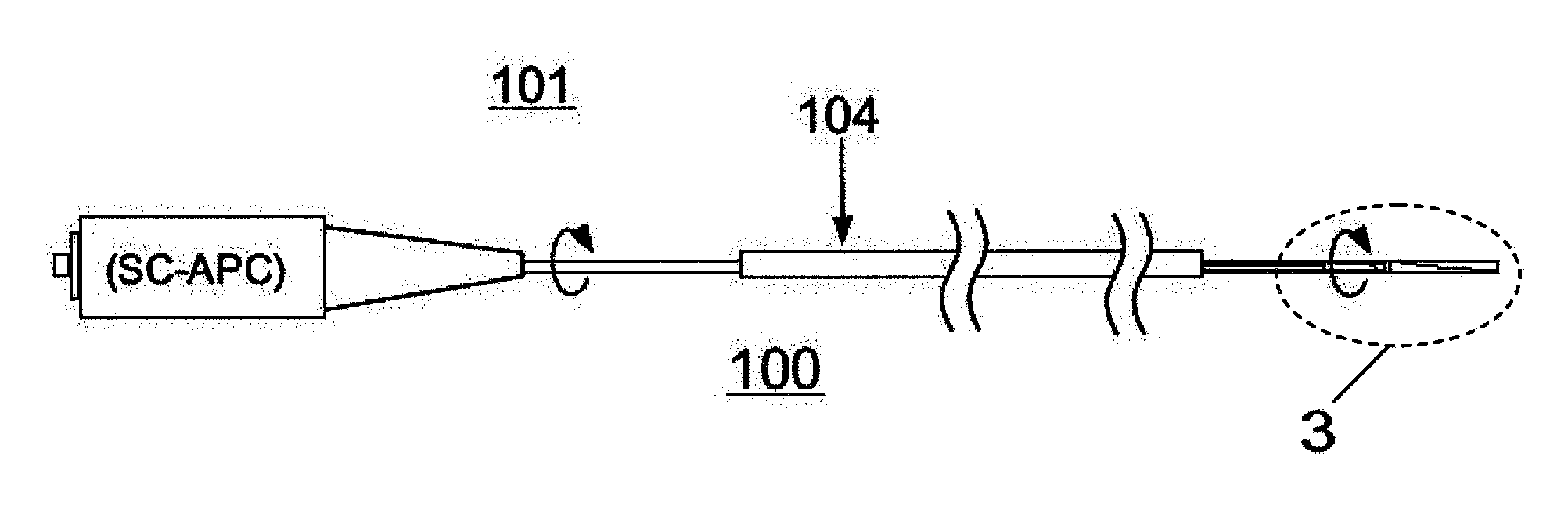
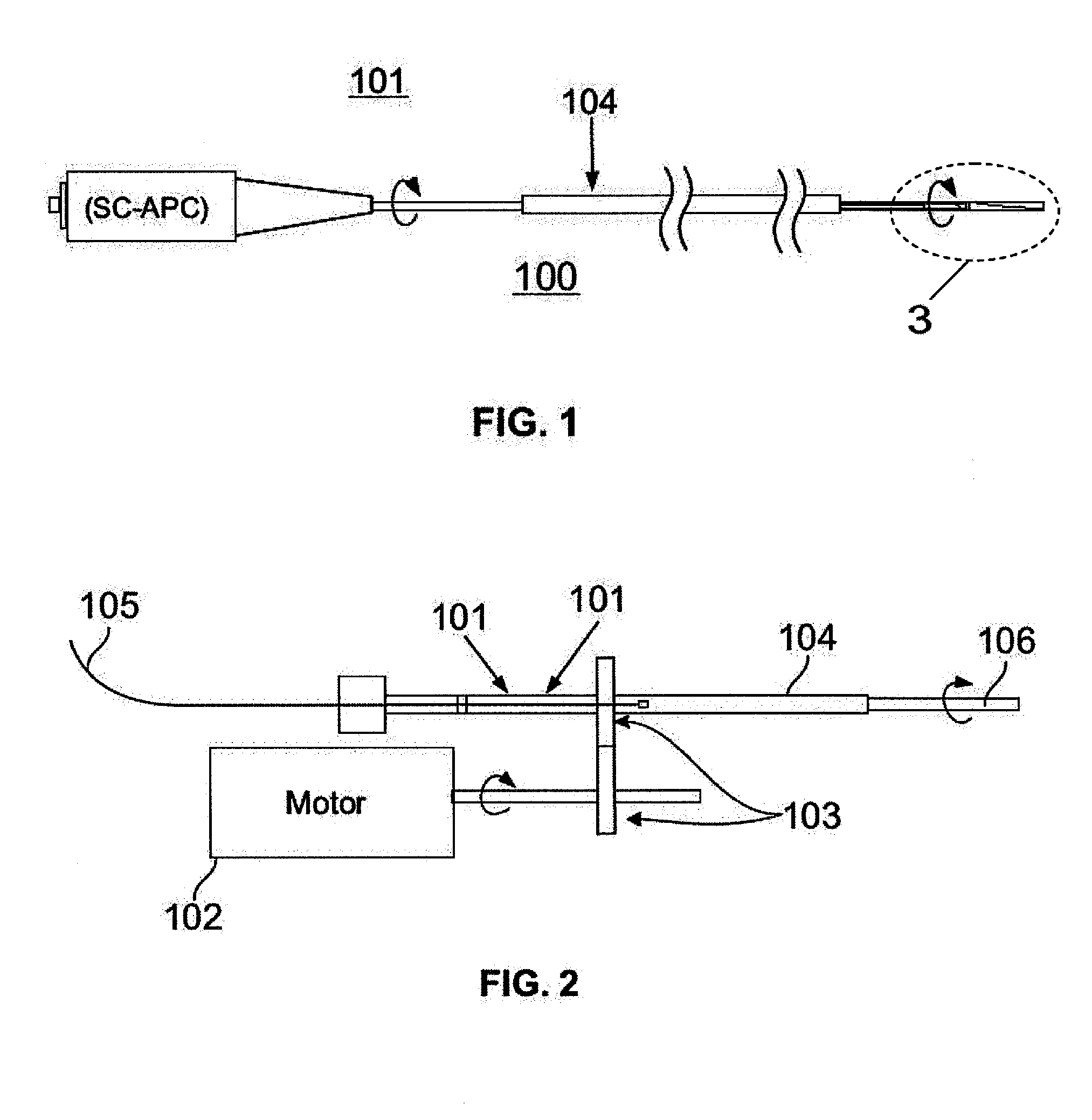
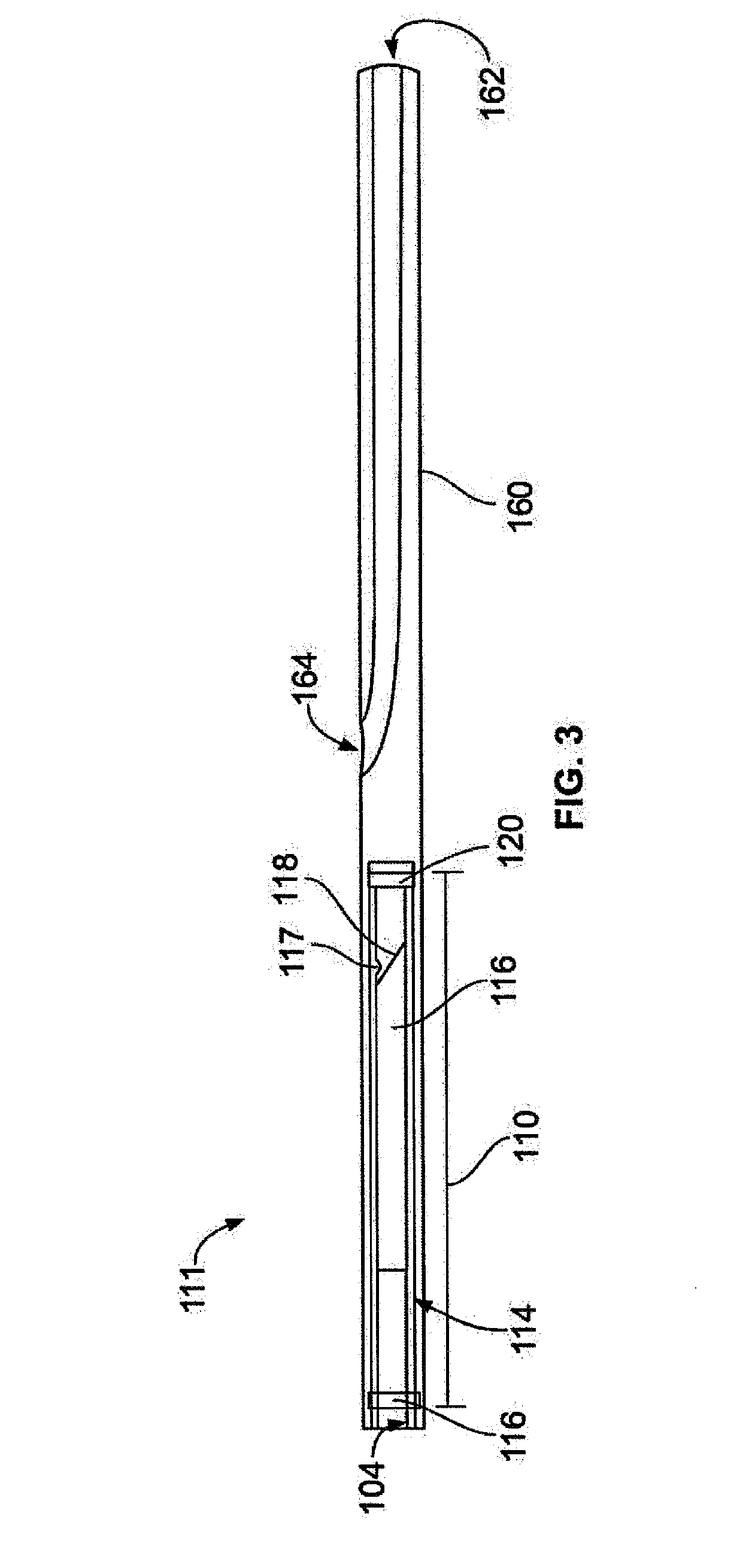
[0026]In the present invention, a distal end assembly including an ultrasound transducer 120 and an optical coherence tomography (“OCT”) optical assembly (as hereinafter described) are positioned longitudinally adjacent or in close proximity to each other at or near a distal end of a catheter assembly 111. Both the ultrasound transducer 120 and the OCT optical assembly are coupled to a rotary drive system (as hereinafter described) that rotates both the OCT optical assembly and ultrasound transducer 120 about their longitudinal axis and within a catheter sheath. In use, the OCT-IVUS catheter assembly 111 is carefully maneuvered through a patient's body to a point of interest such as within a blood vessel to position the distal end in imaging proximity with the point of interest.

[0027]The ultrasound transducer 120 may be a single-...


PUM


Abstract

The invention relates to an apparatus for in vivo imaging. More specifically, the present invention relates to a catheter that incorporates an Optical Coherence Tomography (OCT) system and an Intravascular Ultrasound (“IVUS) system for concurrent imaging of luminal systems, such as imaging the vasculature system, including, without limitation, cardiac vasculature, peripheral vasculature and neural vasculature.

Description

CROSS-REFERENCE TO RELATED INVENTIONS[0001]This application claims priority pursuant to 35 U.S.C. §119(e) from U.S. Provisional Application Ser. Nos. 60 / 949,472 and 60 / 949,511, both filed Jul. 12, 2007 and incorporated by reference herein.BACKGROUND OF THE INVENTION[0002]The present invention relates generally to an apparatus for in vivo imaging. More particularly, the present invention relates to a catheter that incorporates an Optical Coherence Tomography (OCT) system and an Intravascular Ultrasound (IVUS) system for concurrent imaging of luminal systems, such as imaging the vasculature system, including, without limitation, cardiac vasculature, peripheral vasculature and neural vasculature.[0003]Myocardial infarction or heart attack remains the leading cause of death in society. Until recently, many investigators believed that the primary mechanism for myocardial infarction was coronary arteries critically blocked with atherosclerotic plaque that subsequently progressed to total ...

Claims


Application Information

Patent Timeline
Patent Type & Authority Applications(United States)
IPC IPC(8): A61B5/05
CPCA61B5/0066A61B5/02007A61B8/486A61B8/12A61B5/6852A61B5/0084
Inventor CASTELLA, PAULKEMP, NATHANIEL J.MILNER, THOMAS E.ROSENBAUM, DAVID G.
Owner VOLCANO CORP
Why Eureka
Try Eureka