
Devices for cell assays

Active Publication Date: 2009-02-26
PLATYPUS TECH
View PDF45 Cites 21 Cited by
  • Summary
  • Abstract
  • Description
  • Claims
  • Application Information

AI Technical Summary

Benefits of technology

[0011]The present invention relates to the fields of molecular biology, cellular biology, immunology, oncology, developmental biology, stem cell differentiation, general laboratory sciences and microbiology, and in particular to methods and compositions based on liquid crystal assays and other biophotonically based assays for detecting and quantifying the number of cells present on a substrate (allows for the quantit

Problems solved by technology

The populations of many of the heavily industrialized countries are particularly susceptible to cancer induced morbidity and mortality.
Metastasis, the formation of secondary tumors in organs and tissues remote from the site of the primary tumor, is the main cause of treatment failure and death for cancer patients.
Unfortunately, the molecular mechanisms that promote and restrain the metastatic spread of cancer cells have yet to be clearly identified.
Previously described cell migration assays suffer from several problems.
In particular, the assays are not standardized, lack sensitivity and reproducibility, and are not adaptable for conducting large numbers of assays in parallel.

Method used


Image

  • Devices for cell assays
  • Devices for cell assays
  • Devices for cell assays

Examples


example 1

Demonstration of Mask Functionality

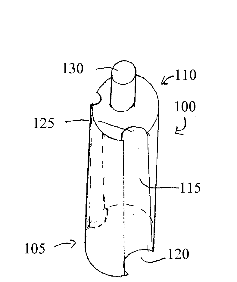
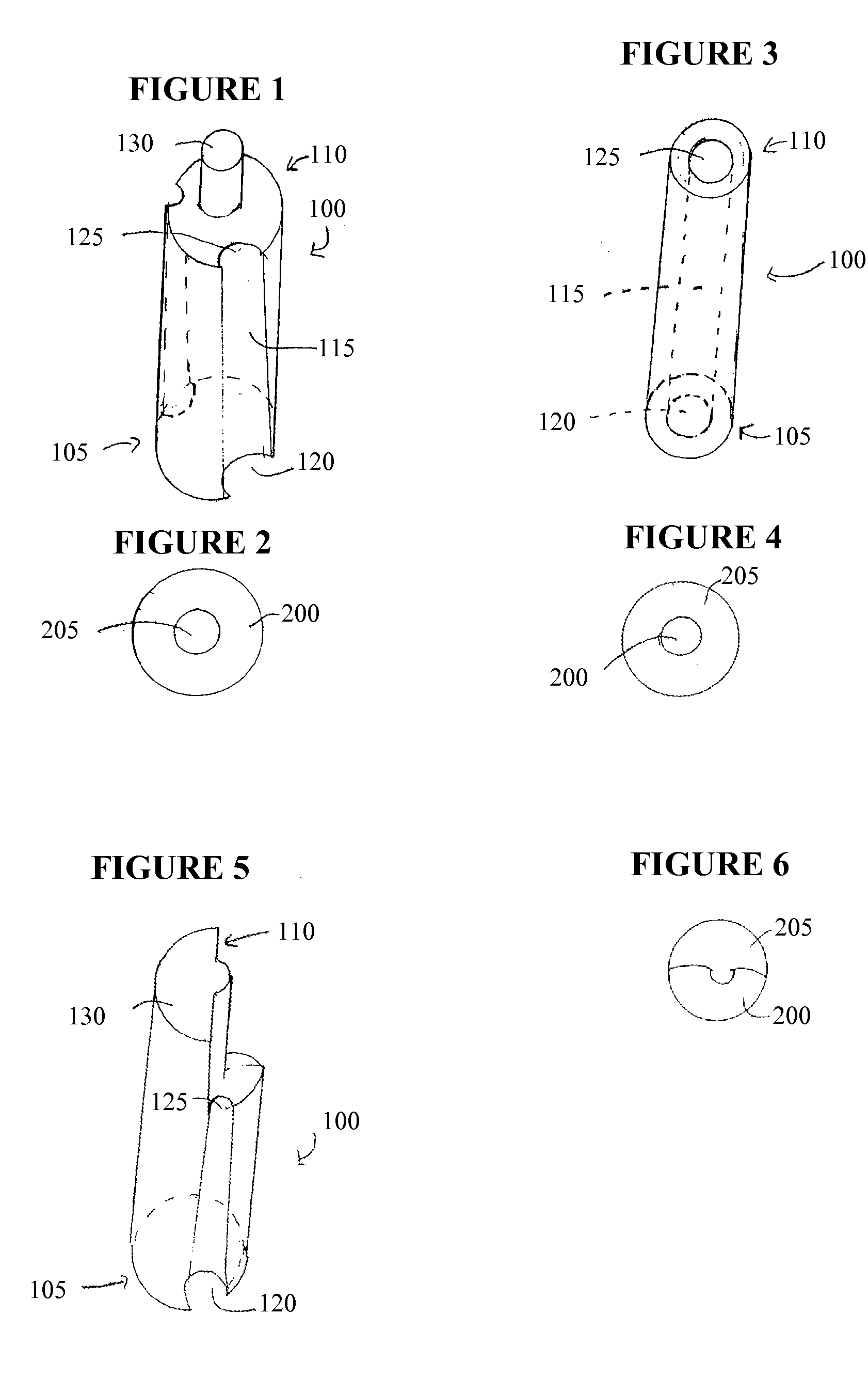
[0176]A 100 ul portion of 3T3 fibroblasts (at 25,000 cells per well and treated with mitomycin C to inhibit proliferation) were seeded into wells of a Greiner 96-well flat bottom plate that contained cell seeding inserts. The fibroblasts were allowed to adhere for four hours at 37° C., 5% CO2. The inserts were then removed from the test wells and the wells were washed with PBS to remove non-adhered cells. A 100 ul volume of cell culture media (MEM containing 10% FBS) was then introduced into each well. In negative control wells, the seeding inserts remained in place for the duration of the incubations. The seeded plate was incubated overnight (˜21 hours) to permit migration of the cells in the test wells. Following incubation, the inserts were removed from the control wells. All wells were washed with PBS and the cells were stained with a fluorescent Calcein AM dye using standard methods per manufacturer instructions. The well contents were observe...

example 2

Influence of Mask Aperture Size

[0178]An analysis of migration assay performance was performed using a set of machined masks, each having 96 apertures of a defined diameter. The aperture diameters tested ranged from 1.8 to 2.3 mm in 0.1 mm increments.

[0179]Cells were seeded into four assay plates containing the silicone inserts and cultured overnight to allow the cells to attach. At this point, the inserts were removed from two of the four plates. The inserts were left in place in the other two plates to serve as controls. Two migration intervals (6 and 22 hours) were evaluated. Following each time interval one test and one control plate was stained with Calcein AM. After 22 hrs over 90% of the analytical area in the wells of the test plate contained cells.

[0180]To determine the extent of cell migration into the exclusion zone, each of the six masks with apertures ranging from 1.8-2.3 mm were fit to the bottom of the stained plates. The prototype masks do not have a complete set of r...

example 3

Photoimmobilized Hyaluronic Acid Transiently Disrupts Cell Adherence to Tissue Culture Plate Surfaces

[0184]This example describes the ability to block adhesion of HT-1080 cells to a surface coated with hyaluronic acid (HA). The HA was functionalized with a photoactive linker and immobilized to the bottom of a well in a tissue culture plate. The material was prepared by reacting the carboxyl group of HA disaccharides with the amine group of a heterobifunctional crosslinker (4-[p-azidosalicylamido]butylamine, ASBA) via carbodiimide chemistry. The other end of the ASBA crosslinker contains a photoactive group, so this reaction renders the HA photoactive. Carboxylate modifications of the HA do not affect its degradability by hyaluronidase (HA-ase). Briefly, the HA was dissolved in MES buffer and reacted with EDC and Sulfo-NHS for 15 min at room temperature. The pH of the buffer was adjusted to ca. 7.0 with concentrated PBS and ASBA was added to the solution and allowed to react for 2 h ...


PUM


Abstract

The present invention relates to the field of molecular diagnostics. In particular, the present invention provided improved substrates and methods of using liquid crystals and other biophotonically based assays for quantitating the amount of an analyte in a sample. The present invention also provides materials and methods for detecting non-specific binding of an analyte to a substrate by using a liquid crystal or other biophotonically based assay formats.

Description

[0001]The Application claims the benefit of U.S. Prov. Appl. 60 / 965,446, filed Aug. 20, 2007, which is incorporated by reference in its entirety.[0002]This application was made with the support of Nat'l Institute of General Medical Sciences (NIGMS) grant 2R44GM069026-03. The government may have certain rights in this invention.FIELD OF THE INVENTION[0003]The present invention relates to the fields of molecular biology, cellular biology, developmental biology, stem cell differentiation, immunology, oncology, general laboratory sciences and microbiology, and in particular to methods and compositions based on liquid crystal assays and other biophotonic based assays for detecting and quantifying the number of cells present on a test surface or within a test substrate and the proliferation, death or movement of cells under controlled conditions and in response to chemotactic and other cytoactive (including compounds that are chemokinetic but not chemotactic and agents that inhibit cell m...

Claims


Application Information

Patent Timeline
IPC IPC(8): C40B30/10C12M1/00G01N33/53C40B60/12C40B50/14
CPCB01L3/5085B01L2300/0851B01L2300/0829B01L2200/0668
Inventor ABBOTT, NICHOLASMURPHY, CHRISTOPHERISRAEL, BARBARASOTOS, JOSHHANSMANN, DOUGHERBER, RENEEBURKHOLDER, JOSEPHHULKOWER, KARENBONDS, MICHAEL
Owner PLATYPUS TECH
Learn More