Focused Ultrasound Therapy System

Inactive Publication Date: 2009-02-26
CHONGQING HAIFU MEDICAL TECH CO LTD
View PDF16 Cites 39 Cited by
  • Summary
  • Abstract
  • Description
  • Claims
  • Application Information

AI Technical Summary

Benefits of technology

[0030]Through the real-time B-mode ultrasound images and diagnosis images, such as the registration of CT or MRI images, the operator can accurately find out the target area to be treated. On the basis of images registration, the present invention makes the registered images, CT or MRI images fusion with real-time B-mode ultrasound images so as to better guide the operator to perform the therapy. On the basis of image registration and fusion and thr

Problems solved by technology

But, the ultrasound image is limited on the depth of observation and it almost cannot display the tissue behind the bone because the bone influences the image greatly, and the serious noises on the images exist during monitoring treatment.
Further, the ultrasound image has poor capacity to identify the tissue boundary and its resolution for tumors is not ideal, particularly B-mode ultrasound image almost can not identify the small tumors and deep-bedded tumors, therefore, it is very difficult for an operator to determine the boundary of tumor and sometimes the tumor even completely can not be determined.
Such therapy system quite depends on the clinical experiences of the operator, the implementation of therapy becomes more complex and the uncertainty of therapeutic results becomes larger.
Or, under relatively complex conditions, it is very difficult to implement the therapeutic procedures and the safety and effects of the therapy cannot be ensured.
The influences on the target area to be treated from this kind of movements can not be learned because of no monitoring, the treatment may st

Method used


Image

  • Focused Ultrasound Therapy System
  • Focused Ultrasound Therapy System
  • Focused Ultrasound Therapy System

Examples


embodiment 1

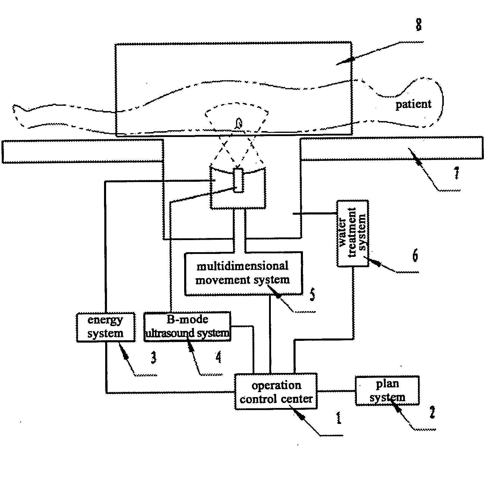
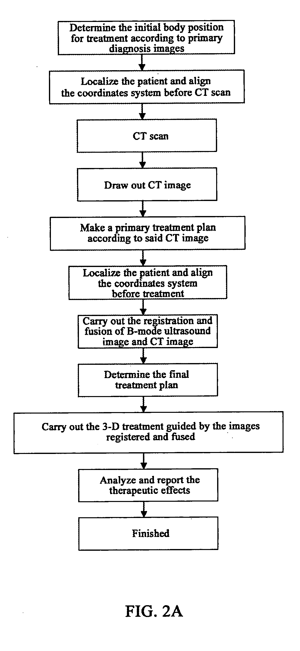
[0041]As shown in FIG. 1 and FIG. 2A, the embodiment of the present invention includes operation control system 1, 3-D treatment plan system 2, energy controller 3, B-mode ultrasound system 4, multidimensional movement system 5, water treatment system 6, treatment bed 7 and locating means 8 and etc.

[0042]In embodiment 1 of the present invention, the diagnosis image is CT image with a relatively high resolution. There are many kinds of existing CT scanners in the market and the products of GE, Philips, Siemens, Toshiba and etc., for example, LightSpeed 16 from GE, can be selected. For the relative information, refer to http: / / www.gehealthcare.com / cnzh / rad / ct / products / light-series / index.html.

[0043]The CT image and B-mode ultrasound image of the patient (the B-mode scanner may adopt ESAOTE DU4, see http: / / www.esaote.com.cn / product.asp) are registered and fused so as to guide the operator to perform the treatment. Or, according to the registered and fused image, 3-D treatment plan is ma...

embodiment 2

[0046]For embodiment 2 of the present invention, refer to FIG. 1 and FIG. 2B. The MRI image for diagnosis with a relatively high resolution and B-mode ultrasound image of the therapy system are registered and fused so as to guide the operator to perform the treatment. Or, according to the registered and fused image, 3-D treatment plan is made and then the automatic treatment under the monitoring by operator is carried out.

[0047]This MRI image can be gained from the equipments sold in the market, for example, Signa MR / i 1.0 / 1.5T from GE (referring to http: / / www.gehealthcare.com / cnzh / rad / mri / products / mri / mri.html).

embodiment 3

[0048]For embodiment 3 of the present invention, refer to FIG. 1 and FIG. 2C. The fused image of CT and MRI with a relatively high resolution and B-mode ultrasound image of the therapy system are registered and fused so as to guide the operator to perform the treatment. Or, according to the registered and fused image, 3-D treatment plan is made and then the automatic treatment under the monitoring by operator is carried out.

[0049]Before fusion of CT and MRI images, CT scan and MRI scan on patient are respectively performed. When scanning, the locating means 8 shall be used for localization and immobilization.

[0050]Other procedures are the same as those in embodiment 1.

The Common Working Procedures of the Embodiments of the Invention

[0051]The other processing procedures of the invention are described in detail thereinafter.

Initial Positioning and Pre-Positioning

[0052]The operations of initial positioning and pre-positioning are very simple and may have been used in many treatment mod...


PUM


Abstract

The present invention pertains to an ultrasound therapy system and more specifically, to a high-intensity focused ultrasound (HIFU) therapy apparatus guided by image registration and fusion method. The therapy system of the present invention comprises a central control means; an acoustic energy applicator; a mechanical driving and locating means of said acoustic energy applicator; a real-time B-mode ultrasound image guiding device. Further the therapy system of the present invention comprises an immobilization means for body position. By the help of this immobilization means for body position, real-time B-mode ultrasound images can be registered with diagnosis images, and then on the basis of registration, said B-mode ultrasound images are fused with said diagnosis images for guiding said therapy. Comparing to the existed technical solutions in this field, the present invention has effectively solved the difficult problems in high-intensity focused ultrasound therapy and particularly for tumor treatment with a low cost. And it provides a very practical technical solution, which can be easily applied to clinical treatment of tumors.

Description

FIELD OF THE INVENTION[0001]The present invention pertains to an ultrasound therapy system and more specifically, to a high-intensity focused ultrasound (HIFU) therapy apparatus guided by using image registration and fusion method.BACKGROUND OF THE INVENTION[0002]In recent years, the using scope of the ultrasound therapy has become wider increasingly. With the development of ultrasound therapy, the special ultrasound therapy, in particular, the High-intensity focused ultrasound (HIFU) applies a damaging dose to effectively treat many kinds of diseases, particularly the tumor. Comparing to the conventional surgery and chemotherapy, HIFU therapy brings the patient less trauma and can realize a non-invasive treatment. Therefore its clinical applications have been developed rapidly. Its indications include liver cancer, bone sarcoma, breast cancer, pancreas cancer, kidney cancer, soft tissue tumor, pelvic tumor and etc.[0003]The ultrasound tumor therapy devices normally adopt the sphere...

Claims


Application Information

Patent Timeline
IPC IPC(8): A61B8/00A61N7/00
CPCA61B19/203A61N7/02A61B2019/5276A61B90/14A61B2090/378A61N7/00
Inventor LIN, TAOCHEN, WENZHIWEN, YINGANGWANG, ZHIBIAOWANG, ZHILONG
Owner CHONGQING HAIFU MEDICAL TECH CO LTD
Why Eureka
Try Eureka