Balloon cannula system for accessing and visualizing spine and related methods

a cannula system and ball cannula technology, applied in the field of ball cannula system for accessing and visualizing spine, can solve the problems of lack of real-time visualization capabilities, difficulty in accurately diagnosing back pain, and lack of tactile sensation of the device for the surgeon or allower, so as to enhance direct visualization and enhance direct visualization

Inactive Publication Date: 2009-03-05
SPINE VIEW INC
View PDF71 Cites 91 Cited by
  • Summary
  • Abstract
  • Description
  • Claims
  • Application Information

AI Technical Summary

Benefits of technology

[0006]In one embodiment, a balloon cannula system (access system) is fitted with an extrusion (e.g. deflated balloon material) that is terminally bonded. Following positioning of the balloon cannula system at the targeted site to be treated, the balloon may be inflated and may be used as an atraumatic tool for dissection and / or an atraumatic tool to create 
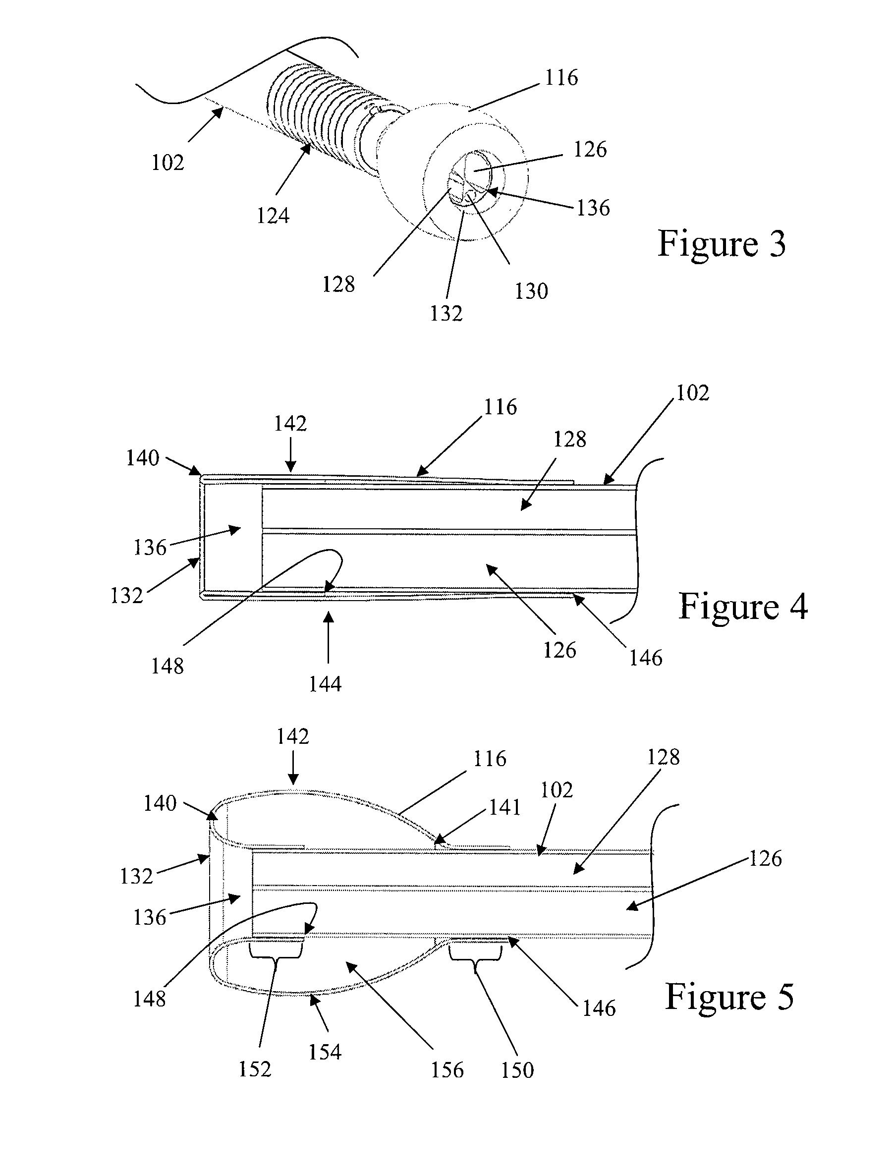
working space, thereby enhancing visualization of the surrounding structures. In one embodiment, the balloon is a forward-looking structure so that the distal tip of the balloon may push obstructive tissue away from the scope, and the distal tip of the balloon may provide a depth of view between the scope and the targeted sites to be treated.
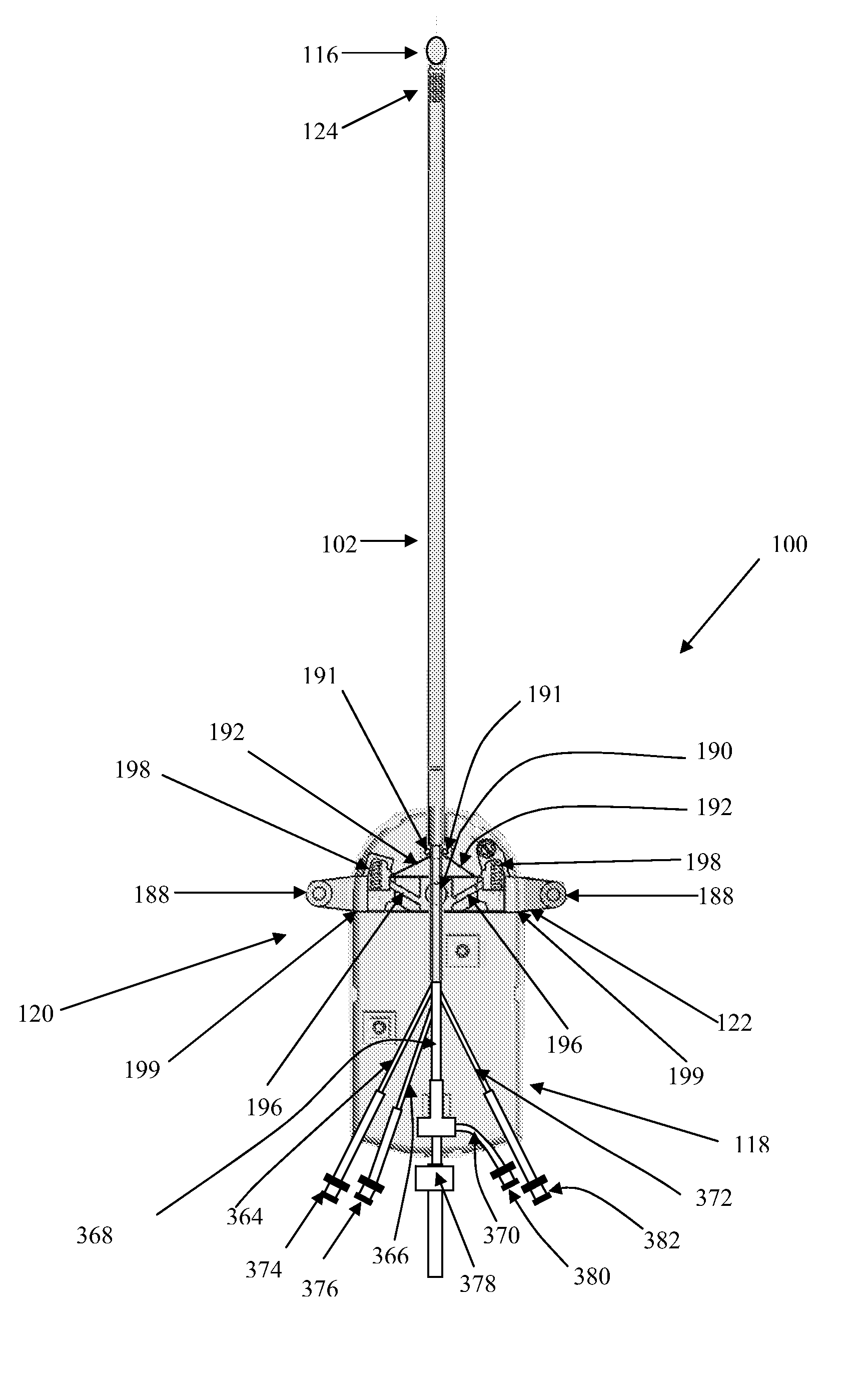
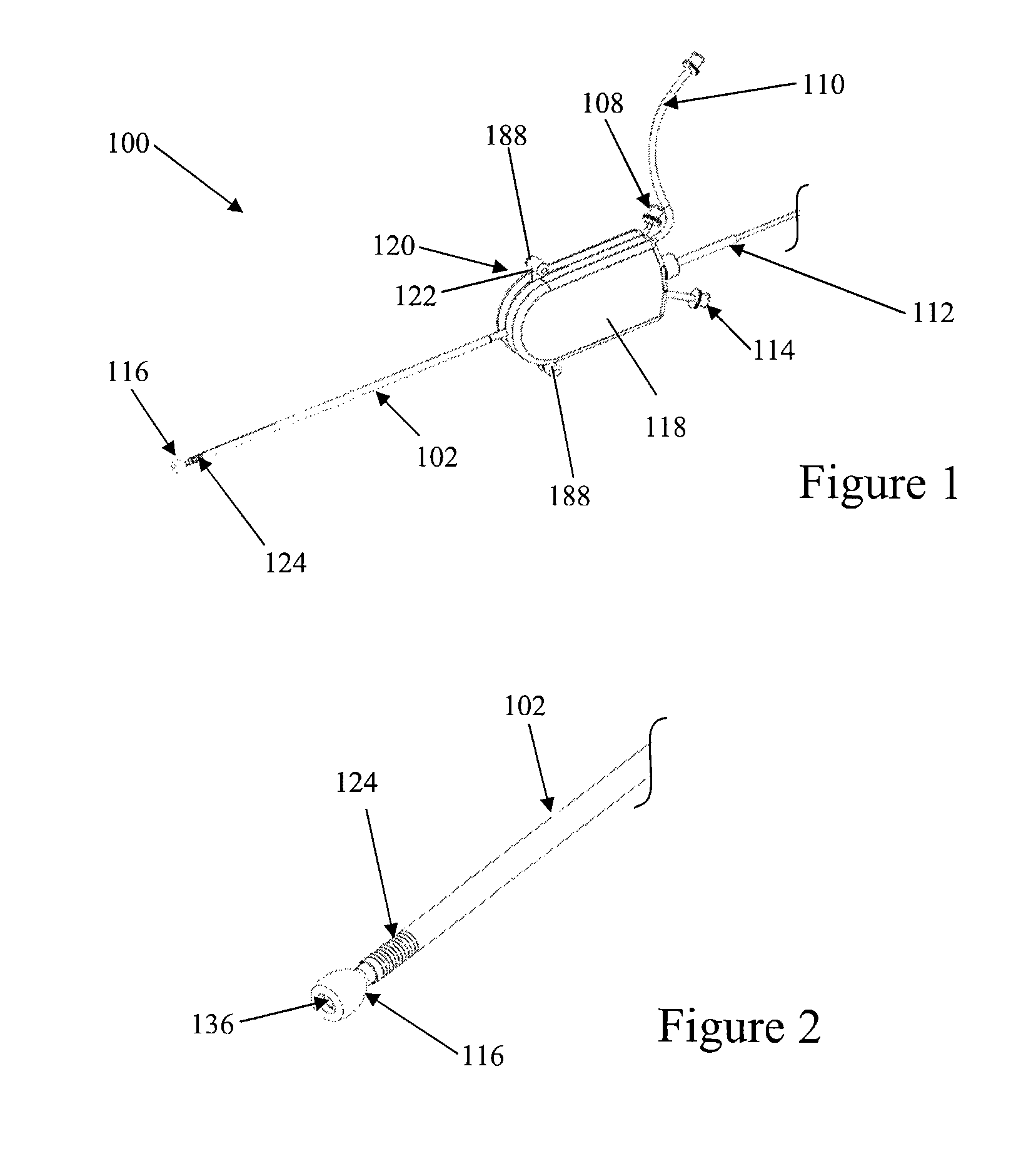
[0007]One embodiment is directed to a balloon cannula device comprising a multi-lumen elongated shaft, a balloon attached at its distal end of the shaft, wherein the proximal end of the balloon and distal end of the balloon are attached to the outer surface of the same elongated shaft, and wherein the balloon is constructed such that following inflation of the balloon, said balloon is forward-looking to create a working space distally to the viewing scope to enhance direct visualization. In another embodiment, the balloon of the balloon cannula system includes at least one portion that is elastically expandable. The expandable balloon may be inflated with air, sterile saline, contrasting agent, or other agents that may be delivered via a syringe or a pump. In some embodiments, the balloon is able to simultaneously undergo radial expansion and keep the forward-looking feature of the balloon cannula system. In one or more of the embodiments described herein, the distance between the points of attachment of the balloon to the same outer shaft of the elongated shaft is between about 1 mm and about 15 mm. In another embodiment, one end of the balloon is attached to a distal end of the balloon catheter in a flipped manner (e.g. everted or inverted), such that the internal surface of the balloon is in contact with the elongated shaft distally, and the outer surface of the balloon is in contact with the same elongated shaft proximally. In yet another embodiment, the balloon includes at least one elastically deformable portion. In yet another embodiment, the deformable portion is constructed of polyurethane.
[0010]In one embodiment, a method for treating intervertebral disc degeneration in a spine includes introducing a balloon cannula device that permits direct visualization capability into a portion of the spine, inflating the balloon cannula to create a forward-looking capability to enhance visualization and displacement of tissues, and introducing a therapy device into the balloon cannula device to treat disc degeneration.
[0011]In another embodiment, a method for treating intervertebral disc degeneration in a spine of a body includes making an incision into a skin of the body, introducing a balloon cannula device that permits direct visualization into a portion of the spine, inflating the balloon cannula to create a forward-looking capability that enhances visualization and displacement of tissues, introducing a therapy device into balloon cannula device to treat disc degeneration, and treating the disc degeneration.
[0017]In another embodiment, a balloon cannula device for assessing a target site within the body may include a multi-lumen elongated shaft and a balloon attached at a distal end of the shaft, wherein the proximal end and distal end of the balloon are attached to the outer surface of the elongated shaft and wherein the balloon is constructed such that following inflation of the balloon, the balloon is forward-looking and create a working space distally to the viewing scope to enhance direct visualization.
[0018]In another embodiment, a balloon cannula device for visualizing a target site within body may include a proximal portion and a distal portion, at least three lumens positioned within the proximal portion, wherein at least one lumen is suitable for allowing the passage of endoscope, at least one lumen is suitable for inflation of a balloon, and at least one lumen is suitable for allowing passage of therapeutic instruments or injection of medications. In some embodiments, at least two lumens may be positioned within the distal end, and at least one of the lumens permits visualization of therapeutic instruments or injected medications. A balloon may be attached to an outer surface of the distal portion of the balloon cannula device, and at least part of the distal portion of the balloon cannula device may be constructed such that following inflation of the balloon, the balloon is forward-looking to create a working space distally to enhance direct visualization. In one or more of the embodiments described herein, the balloon is constructed of polyurethane, but in other embodiments may be constructed of a polymer material other than polyurethane.

Problems solved by technology

However, these devices also provide little in the form of tactile sensation for the surgeon or allow the surgeon to atraumatically manipulate surrounding tissue.
In general, these conventional systems rely on external visualization for the approach to the disc and thus lack any sort of real time, on-board visualization capabilities.
Furthermore, accurately diagnosing back pain is often more challenging than many people expect and often involves a combination of a thorough patient history and physical examination, as well as a number of diagnostic tests.
A major problem is the complexity of the various components of the spine, as well as the broad range of physical symptoms experienced by individual patients.
These anatomical elements make it difficult to treat or diagnose conditions within the epidural area because they tend to collapse around any instrument or device inserted therein.
This may reduce visibility in the epidural space, and may cause inadvertent damage to nerve roots during device insertion.
Also, the insertion of a visualization device may result in blocked or reduced viewing capabilities.
As such, many anatomical elements within the epidural space may limit the insertion, movement, and viewing capabilities of any access, visualization, diagnostic, or therapeutic device inserted into the epidural space.

Method used


Image

  • Balloon cannula system for accessing and visualizing spine and related methods
  • Balloon cannula system for accessing and visualizing spine and related methods
  • Balloon cannula system for accessing and visualizing spine and related methods

Examples


Embodiment Construction

[0059]Conventional systems often rely on external visualization such as fluoroscopy and CT scanning for the approach to the disc, and thus lack any sort of real time, on-board visualization capabilities. Also, existing devices provide little in the form of tactile sensation for the surgeon and do not allow the surgeon to atraumatically manipulate surrounding tissue.

[0060]There is a need, therefore, for minimally invasive techniques and systems that provide the capability to diagnose or repair the spine using direct visualization while minimizing damage to surrounding anatomical structures and tissues. There is also a need for a method and device that allows a physician to effectively enter the epidural space of a patient, clear an area within the space to enhance visualization and use the visualization capability to diagnose and treat the disc injury.

[0061]The embodiments disclosed herein will be more clearly understood and appreciated with respect to the following Detailed Descript...


PUM

PropertyMeasurementUnit
inflation pressureaaaaaaaaaa
distanceaaaaaaaaaa
distanceaaaaaaaaaa

Abstract

Balloon cannula systems may be used for accessing and visualizing the spine and related methods of treatment, including a forward-looking balloon system for creating a working space and the balloon system having atraumatic dissection capability to allow visualization in spine. The devices and methods described may be used, for example, to perform annulus repair, herniated disc excision, and denervation of neurological tissue; to dispense pharmacological agents and / or cell or tissue therapy agents; to diagnose disc degeneration and bony degeneration, spinal stenosis, and nucleus decompression, and to perform disc augmentation.

Description

CROSS-REFERENCE TO RELATED APPLICATIONS[0001]The present application claims priority under 35 U.S.C. §119(e) to U.S. Provisional Application Ser. No. 60 / 968,086, filed Aug. 27, 2007, and U.S. Provisional Application Ser. No. 61 / 045,919, filed Apr. 17, 2008, all of which are hereby incorporated by reference in their entirety.BACKGROUND OF THE INVENTION[0002]Injured intervertebral discs are generally treated with bed rest, physical therapy, modified activities, and pain medications for substantial treatment durations. There are also a number of treatments that attempt to repair injured intervertebral discs and to avoid surgical removal of injured discs. For example, disc decompression is a procedure used to remove or shrink the nucleus, thereby decompressing and decreasing the pressure on the annulus and nerves. Less invasive procedures, such as microlumbar discectomy and automated percutaneous lumbar discectomy, remove the nucleus pulposus of a vertebral disc by aspiration through a ...

Claims


Application Information

Patent Timeline
Patent Type & Authority Applications(United States)
IPC IPC(8): A61B17/70A61B1/32B23P11/00A61B1/018A61F2/958
CPCA61B1/00082A61B1/0051A61B1/012A61B1/3135A61B17/0218A61B2017/003Y10T29/49826A61B2017/320048A61B2019/481A61B2217/005A61M25/0054A61M25/0138A61M25/10A61B2017/00557A61B2090/08021
Inventor CHIN, SINGFATTTO, JOHN T.JANSEN, LEX P.FABRO, MYRA I.L.
Owner SPINE VIEW INC
Why Eureka
Try Eureka