System using a helical retainer in the direct plication annuloplasty treatment of mitral valve regurgitation

a technology of mitral valve and helical retainer, which is applied in the field of direct plication annuloplasty can solve the problems of imperfect valve closure, problem of mitral valve gap, and no commercially successful device for the treatment of mitral valve regurgitation in such a manner currently exists, so as to reduce the possibility of clot development, reduce interference with leaflet motion or

Inactive Publication Date: 2009-04-23
CORDIS CORP
View PDF107 Cites 141 Cited by
  • Summary
  • Abstract
  • Description
  • Claims
  • Application Information

AI Technical Summary

Benefits of technology

[0021]Current tissue retainers are clips that have a profile outside the plane of the tissue which they plicate or retain . The helical retainer may be driven completely into the tissue with no part of the retainer being left exposed. In the heart, this will reduce the possibility of clots developing around the retainer. At or near the mitral valve, this will also provide the advantage of reducing interference with leaflet motion or the possibility of eroding leaflets over time.

Problems solved by technology

One of the problems that a catheter based device and system could be used to treat in a minimally invasive manner is mitral valve regurgitation, however, no commercially successful device for the treatment of mitral valve regurgitation in such a manner currently exists.
Mitral valve regurgitation is the backflow of blood from the left ventricle into the left atrium due to an improper alignment of the leaflets of the mitral valve thereby causing an imperfect closure of the valve.
The combination of the dilated annulus and the tethering on the leaflets prevents the leaflets from closing properly, thereby causing the problematic gap in the mitral valve.
The resultant backflow through the mitral valve reduces the efficiency of the heart resulting in a need for the heart to beat faster and / or more forcefully in order to produce the same amount of blood flow.
Mitral valve regurgitation may be asymptomatic in some patients but in other patients the reduction in blood flow and the resultant strain on the heart could result in arrhythmias, heart attack and possibly death.
The preferred current treatments for mitral valve regurgitation require open-heart surgery and / or the use of endoscopic techniques that are difficult for the surgeon and potentially dangerous for the patient.
Such heterologous valves may be used in humans but often wear-out prematurely and additional open-heart surgery is required to replace such valves with additional heterologous or mechanical valves.
Long-term anticoagulant treatment causes other problems such as unwanted internal and external bleeding and possibly strokes.
Once implanted, tissue generally grows over the annuloplasty ring, and a line of contact between the annuloplasty ring and the mitral valve will essentially enable the mitral valve to appear and function as a normal mitral valve by reestablishing coaptation of the mitral valve leaflets but the durability of the effect is variable and may decline within six months after the procedure.
Although a patient who receives the annuloplasty ring may be subjected to anti-coagulant therapies, the therapies are not extensive, as a patient is only subjected to the therapies for a matter of weeks, e.g., until tissue grows over the annuloplasty ring.
Such devices are expensive and difficult to implant and require the patient to use anti-coagulant therapy indefinitely.
Long-term use of anti-coagulant therapy may result in unnecessary bleeding and strokes.
While such invasive surgical procedures have under certain circumstances been shown to be effective in the treatment of mitral valve leakage, invasive surgical procedures often have significant drawbacks.
Any time a patient undergoes open-heart surgery, there is a risk of infection.
Opening the sternum and using a cardiopulmonary bypass machine has also been shown to result in a significant incidence of both short and long term neurological deficits.
Some minimally invasive procedures have been developed to treat mitral valve regurgitation but, to date, none have become commercially successful standard procedures.
In this system the number of permanent implants required in order to achieve an initial effect, and commitment to these implants before success of effect is able to be determined are serious drawbacks.

Method used


Image

  • System using a helical retainer in the direct plication annuloplasty treatment of mitral valve regurgitation
  • System using a helical retainer in the direct plication annuloplasty treatment of mitral valve regurgitation
  • System using a helical retainer in the direct plication annuloplasty treatment of mitral valve regurgitation

Examples


Embodiment Construction

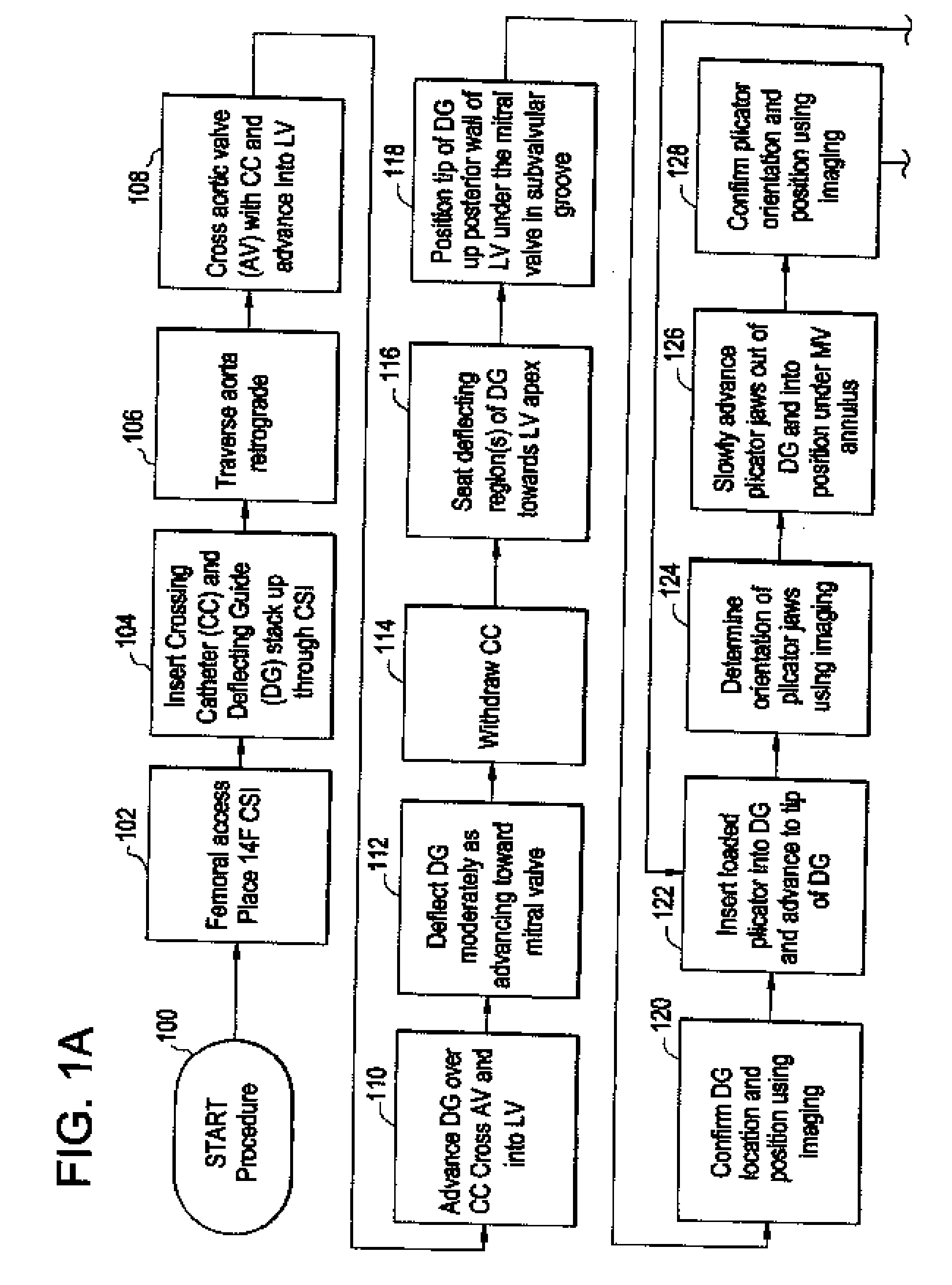
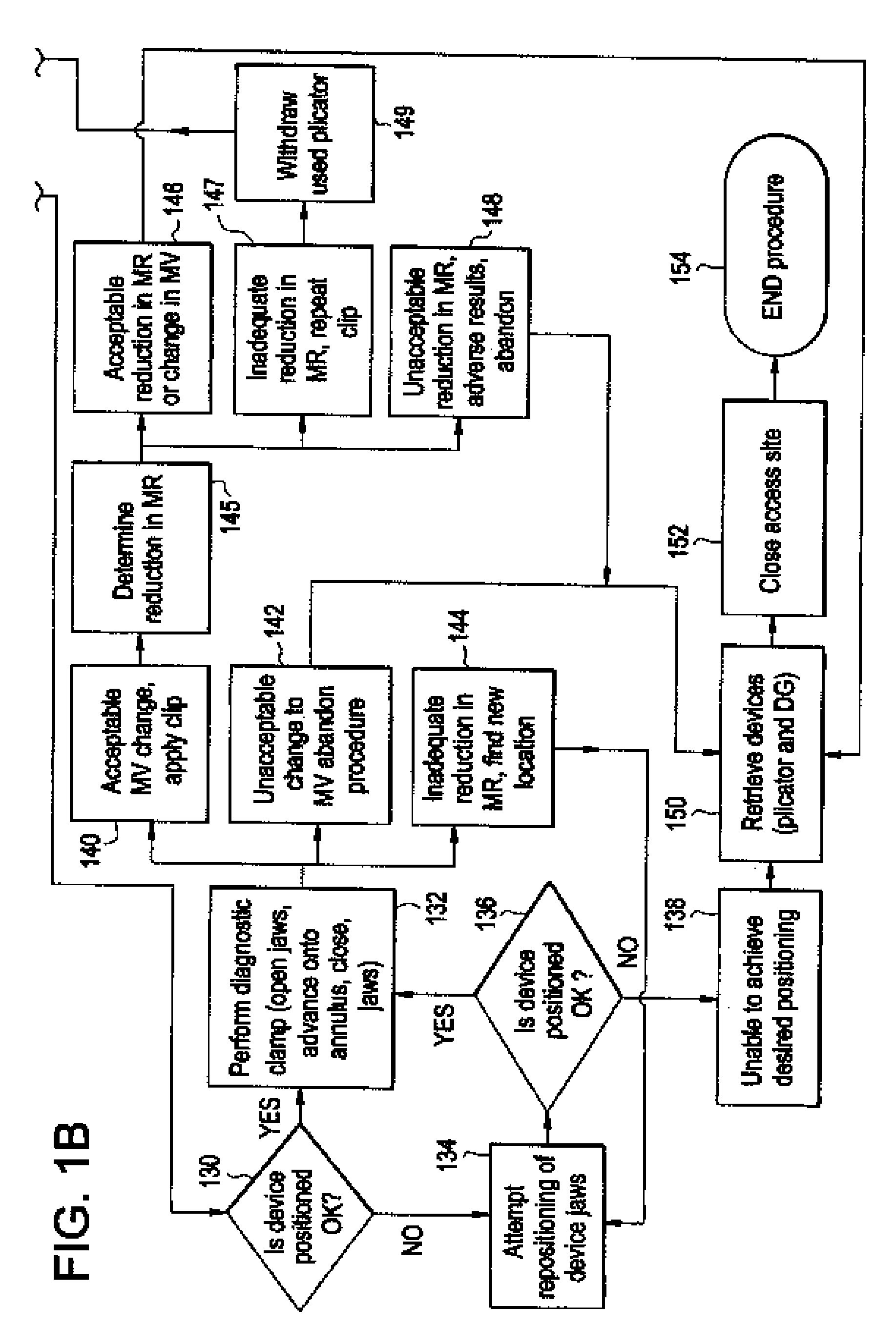
[0056]FIG. 1 is a flow diagram depicting a method of providing direct plication annuloplasty to the mitral valve in a heart such as that depicted in FIG. 2A in accordance with the present invention. At step 100 the procedure begins with a puncture for access to the femoral artery using standard techniques. At step 102 the physician or other practitioner places a catheter sheath introducer (CSI) into the femoral access point using standard techniques. Any known CSI may be used in the procedure with the preferable size being approximately 14 french. At step 104 a crossing catheter, preferably prolapseable or having a curved tip, and a deflecting guide catheter are inserted together in a “stack” formation through the CSI. Alternatively, the deflecting guide catheter is inserted through the CSI without a crossing catheter although the use of a crossing catheter is the preferred method. The crossing catheter is described herein in greater detail with respect to FIGS. 4 and 5 below and th...


PUM


Abstract

A system for using a helical retainer in the treatment of mitral valve regurgitation by reshaping the mitral valve annulus using one or more plications of annular tissue each fixed by a retaining clip is described. The system includes four devices to achieve such percutaneous direct plication annuloplasty. The first is a preferably prolapsable crossing catheter. Second, a deflecting guide catheter is used to provide a means for guiding the plication device into proper position at the subvalvular region of the mitral valve annulus. Third, the plication device is then used to make plications in at or near the subvalvular region of the mitral valve annulus. Fourth, a helical retainer clip deployed by the plication device in order to retain the plicated tissue in the plicated form. Alternatively, the fourth device is a retainer delivery catheter that enables delivery of a helical retainer over the outside of the plication device.

Description

CROSS-REFERENCE TO RELATED APPLICATIONS[0001]This application claims the benefit of U.S. Provisional Patent Application Ser. No. 60 / 981,303 filed Oct. 19, 2007 which is herby incorporated by reference in its entirety.FIELD OF THE INVENTION[0002]The present invention relates to a device and method for treating the vasculature and internal organs of a patient. Particularly, the present invention is directed to a system and method for treating mitral valve regurgitation in the heart of a patient using a plication device to insert a helical fastener in plicated tissue.BACKGROUND OF THE INVENTION[0003]Catheter based devices are used to treat a wide variety of medical problems in a minimally invasive manner. Catheters are used to place and expand angioplasty balloons used to widen veins and arteries narrowed by plaque. Small scaffolds called stents have been introduced into the vasculature using catheter-based systems in order to prevent the restenosis of such vessels. One of the problems...

Claims


Application Information

Patent Timeline
Patent Type & Authority Applications(United States)
IPC IPC(8): A61F2/24
CPCA61B17/00234A61B17/29A61B2017/00243A61B2017/00292A61B2017/2905A61F2/2466A61B2017/2925A61B2017/2927A61B2017/2939A61B2017/2946A61B2017/2923
Inventor OLSEN, DANIEL H.KREVER, MATTHEWCEDRO, RUDOLPHBLACK, ERINMACON, NATALIE D.
Owner CORDIS CORP
Why Eureka
Try Eureka