
Ultrasonic vascular closure device

a technology of vascular closure and ultrasonic sound, which is applied in the field of low frequency medical devices and methods to achieve the effect of preventing foam sealant leakage and reducing the volume of the chamber

Inactive Publication Date: 2009-09-03
BACOUSTICS LLC
View PDF58 Cites 9 Cited by
  • Summary
  • Abstract
  • Description
  • Claims
  • Application Information

AI Technical Summary

Benefits of technology

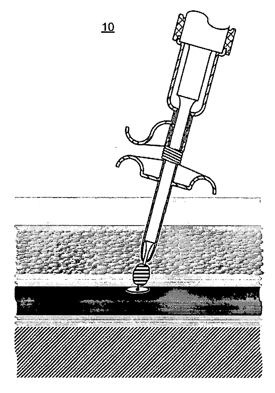
[0009]The present inventions provide an ultrasonic vascular closure device for sealing a patient's tissue wound with a collagen material. A specific application for use in vascular sealing is described in detail as an exemplary embodiment of the invention. However, the device has applicability to the sealing of tissue puncture wounds generally. The present invention does not rely on the patient's blood to provide the seal between the plug and the tissue. Rather, the application of ultrasound to the collagen creates a matrix between the tissue and the plug. Therefore, extending hospital stay time for anticoagulant dissipation is not required. Furthermore, the time required to complete the procedure of the present invention can be completed in 5 to 10 minutes or less, rather than the 30 minutes to several hours required for compression techniques. The reduction in patient stay length and staff time can result in considerable cost savings. Furthermore, there is considerable less likelihood of the wound reopening after the patient is released, reducing costs as well as reducing risks to patient recovery from the procedure.
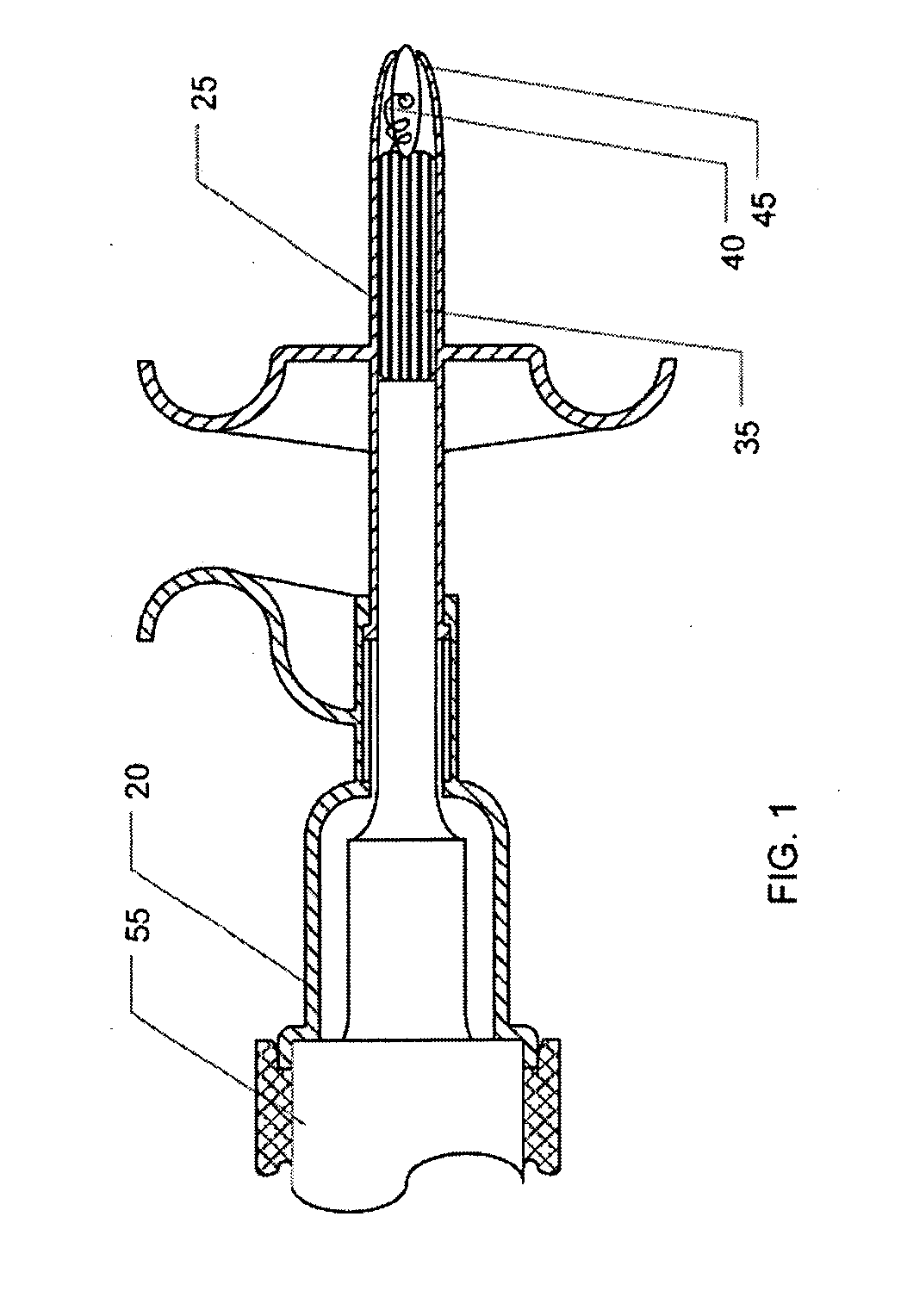
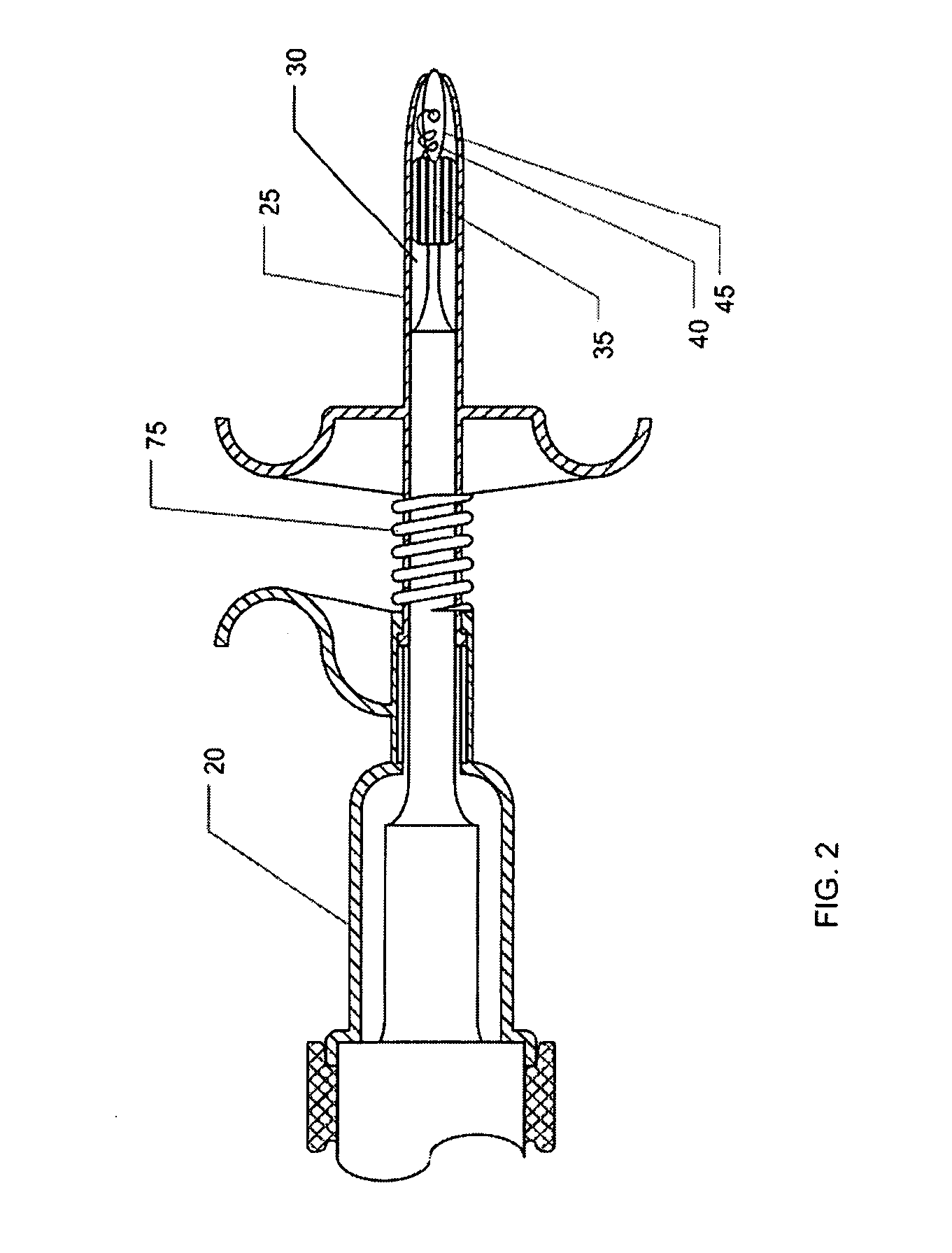
[0011]The ultrasonic device also has a body to contain the transducer housing and proximal portions of the transducer tip; the body is slideably attached to a dispenser portion for ejecting the foam sealant from the chamber, through an orifice and into a tissue puncture wound. A dispenser portion forms the chamber that contains at least portions of the transducer tip including a radiation surface inside the chamber. Sliding the dispenser portion into the body reduces the volume of the chamber. The interface between the body and the dispenser portion is designed with a sealing function to prevent leakage of the foam sealant.

Problems solved by technology

However, the device has applicability to the sealing of tissue puncture wounds generally.

Method used


Image

  • Ultrasonic vascular closure device
  • Ultrasonic vascular closure device
  • Ultrasonic vascular closure device

Examples


Embodiment Construction

[0019]The figures generally illustrate embodiments of an ultrasonic device 10 including aspects of the present inventions. The particular exemplary embodiments of the ultrasonic device 10 illustrated in FIGS. 1-4 have been chosen for ease of explanation and understanding of various aspects of the present invention. These illustrated embodiments are not meant to limit the scope of coverage but instead to assist in understanding the context of the language used in this specification and the appended claims. Accordingly, many variations from the illustrated embodiments may be encompassed by the appended claims.

[0020]The present invention provides an ultrasonic device 10 for sealing a patient's tissue puncture wounds using ultrasound radiation. As illustrated in FIGS. 1 and 2, the ultrasonic device 10 includes an ultrasound generator connected to an ultrasound transducer 50 through a releasable connector 70.

[0021]A transducer housing 55 is fixedly attached to the ultrasound transducer 5...


PUM


Abstract

The present invention relates to the use of a low frequency ultrasonic device for the delivery and activation of collagen based foam sealants to a human and / or animal patient for sealing puncture wounds in vascular tissues. The ultrasonic vascular closure device comprises an ultrasonic generator, an ultrasound transducer, a chamber containing a foam sealant, a transducer tip, a radiation surface, an orifice located at the distal end of the chamber. The foam sealant is ejected into a puncture wound and activated with ultrasonic waves emitting from the radiation surface. The ultrasonic waves induce vibrations within the foam sealant, slightly warming the foam sealants to assist the rapid sealing the puncture. The ultrasonic waves also provide and anesthetic effect for the pain and discomfort from the puncture site.

Description

BACKGROUND OF THE INVENTION[0001]The present invention relates to a low frequency medical device and method for the sealing of tissue puncture wounds after surgical procedures.[0002]With improvement in medical devices and procedures, it is increasingly becoming possible to avoid the trauma and complications caused by open surgery using various transcatheter techniques. Some examples of these procedures include; angiography, percutaneous transluminal coronary angioplasty, stenting, atherectomy and catheter ablation. These new methods are being developed because they offer significant benefits with respect to patient recovery, potential complications and cost. The early discharge of patients undergoing these elective and interventional procedures hinges on the lack of bleeding complications at the access site after the procedure sheath is removed from the artery. The size of the access route, coupled with the routine administration of anticoagulants, creates a strong need to stop blee...

Claims


Application Information

Patent Timeline
IPC IPC(8): A61B17/03
CPCA61B17/0057A61B17/32A61B2017/00004A61M35/00A61B2017/00601A61B2017/00654A61B2017/005
Inventor BABAEV, EILAZMENENDEZ, ADOLFO
Owner BACOUSTICS LLC
Learn More