Allograft osteochondral plug combined with cartilage particle mixture

a cartilage particle and osteochondral plug technology, applied in the direction of biocide, peptide/protein ingredients, prosthesis, etc., can solve the problems of articular cartilage lesions, general non-healing or healing, pain in joint movement or severe restriction, etc., to promote cartilage cell migration into and proliferation, and enhance tissue integration , the effect of increasing chondrocyte (and/or other cellular) migration

Inactive Publication Date: 2009-11-26
MUSCULOSKELETAL TRANSPLANT FOUND INC
View PDF100 Cites 59 Cited by
  • Summary
  • Abstract
  • Description
  • Claims
  • Application Information

AI Technical Summary

Benefits of technology

[0007]A combination for repairing articular cartilage defects includes (i) a mixture including lyophilized, freeze-milled cartilage particles, and (ii) a sterile allograft osteochondral plug. The plug includes a subchondral bone portion and an integral overlying cartilage cap which has been treated to remove cellular debris, chondrocytes and proteoglycans. At least a portion of the plug has a lateral dimension (e.g., diameter) selected to form an interference fit against a tissue layer exposed as a result of a bore formed in a defect area in articular cartilage of a host. The cartilage particle mixture is placed adjacent at least a portion of the plug for promoting cartilage cell migration into and proliferation in the bore and for enhancing tissue integration between the plug and host tissue, when the plug is inserted into th...

Problems solved by technology

If the lining becomes worn or damaged, lesions may form in the lining, and as a result, joint movement may become painful or severely restricted.
Whereas damaged bone typically can regenerate successfully, articular cartilage regeneration is quite limited because of its limited regenerative and reparative abilities.
Articular cartilage lesions, and other defects, generally do not heal, or heal only partially under certain biological conditions, due to the lack of nerves, blood vessels and a lymphatic system.
The limited reparative capabilities of articular cartilage usually results in the generation of repair ti...

Method used


Image

  • Allograft osteochondral plug combined with cartilage particle mixture
  • Allograft osteochondral plug combined with cartilage particle mixture
  • Allograft osteochondral plug combined with cartilage particle mixture

Examples


example 1

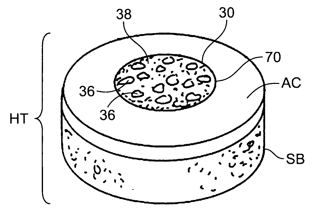
Complete Interference-Fit Osteochondral Plug

[0114]A non-viable or decellularized osteochondral plug including a subchondral bone base and an overlying cartilage cap, according to one of the embodiments disclosed herein and illustrated in FIGS. 2-9, was treated with a solution or variety of solutions such as hyaluronidase (type IV-S), trypsin and a chloroform / methanol solution to remove the cellular debris and the proteoglycans, as noted in the treatment described above. It is believed that this removal provides signaling to stimulate the surrounding chondrocytes to proliferate and form new proteoglycans and other factors producing new cartilage matrix. The plug is then subjected to an antibiotic soak and milled to a desired configuration (e.g., as illustrated in FIG. 3, 5, 7 or 9) to have an interference fit with the blind bore to be cut in the patient's tissue.

example 2

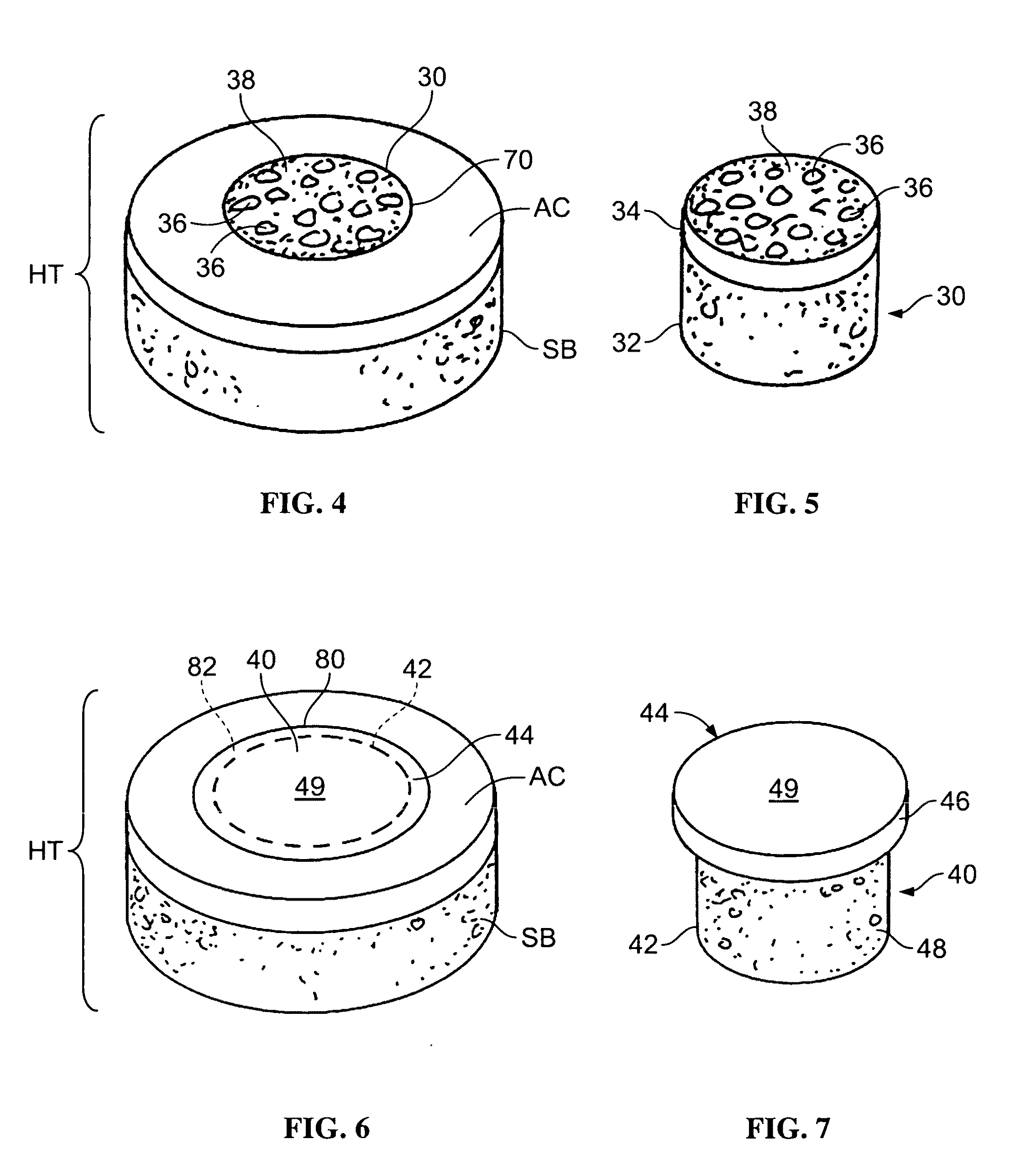
Partial Interference-Fit Osteochondral Plug

[0115]A non-viable or decellularized osteochondral plug including a subchondral cylindrical bone base and an overlying smaller diameter cylindrical cartilage cap cut from the original plug, according to one of the embodiments disclosed herein and illustrated in FIGS. 10-13, was treated with a solution or variety of solutions such as hyaluronidase (type IV-S), trypsin and a chloroform / methanol solution to remove the cellular debris as well as the proteoglycans, as noted in the treatment described above. It is believed that this removal provides signaling to stimulate the surrounding chondrocytes to proliferate and form new proteoglycans and other factors producing new matrix. The plug is then subjected to an antibiotic soak and milled to a desired configuration (e.g., as illustrated in FIG. 11, 12 or 13) so that the subchondral bone portion will have an interference fit with the blind bore to be cut in the patient's tissue.

example 3

Tissue Extraction and Particularization

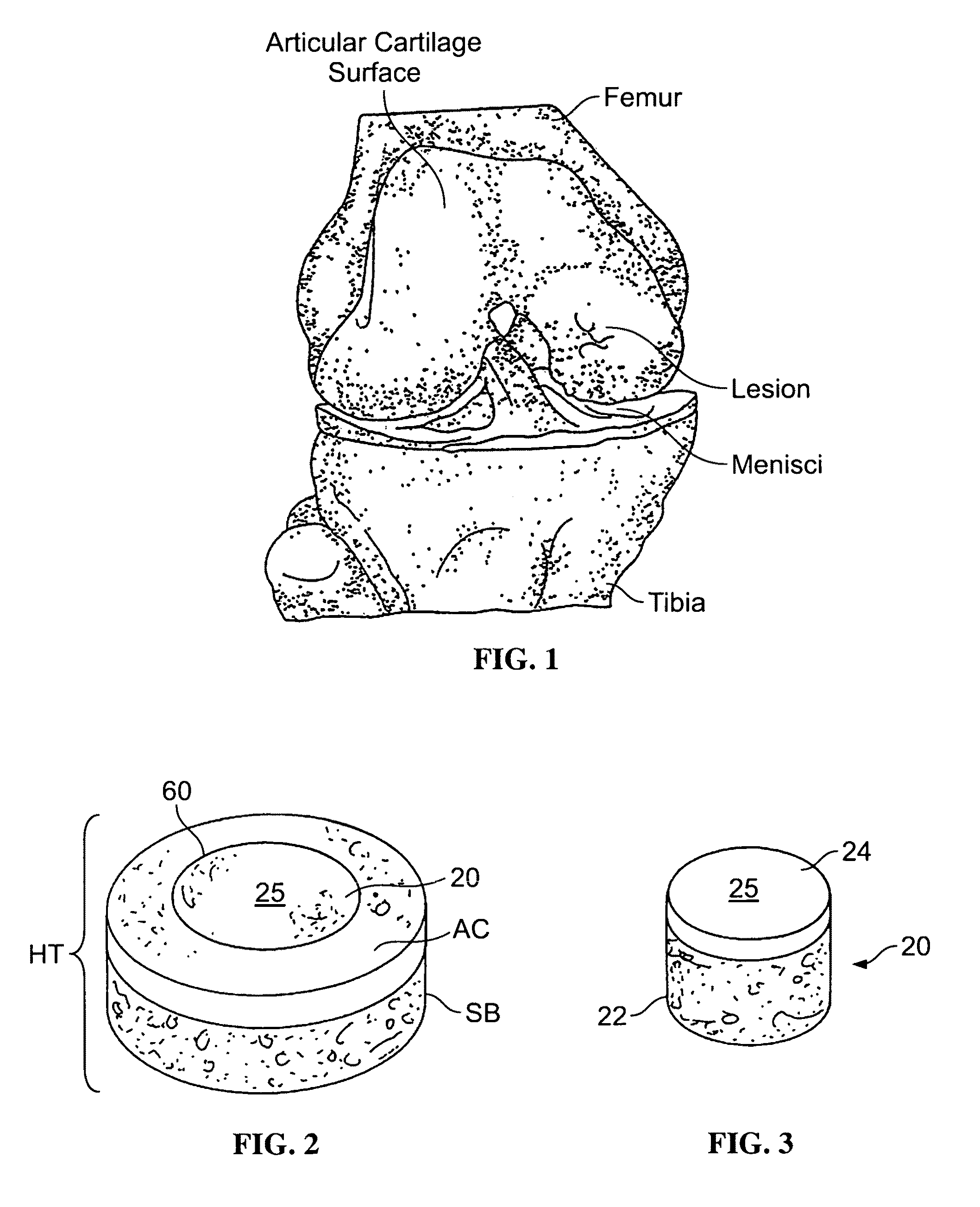
[0116]A process of cartilage particle extraction may be applied to any of a number of different soft tissue types (for example, meniscus tissue). In one embodiment, cartilage is recovered from deceased human donors, and the tissue is treated with a soft tissue process.

[0117]Fresh articular cartilage is removed from a donor using a scalpel, taking care to remove the cartilage so that the full thickness of the cartilage is intact (excluding any bone). Removed cartilage is then packaged in double kapak bags for storage until ready to conduct chemical cleaning of the allograft tissue. In one example, the cartilage can be stored in the refrigerator for 24-72 hours or in the freezer (e.g., at a temperature of −70° C.) for longer-term storage.

[0118]Chemical cleaning of cartilage tissue is then conducted according to methods known by those skilled in the art. Subsequent to chemical cleaning, the cartilage is lyophilized, so as to reduce the water conte...


PUM

PropertyMeasurementUnit
Lengthaaaaaaaaaa
Lengthaaaaaaaaaa
Lengthaaaaaaaaaa

Abstract

An allograft osteochondral plug is combined with a mixture that includes freeze-milled cartilage particles, and such combination is used to repair defects in articular cartilage. The plug includes an subchondral bone portion and an integral overlying cartilage cap which is treated to remove cellular debris and proteoglycans. At least a portion of the plug has a lateral dimension selected to form an interference fit against a tissue layer exposed as a result of a bore formed in a defect area in articular cartilage of a host. The cartilage particle mixture is placed adjacent at least a portion of the plug for promoting cartilage cell migration into (i.e., from the adjacent host cartilage) and proliferation in the bore, and for enhancing tissue integration between the plug and patient (i.e., host) tissue when the plug is inserted into the bore. Methods for surgical implantation of the plug into a patient are also disclosed.

Description

RELATED APPLICATIONS[0001]This application is a continuation-in-part of (i) U.S. patent application Ser. No. 10 / 438,883 filed May 16, 2003, (ii) U.S. patent application Ser. No. 11 / 151,270 filed Jun. 14, 2005, and (iii) U.S. patent application Ser. No. 12 / 179,034 filed Jul. 24, 2008, and also claims priority to (iv) U.S. Provisional Patent Application Ser. No. 61 / 189,252 filed Aug. 15, 2008, and (v) U.S. Provisional Patent Application Ser. No. 61 / 205,433 filed Jan. 15, 2009. All of the foregoing related applications are incorporated by reference herein in their entirety.FIELD OF INVENTION[0002]The present invention is generally directed toward allograft tissue plugs and a cartilage particle mixture for use in repairing defects in articular cartilage. More particularly, the present invention is directed toward allograft osteochondral plugs, each of which includes a subchondral bone base and a cartilage cap, and a cartilage mixture containing freezer-milled (i.e., freeze-milled) carti...

Claims


Application Information

Patent Timeline
IPC IPC(8): A61F2/28A61K38/18A61K38/00A61K35/32A61K35/28A61K38/28
CPCA61F2/28A61F2/30756A61L2430/06A61F2/3859A61F2002/2817A61F2002/2839A61F2002/30004A61F2002/30062A61F2002/30136A61F2002/30224A61F2002/30233A61F2002/3024A61F2002/30299A61F2002/30327A61F2002/30764A61F2002/30785A61F2002/4645A61F2210/0004A61F2230/0004A61F2230/0069A61F2230/0093A61F2250/0014A61F2250/0039A61F2310/00365A61F2310/00383A61L27/3608A61L27/3612A61L27/3654A61L27/3804A61L27/48C08L89/00A61F2002/30235
Inventor TRUNCALE, KATHERINE G.GERTZMAN, ARTHUR A.SUNWOO, MOON HAETOMFORD, WILLIAM W.YANNARIELLO-BROWN, JUDITH I.HUANG, YEN-CHEN
Owner MUSCULOSKELETAL TRANSPLANT FOUND INC
Why Eureka
Try Eureka