Nanoparticles in diagnostic tests

a technology of nanoparticles and diagnostic tests, applied in the field of nanoparticles in diagnostic tests and immunoassays, can solve the problems of limited temperature range of detection, limited use of immunoassays, and limited cost of producing antibodies, so as to avoid the problem of antibody degradation, eliminate sample preparation steps, and prolong the effect of tim

Inactive Publication Date: 2009-11-26
RAPID PATHOGEN SCREENING INC
View PDF24 Cites 67 Cited by
  • Summary
  • Abstract
  • Description
  • Claims
  • Application Information

AI Technical Summary

Benefits of technology

[0042]Another important advantage of the invention is that nanoparticles are highly stable in both solution and solid phases, with a shelf-life that is much longer (5 to 20 years) than currently available diagnostic tests because there is no need to use antibodies which tend to degrade over short periods of time, typically less than 1 to 3 years. Therefore, the disclosed method is more stable and longer lasting than the immunoassay methods of the prior art. Furthermore, due to the thermal stability of the nanoparticles, such tests can be conducted at wider temperature ranges than the methods of the prior art. The test kits of the present invention can also be produced, transported and stored in a wider variety of temperatures and conditions than antibody-based test kits.
[0043]The nanoparticles are designed to bind to specific surface markers on the targets. Thus, a further advantage of the present invention includes elimination of sample preparation steps since bacteria and viruses in a sample do not need to be lysed prior to detection. Furthermore, since antibodies are not used in the assay, the cost of producing antibodies, as well as the problem of antibody degradation, is avoided. In addition, while only a few biological fluids are practicable matrices for antibody-based immunoassays, the present invention can detect targets in any matrix where the target is found. Additional cost savings are realized by manufacturing single or multiple formulations of the specially designed nanoparticles like those in a panel such as Hepatitis panel (Hepatitis A, B, C etc,), cardiac panel, sexually transmissible infection panel, neurological panel, respiratory panel etc. where different members of the panel or closely related members are simultaneously detected and / or differentiated.
[0044]In a preferred embodiment, the present invention provides for the reduction of interfering substances that might be present in the sample to be tested. Since an interfering substance, e.g. a human anti-mouse antibody (HAMA), may also be capable of forming a complex with the labeled, non-immobilized reagent of the reagent zone and the immobilized binding partner of the detection zone, thus indicating a positive test result in the immunoassay, the carrier may further comprise at least one capturing zone. Each capturing zone contains an immobilized capturing reagent specifically binding to a certain interfering substance, thereby immobilizing the interfering substance in the capturing zone. As the capturing zone is separated from the detection zone by space, and the sample starts to migrate over the reagent zone and the capturing zone before reaching the carrier's detection zone, the method allows a separation of the interfering substance or substances from the analyte or analytes of interest. Preferably, the capturing zone is located between the reagent zone and the detection zone. However, the capturing zone may also be located between the application zone and the reagent zone.
[0045]The invention also discloses a point-of-care method for detection of targets. The method is suitable for diagnosis in human beings, plants and animals, e.g. pets or livestock animals. A preferred application is the detection of pathogens in a biological fluid. For example, the pathogen is selected from the group of viruses, fungi, and bacteria and combinations thereof.
[0046]Examples of viral pathogens include, but are not limited to, retroviruses, adenoviruses, herpesviruses, cytomegaloviruses, hepatitis viruses, dengue, influenza viruses, parainfluenza, papilloma viruses, rotavirus, human immunodeficiency virus (HIV), feline immunodeficiency virus (FIV) coxsackie, enterovirus, marburg, ebolavirus, Epstein Barr Virus, respiratory syncytial virus, echovirus, meningitis, lyssavirus, foot and mouth disease virus, rabies, pseudorabies, viral pneumonia, West Nile virus, parvoviruses, feline leukemia virus (FeLV), Rous sarcoma virus (RSV), Norwalk virus, rhinoviruses, Rubella, Astrovirus, Varicella-Zoster, and Metapneumovirus.
[0047]Examples of bacterial pathogens which can be detected by the invention include, but are not limited to, Chlamydia species, Mycoplasma species, Proteus, Anthrax, Clostridium species, Salmonella species, Pseudomonas species, Shigella species, Hemophillus species, Campylobacter species, Mycobacterium and Atypical Mycobacterium species including leprosy, avium, chelonae and tuberculosis, Streptococcal species, Staphylococcal species, Neisseria species, helicobacter, Escherichia coli species, Brucella species, Rickettsial species, Gardenella, Borrelia species, Diphtheria species, trichomonas, Toxoplasma species, Moraxella species, Bordetella species, Treponema species, and Legionella species.

Problems solved by technology

Immunoassays take advantage of the specific binding of an antibody to its antigen, however the use of immunoassays is limited by the availability of antibodies for the specific target, degradation of the antibodies, strength of binding to the antigen, and the cost of producing antibodies.
Further, the temperature range of detection is limited by the thermal stability of the antibody.
The sample matrix is often limited to a few biological fluids that are suitable for maintaining both the stability of the antibody and the affinity of the antibody for the antigen.

Method used


Image

  • Nanoparticles in diagnostic tests
  • Nanoparticles in diagnostic tests
  • Nanoparticles in diagnostic tests

Examples


example 1

[0073]As shown in FIGS. 7A and 7B, and referring back to FIG. 6, in one example, the sample zone (101) of a sample analysis device is composed of nanoparticles (110) that mimic the action of T cells in the immune system (T cell Mimics). The “T cell mimics” preferably only include the active site (the binding site) for the viral particle of interest (H5N1 in this example). Since there are multiple binding sites, the device has a higher binding affinity and higher sensitivity for the target (a virus in this example) than in prior art devices. The “T cell mimics” are dyed with a visible color to create color at the test line for a positive test. The sample zone “T cell Mimics” may be specific to a single serotype or general to bind to all viruses.

[0074]At the test line (102), additional nanoparticles (112), which are also “T cell mimics”, are immobilized on the sample analysis device. These nanoparticles (112) are preferably colorless. The test line “T cell mimics” may be specific to b...

example 2

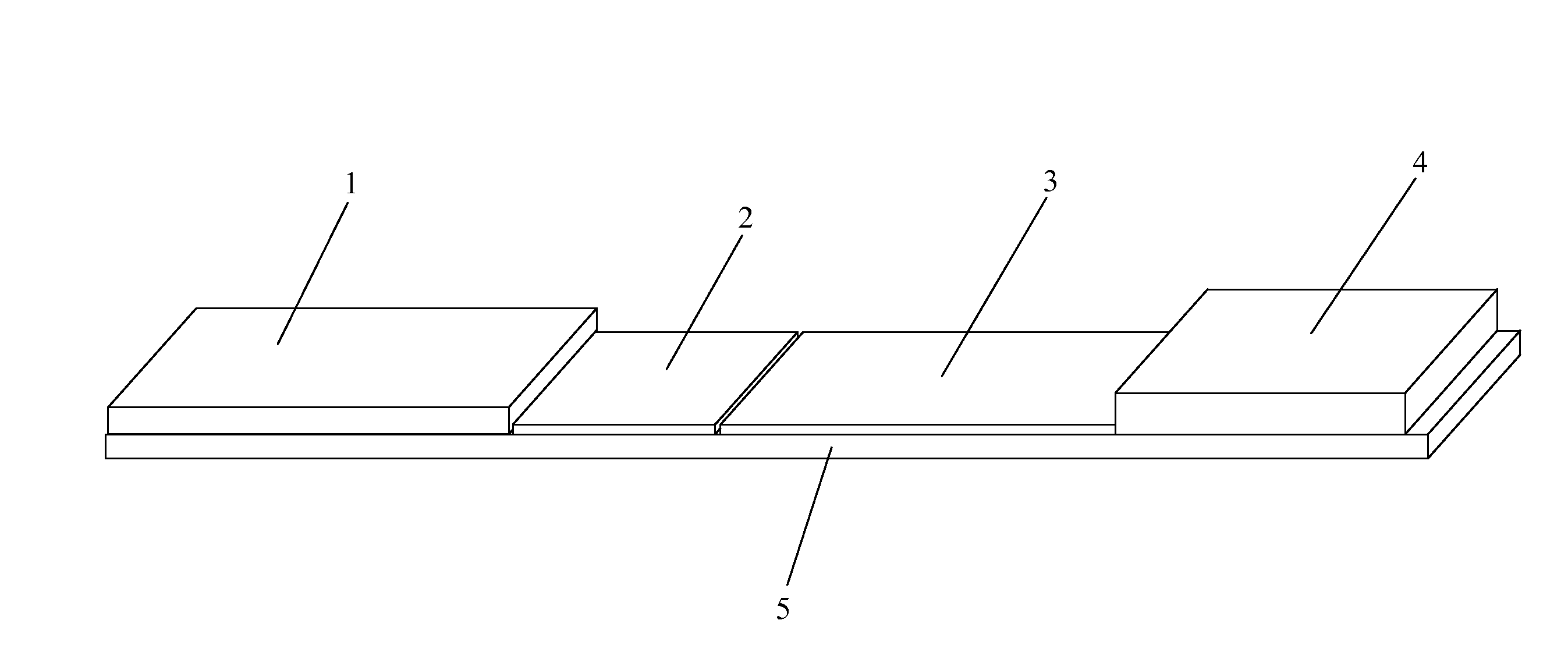
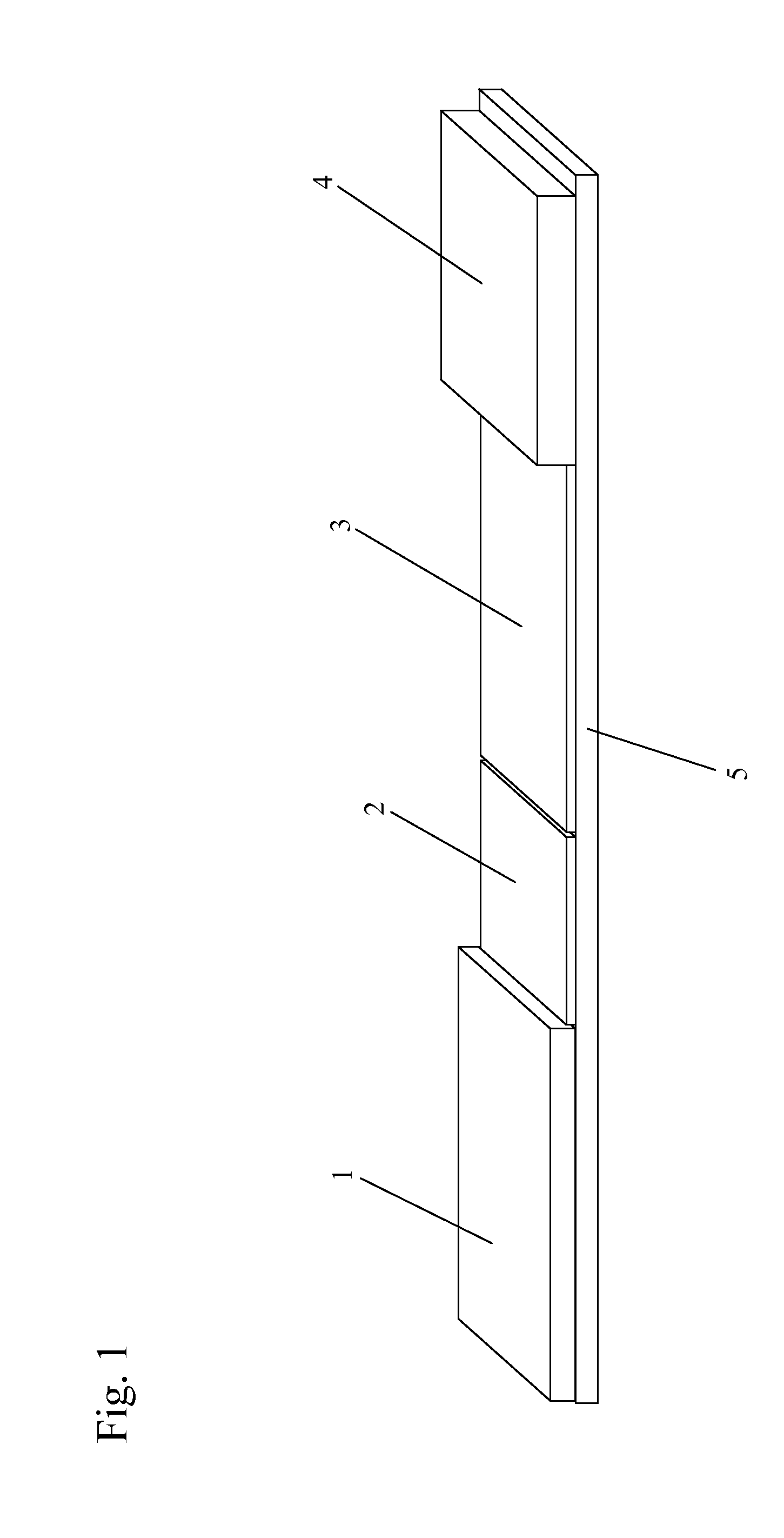
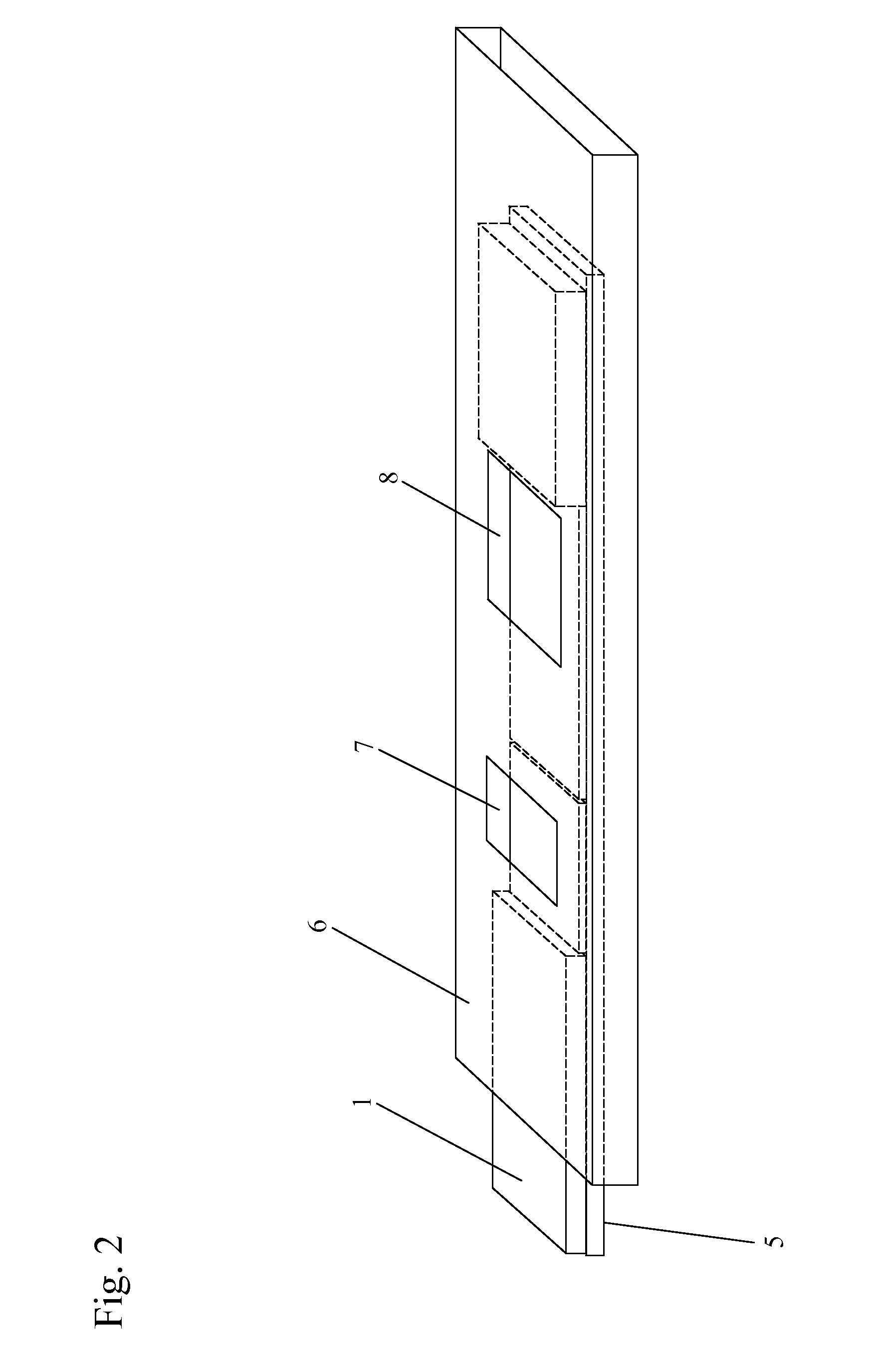
[0078]A test kit includes a test strip for the detection of Adeno virus in a tear sample from a patient. The test strip in this example includes a nitrocellulose protein binding membrane. The application zone includes an accessible portion of the test strip upstream of a test line. The test line is upstream of a control line.

[0079]The detection zone includes a nitrocellulose (NC) membrane with a nominal pore size of 8 pm and a thickness of 100 pm produced by Schleicher & Schuell, Germany. The test line contains an immobilized nanoparticle and a ligand rationally designed to bind to Hexon protein or any specific viral surface marker and is hence specific for the surface marker epitope on the Adeno virus membrane. As long as the test is working, the control line will appear regardless of whether the sample is positive or negative and thus indicates the correct flow characteristics of the immune-chromatography test. The chromatographic zones are in fluid communication with each other i...


PUM

PropertyMeasurementUnit
sizeaaaaaaaaaa
sizeaaaaaaaaaa
sizeaaaaaaaaaa

Abstract

The present invention includes methods and devices that detect target molecules in a biological sample. The sample analysis device of the present invention includes nanoparticles. In one embodiment, the nanoparticles are directly immobilized on the surface of the sample analysis device. In another embodiment, the nanoparticles are indirectly immobilized on the surface of the sample analysis device by incorporating them in appropriate media and immobilizing the nanoparticles within a matrix.

Description

REFERENCE TO RELATED APPLICATIONS[0001]This application claims one or more inventions which were disclosed in Provisional Application No. 61 / 071,833, filed May 20, 2008, entitled “NANOPARTICLES IN DIAGNOSTIC TESTS”. Provisional Application No. 61 / 060,258, filed Jun. 10, 2008, entitled “COMBINED VISUAL / FLUORESCENCE ANALYTE DETECTION TEST”, Provisional Application No. 61 / 098,935, filed Sep. 22, 2008, entitled “1N SITU LYSIS OF CELLS IN LATERAL FLOW IMMUNOASSAYS, and Provisional Application No. 61 / 179,059, filed May 18, 2009, entitled “METHOD AND DEVICE FOR COMBINED DETECTION OF VIRAL AND BACTERIAL INFECTIONS”. The benefit under 35 USC §119(e) of the United States provisional applications is hereby claimed, and the aforementioned applications are hereby incorporated herein by reference.BACKGROUND OF THE INVENTION[0002]1. Field of the Invention[0003]The invention pertains to the field of immunoassays. More particularly, the invention pertains to immunoassays that include nanoparticles t...

Claims


Application Information

Patent Timeline
Patent Type & Authority Applications(United States)
IPC IPC(8): G01N33/543B01J19/00
CPCB01J2219/00576B01J2219/00648B01J2219/00725G01N33/54346B01J2219/00743B82Y30/00B01J2219/0074
Inventor BABU, UMA MAHESHVANDINE, ROBERT W.SAMBURSKY, ROBERT P.
Owner RAPID PATHOGEN SCREENING INC
Why Eureka
Try Eureka