

Methods and reagents for in vivo imaging of cancer cell lines
a cancer cell line and in vivo imaging technology, applied in the field of in vivo imaging of cancer cell lines, can solve the problem that fluorescence based imaging has only been used in biopsied tissues
- Summary
- Abstract
- Description
- Claims
- Application Information
AI Technical Summary
Problems solved by technology
Method used
Image

Examples
example 1
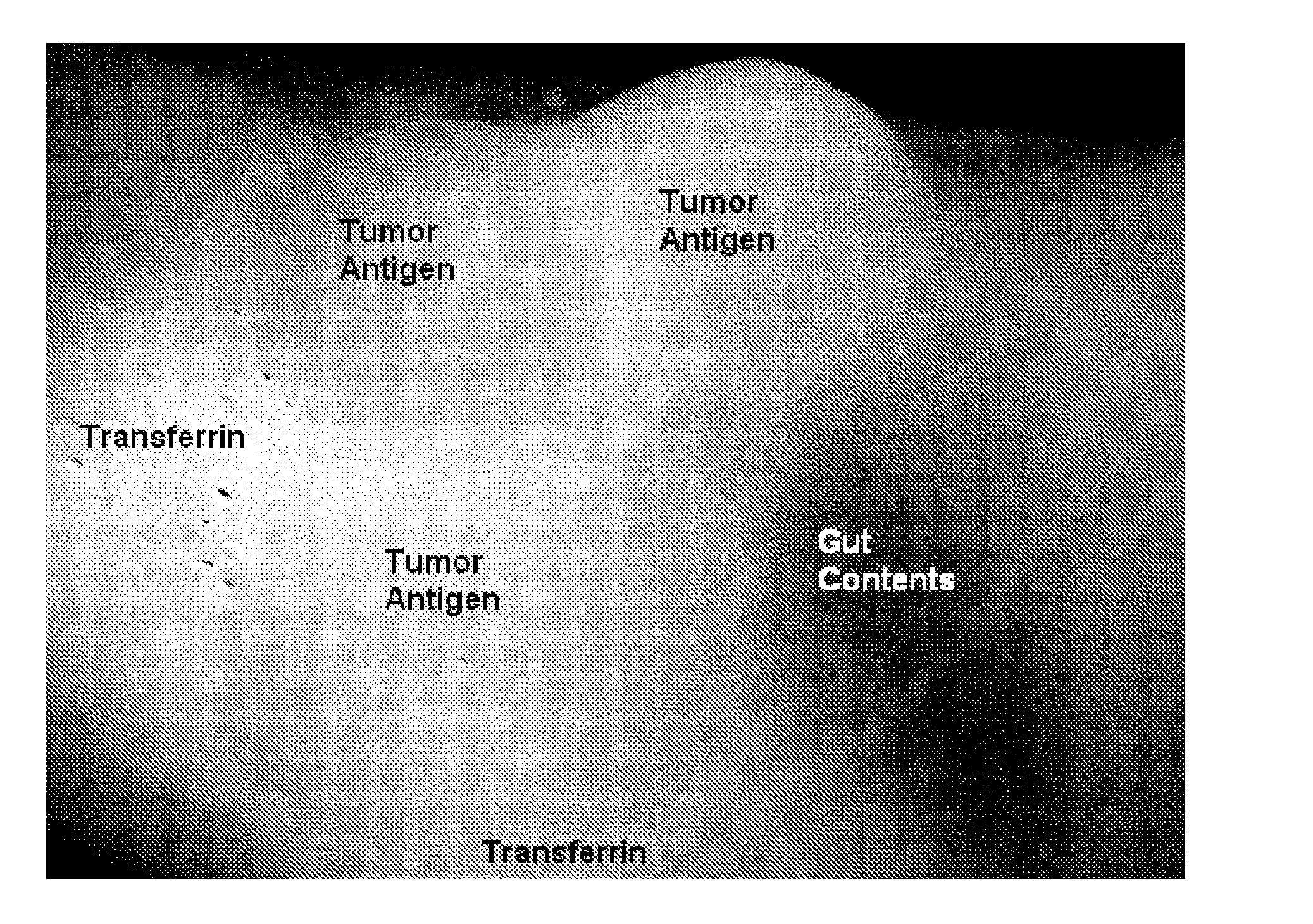
[0239]Preparation of the Monoclonal Antibody: anti-CEA Alexa Fluor® 680
[0240]The mouse monoclonal antibody, anti-Carcinoembryonic Antigen (CEA), clone Col-1, (MPX18-0057, Invitrogen Corp, Carlsbad, Calif.) was conjugated with Alexa Fluor® 680 dye according to the procedure in the SAIVI™ Alexa Fluor® 680 Antibody / Protein 1 mg labeling kit (S30039, Invitrogen Corp, Eugene, Oreg.). Briefly, 500 μl of 2 mg / ml of antibody protein in PBS was combined with 50 μl 1 M sodium bicarbonate, pH 8.3, and 45 μl 15 mM lysine. This mixture was added to a reaction tube containing 96 μg lyophilized Alexa Fluor 680 carboxylic acid, succinimidyl ester. The reactive dye was dissolved and fully mixed, and the reaction was incubated for 60 minutes at ambient temperature (18-20° C.), protected from light. The dye-conjugated antibody was purified by size exclusion chromatography. The degree of labeling (mole fluorophore / mole antibody), determined spectrally, was 2.1
[0241]The dye conjugated antibody was adjus...
example 2
Preparation of the Monoclonal Antibody: Anti-CEA Alexa Fluor® 750
[0242]The mouse monoclonal antibody, anti-Carcinoembryonic Antigen (CEA), clone Col-1, (MPX18-0057, Invitrogen Corp, Carlsbad, Calif.) was conjugated with Alexa Fluor® 750 dye according to the procedure in the SAIVI™ Alexa Fluor® 750 Antibody / Protein 1 mg labeling kit (S30040, Invitrogen Corp, Eugene, Oreg.). Briefly, 500 μl of 2 mg / ml of antibody protein in PBS was combined with 50 μl 1 M sodium bicarbonate, pH 8.3, and 40 μl 15 mM lysine. This mixture was added to a reaction tube containing 90 μg lyophilized Alexa Fluor 750 carboxylic acid, succinimidyl ester. The reactive dye was dissolved and fully mixed, and the reaction was incubated for 60 minutes at ambient temperature (18-20° C.), protected from light. The dye-conjugated antibody was purified by size exclusion chromatography. The degree of labeling (mole fluorophore / mole antibody), determined spectrally, was 1.6.
[0243]The dye conjugated antibody was adjusted t...
example 3
Preparation of the Monoclonal Antibody: Anti-CEA Alexa Fluor® 790
[0245]The mouse monoclonal antibody, anti-Carcinoembryonic Antigen (CEA), clone Col-1, (MPX18-0057, Invitrogen Corp, Carlsbad, Calif.) was conjugated with Alexa Fluor® 790 dye in a procedure very similar to that of the SAIVI™ Alexa Fluor® dye Antibody / Protein labeling kits in the previous 2 examples. Briefly, 500 μl of 2 mg / ml of antibody protein in PBS was combined with 50 μl 1 M sodium bicarbonate, pH 8.3, and 40 μl 15 mM lysine. This mixture was added to a reaction tube containing 125 μg lyophilized Alexa Fluor 790 carboxylic acid, succinimidyl ester. The reactive dye was dissolved and fully mixed, and the reaction was incubated for 60 minutes at ambient temperature (18-20° C.), protected from light. The dye-conjugated antibody was purified by size exclusion chromatography. The degree of labeling (mole fluorophore / mole antibody), determined spectrally, was 1.6.
[0246]The dye conjugated antibody was adjusted to 1 mg / ...
PUM
| Property | Measurement | Unit |
|---|---|---|
| Wavelength | aaaaa | aaaaa |
| Wavelength | aaaaa | aaaaa |
| Wavelength | aaaaa | aaaaa |
Abstract
Description
Claims
Application Information