Laser Illumination System in Fluorescent Microscopy

a fluorescent microscopy and laser illumination technology, applied in the field of laser illumination systems in fluorescent microscopy, can solve the problems of lack of evidence, insufficient sensitiveness of other currently available methodologies for accurate classification and typing of rare events such as circulating tumor cells, and lack of prior art systems without preliminary cell enrichment steps. , to achieve the effect of improving detection, enumeration and classification, and avoiding cell loss

a fluorescent microscopy and laser illumination technology, applied in the field of laser illumination systems in fluorescent microscopy, can solve the problems of lack of evidence, insufficient sensitiveness of other currently available methodologies for accurate classification and typing of rare events such as circulating tumor cells, and lack of prior art systems without preliminary cell enrichment steps. , to achieve the effect of improving detection, enumeration and classification, and avoiding cell loss

US20090311734A1Inactive Publication Date: 2009-12-17GREVE JAN +1

Image

  • Laser Illumination System in Fluorescent Microscopy
  • Laser Illumination System in Fluorescent Microscopy
  • Laser Illumination System in Fluorescent Microscopy

Examples


Embodiment Construction

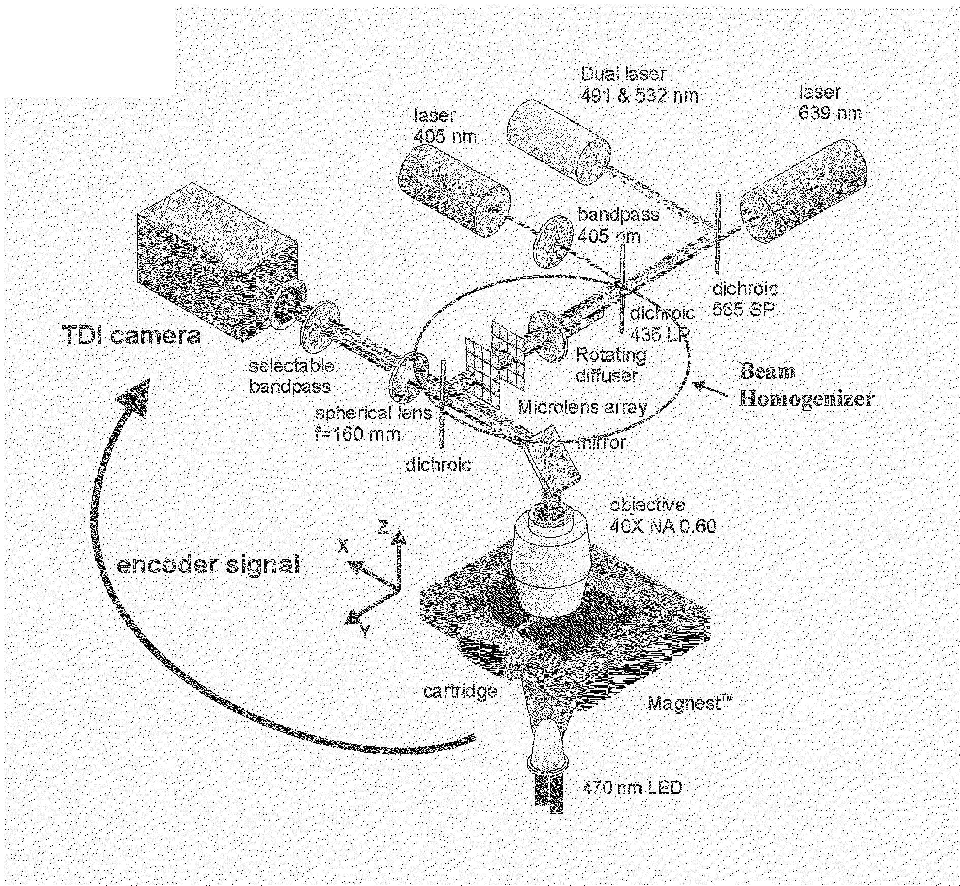
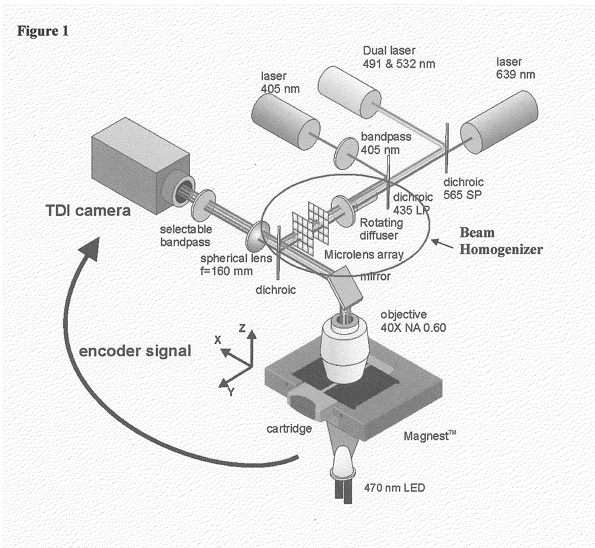
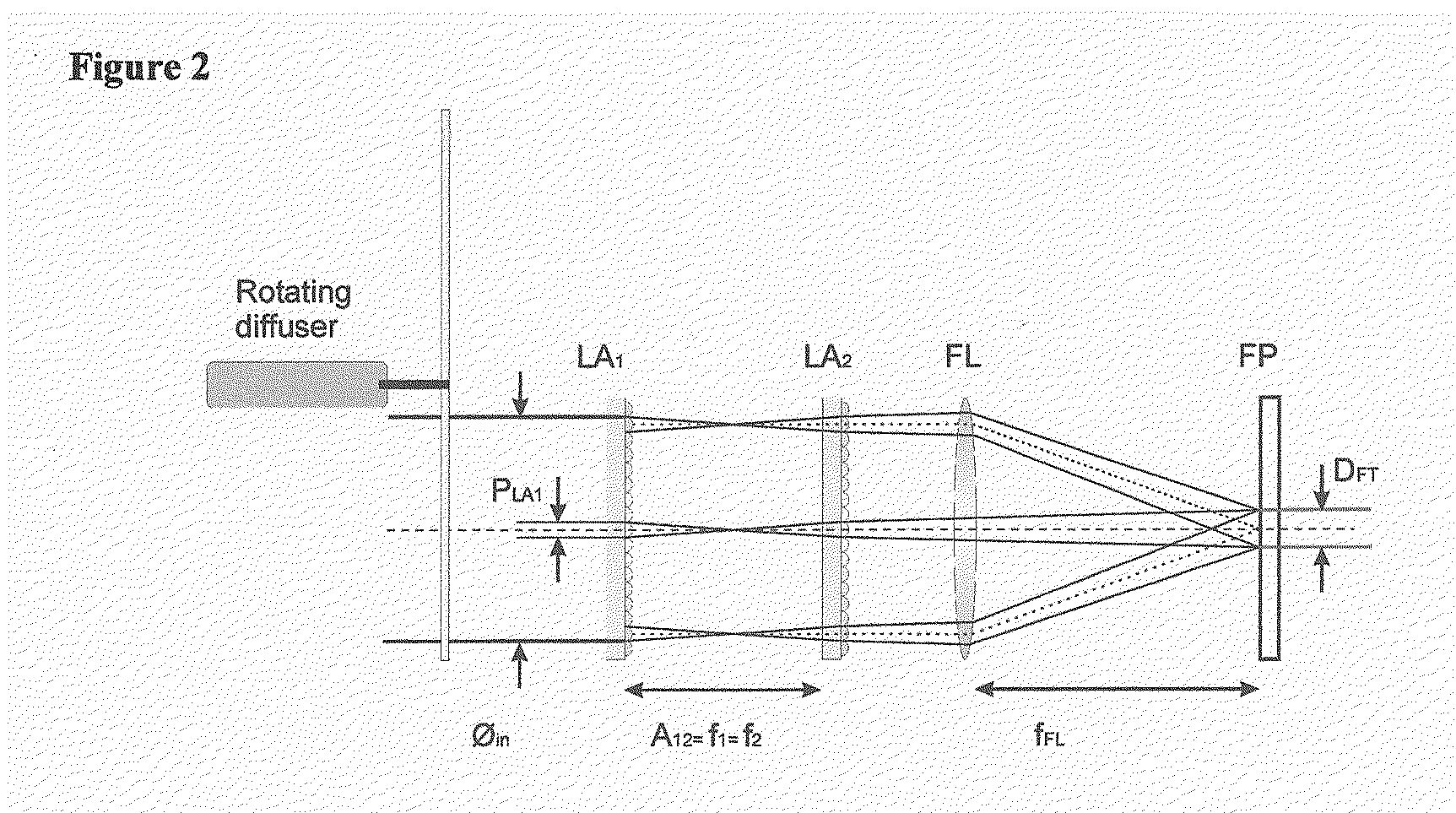
[0019]The present invention combines fluorescent microscopy and a beam homogenizer to illuminate the sample with a laser. The beam homogenizer is particular useful for automated fluorescent microscopy.

[0020]The illumination area can be adapted to a small area that exactly matches the size of the detection area (CCD), without unnecessary bleaching of the sample. (Only the collection area is illuminated)

[0021]The homogeneity of the illumination with this homogenizer is also better than other methods like HBO illumination. The homogenizer can be used for the entire visible wavelength range (300-850 nm). Compared with the HBO a much higher power density can be achieved while the beam homogenizer uses a laser. Another advantage of this system is that the beam profile is not so sensitive to small variations in the positional alignment of the laser. In flow cytometry a small part of a Gaussian profile is used to obtain a homogeneous illumination, therefore most of the illumination power is...


PUM


Abstract

Devices and methods for automated collection and image analysis are disclosed that enable identification or classification of microscopic objects aligned or deposited on surfaces. Such objects, e.g. detectably labeled rare target cells, are magnetically or non-magnetically immobilized and subjected to Time Delay Integration imaging (TDI). Incorporation of TDI technology into image cytometry analysis, like CellTracks®, makes it possible to image moving objects with very high sensitivity and signal-to-noise ratios. Implementation of TDI camera technology with dual excitation and multispectral imaging of enriched rare cells provides a rapid system for detection, enumeration, differentiation and characterization of imaged rare cells on the basis of size, morphology and immunophenotype.

Description

CROSS REFERENCE TO RELATED APPLICATIONS[0001]This application claims priority to U.S. Provisional Application Number 60 / 800,258, filed 12 May 2006.TECHNICAL FIELD[0002]This invention relates generally to devices and methods to obtain to scan and obtain images of objects, and more particularly to images reconstructed from partial sub-images of object such as cells obtained from biological fluids that are distributed in a two-dimensional plane. More specifically, the present invention is applicable in fluorescent microscopy and flow cytometry where the illumination.BACKGROUND OF THE INVENTION[0003]The identification and enumeration of circulating carcinoma cells of epithelial origin in the blood of cancer patients that maybe present at frequencies of less than one carcinoma cell per ml of blood has been detected using magnetic enrichment and image cytometry as in, for example, CellTracks® Systems (Immunicon Corporation, Huntingdon Valley, Pa.). Using a combination of epithelial cell e...

Claims


Application Information

Patent Timeline
17 Dec 2009
Publication
US20090311734A1
IPC
C12Q1/02; G01N33/68; G01N33/48; G01N21/64; C12M1/34
CPC
Y10T436/143333; G01N33/54326
Inventors
GREVE, JAN; SCHREUDER, FREDERIK