

Use of bacterial beta-lactamase for in vitro diagnostics and in vivo imaging, diagnostics and therapeutics
a technology of beta-lactamase and in vitro diagnostics, applied in the field of pathogenic microbiology and imaging technologies, can solve the problems of difficult diagnosis of many of these infections, inability to detect penicillin resistance, and inability tissue culture cells and during infection in animal models and humans, and current methods to quantify and assess the viability of tuberculosis in the laboratory, can only be used for tissue cultur
- Summary
- Abstract
- Description
- Claims
- Application Information
AI Technical Summary
Benefits of technology
Problems solved by technology
Method used
Image

Examples
example 1
Detection of Bla in M. tuberculosis in Culture
[0079]Potential fluoregenic compounds and known compounds, including Nitrocefin (Calbiochem), CENTA Bla substrate (Calbiochem), Fluorocillin Green (Molecular Probes), CCF2-AM (Invitrogen) and CCF4-AM (Invitrogen), are compared for detection of Bla in Mtb using whole cells and whole cell lysates grown to early log-phase. Dilutions are assayed for all of these samples to determine the minimal number of bacteria or amount of lysate that results in significant signal. Titers are carried out to determine the number of actual CFU used, before and after assays with intact cells and before lysis for lysates. Both the sensitivity and reproducibility are evaluated in quadruplicate spectrophotometrically using 96-well plates incubated at 37° C. in bacterial culture medium from 15-120 min. Initially, compounds are used at concentrations recommended by the manufacturer and for CNIR5, 2 nM, i.e., that used for in vivo imaging. Different concentrations...
example 2
Intra-Vital Microscopy Imaging Using the Cell Transplantation Model
[0098]Universal donor Tr, CD8+ T cells, monocytes, macrophages and dendritic cells are transplanted into syngeneic mice infected with BCG, and the distribution of these cells over time are imaged with in vivo bioluminescence imaging (BLI) and image-guided intravital microscopy (IVM). A line of transgenic mice in which luciferase is produced by the beta-actin promoter, provide a source of tissues and cells that will emit light in non-transgenic animals (11-12). This mouse line (L2G85), shows bright bioluminescence from the firefly luciferase (Fluc), but weak GFP fluorescence, so it was mated with a separate line exhibiting strong GFP expression and fluorescence in lymphocytes. The spatial distribution of universal donor stem cells and other cells can thus be followed by BLI in the recipient as they expand, re-distribute or are cleared, and the cells detected can be subsequently visualized by IVM utilizing GFP.
[0099]Th...
example 3
Fluorogenic Substrates for Beta-Lactamase Detection
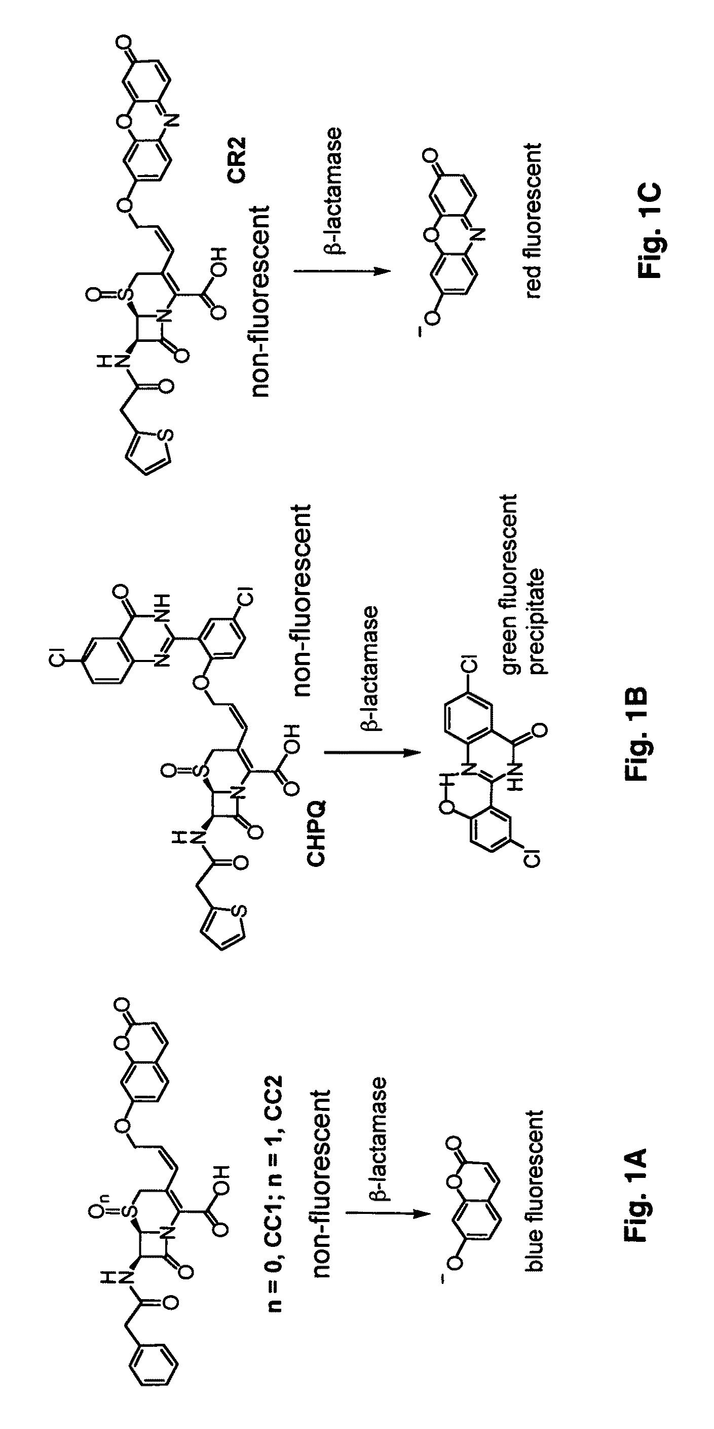
CC1, CC2, CHPQ, and CR2
[0105]Fluorogenic compounds CC1, CC2, CHPQ, and CR2 are effective for detecting Bla activity in vitro and in single cultured cells. These probes are not fluorescent before the hydrolysis by Bla and become fluorescent after the Bla reaction (FIGS. 1A-1B). A range of different fluorescence emissions can be selected as needed to detect Bla: from blue with CC1 and CC2, green with CHPQ to red CR2). These new fluorogenic substrates are smaller than CCF2, easy to make, simple to use, have high sensitivity for detecting Bla activity and facilitate detection of Bla activity in diverse biological samples.
[0106]The insertion of an olefin group between the 3′ carbon of the lactam and the leaving group helps improve the kinetic efficiency of hydrolysis by Bla. For example, for CC1, the value of kcat is 174 s−1, but the value of kcat of its analog without the inserted double bond is just 35 s−1. There is about a 5-fold incr...
PUM
 Abstract
Description
Claims
Application Information