Antigen detection method involving an oligonucleotide enhanced colloidal gold signal

a colloidal gold and antibody detection technology, applied in the field of in vitro diagnostics, can solve the problems of limited suitability and sensitivity, and achieve the effect of enhancing detection sensitivity and enriching signal colour intensity

Inactive Publication Date: 2010-03-18
ARAGEN BIOTECH
View PDF5 Cites 1 Cited by
  • Summary
  • Abstract
  • Description
  • Claims
  • Application Information

AI Technical Summary

Benefits of technology

[0010]Preferably, the first and the second conjugate pads are separated from each other. Such separation may be realized by a divider, preferably a plastic divider, sandwiched between the two pads, or by placing the two pads spaced apart from each other at different locations. The purpose of separating the two pads is to prevent untimely mixing of the conjugates comprised in the first and the second pad.
[0027]Thus, the present invention provides a rapid immunochromatographic detection system for antigen detection comprising a test strip and two conjugate releasing pads with different compositions. The first pad comprises an antibody specific for the antigen to be detected and a protein linked oligonucleotide (“sense” or first oligonucleotide). The antibody and the protein (e.g. bovine serum albumin, BSA) are conjugated with colloidal gold. The second conjugate pad comprises a further protein linked oligonucleotide (“antisense” or second oligonucleotide) also conjugated with colloidal gold. The both oligonucleotides, i.e. the sense and antisense oligonucleotide, are complementary to each other. When the antigen contained in the sample comes into contact with the antibody of the first pad, the antigen is captured by the antibody and thereby a complex is formed comprising the antigen to be detected the conjugated antigen specific antibody and the conjugated protein linked sense oligonucleotide. This complex is carried to the sample zone where further antibody is immobilized. The complex will be captured then by the antigen specific antibody within the sample zone from another site. The conjugated protein linked antisense oligonucleotide which is released from the second conjugate pad also moves to the sample zone where the complementary oligonucleotides, i.e. the sense and the antisense oligonucleotides, bind to each other. Thus, the complex of antigen, antibody and the sense oligonucleotide which is bound within the sample zone serves as a target for the antisense oligonucleotide. Due to sense / antisense oligonucleotide and antigen / antibody interaction, a multi-complex is formed. The binding between the two conjugates will enhance the signal colour intensity and thus enhance the sensitivity of detection.

Problems solved by technology

However, despite the wide use of rapid immunochromatographic test devices, their suitability is still limited with regard to certain applications.
In particular, sensitivity remains a problem.

Method used


Image

  • Antigen detection method involving an oligonucleotide enhanced colloidal gold signal
  • Antigen detection method involving an oligonucleotide enhanced colloidal gold signal
  • Antigen detection method involving an oligonucleotide enhanced colloidal gold signal

Examples


example 1

Preparation of Protein Linked Oligonucleotide

[0033]5 mg of bovine serum albumin (BSA) each was linked to an oligonucleotide (about 20 nucleotides having an amino group at the 5′ terminus) and to a complementary oligonucleotide (about 20 nucleotides having an amino group at the 5′ terminus) according to the method of Duncan et al. (1983)4 which can be illustrated as a procedure comprising the following steps:

example 2

Preparation of Conjugate Pads Comprising Protein Linked Oligo-Nucleotide

[0034]The oligonucleotide and complementary oligonucleotide linked BSA prepared as described in Example 1 are further processed according to a procedure comprising the following steps[0035](a) prepare oligonucleotide linked BSA solution (solution 1);[0036](b) prepare complementary oligonucleotide linked BSA solution (solution 2);[0037](c) prepare 1% aqueous solution of tetrachloroauric acid at room temperature;[0038](d) prepare 4% trisodium citrate aqueous solution at room temperature;[0039](e) prepare 0.05 M potassium carbonate aqueous solution at room temperature;[0040](f) prepare 400 ml of phosphate stabilizing buffer, pH 7.4, containing BSA, Tween 20, sucrose, polyvinylpyrrolidone and preservative (like sodium azide) at room temperature;[0041](g) prepare colloidal gold solution by reduction of 1.7 ml boiling tetrachloroauric acid solution (after dilution in 100 ml) using 1 ml trisodium citrate solution and e...

example 3

Preparation of a Test Device in the Form of a Test Strip

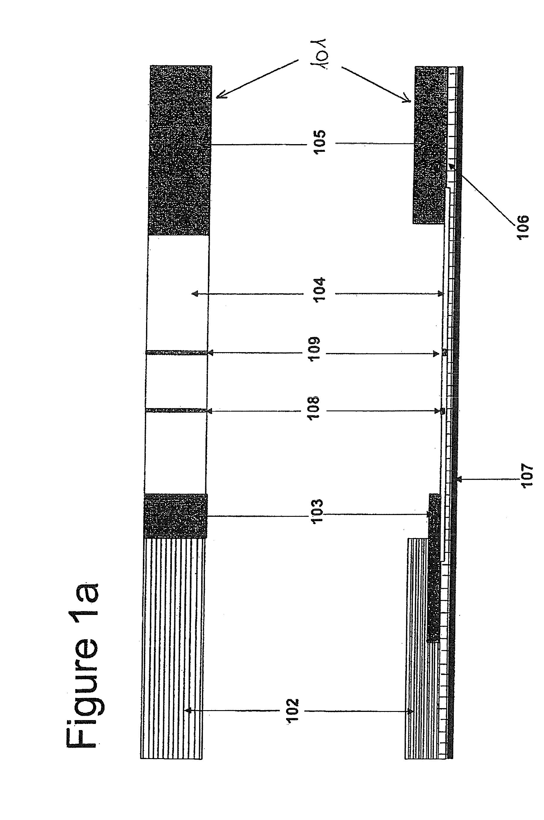
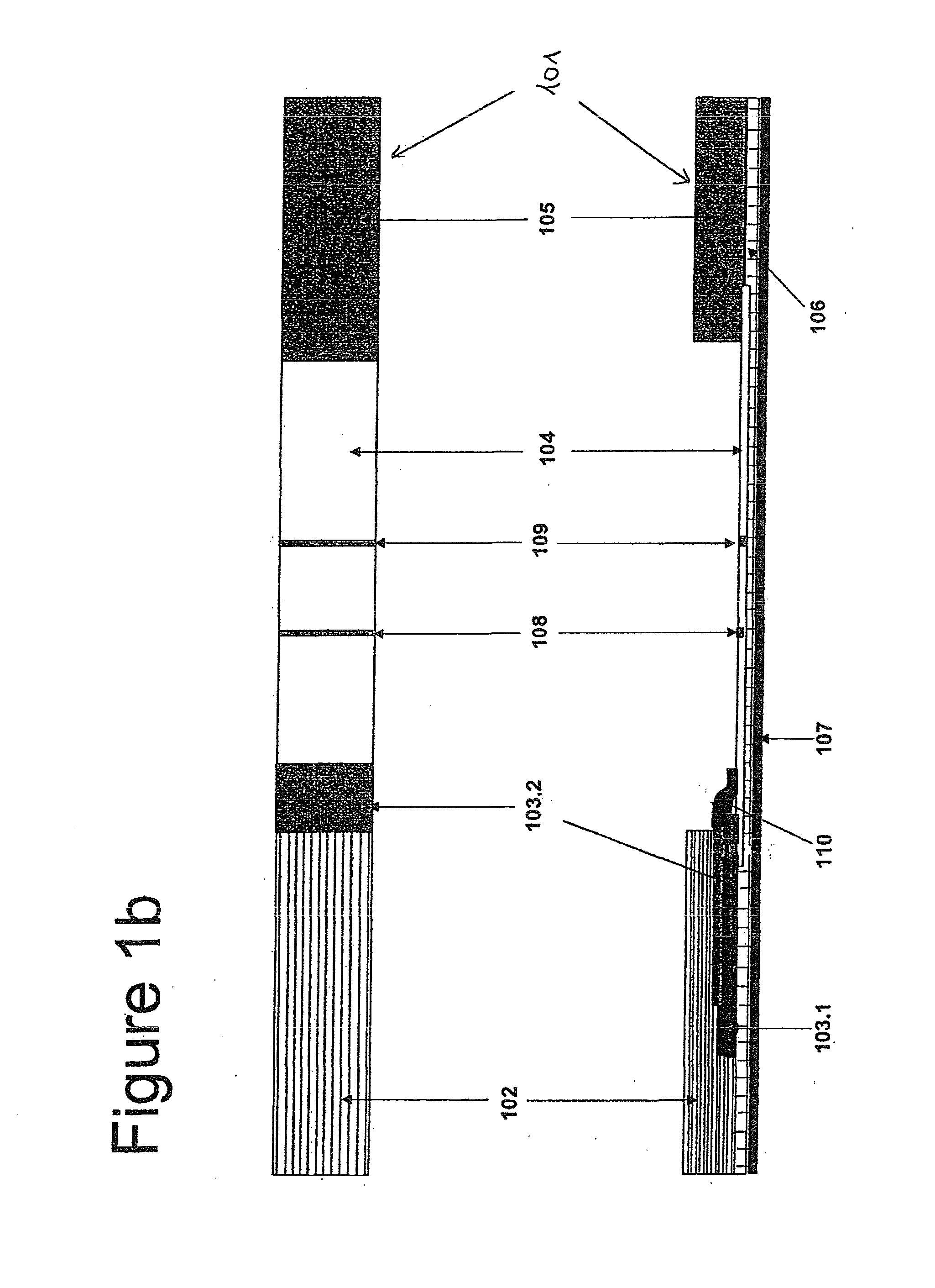
[0054]In case of a test strip, the first conjugate releasing pad 103.1 is prepared by soaking with oligonucleotide linked BSA and the antibody conjugate, while the other pad 103.2 is prepared by soaking with complementary oligonucleotide linked BSA conjugate (see Example 2).

[0055]In more detail, the test device is prepared according to a procedure comprising the following steps:[0056](a) prepare a phosphate sample buffer containing goat serum, ethylenediamine tetraacetic acid (EDTA), non-fat dry milk, preservative (like sodium azide) and Tween 20;[0057](b) soak a sample pad with the phosphate sample buffer and heat dry at a temperature around 50° C.;[0058](c) prepare a colloidal gold solution (see Example 2);[0059](d) conjugate colloidal gold with oligonucleotide linked BSA and the antibody (e.g. antigen specific or non-specific antibody) to prepare the first conjugate (see Example 2), add Tween 20 to this first conjugate solut...


PUM

PropertyMeasurementUnit
temperatureaaaaaaaaaa
pHaaaaaaaaaa
temperatureaaaaaaaaaa

Abstract

The present invention refers to a rapid immunochromatographic test device for antigen detection, comprising a first and a second conjugate releasing pad, wherein the first conjugate pad comprises a gold conjugated protein linked first oligonucleotide and a gold conjugated antigen specific antibody, and the second conjugate pad comprises a gold conjugated protein linked second oligonucleotide, which second oligonucleotide is complementary to the first oligonucleotide. The present invention further refers to a use of such test device for antigen detection in urine or saliva, e.g. human choriogonadotropin (hCG) in urine. Embodiments of the test device are a test strip and a detection cup. The present invention also refers to a method for manufacturing such test device.

Description

[0001]The present invention refers to a rapid immunochromatographic test device in the form of a test strip or a detection cup comprising a first and a second conjugate pad, wherein the first conjugate pad comprises a protein linked oligonucleotide and an antigen specific antibody, and the second conjugate pad comprises a protein linked complementary oligonucleotide, wherein both the protein linked oligonucleotides and the antibody are gold conjugated. The present invention further refers to a use of such test device for antigen detection in urine or saliva, e.g. human choriogonadotropin (hCG) in urine. The present invention also refers to a method for manufacturing such test device.BACKGROUND OF THE INVENTION[0002]In recent years, the in vitro diagnostics (IVD) industry has made enormous efforts to develop immunochromatographic tests. Such tests have found applications in both clinical and non-clinical fields1. A clinical utility of this test format has been shown for more than 150...

Claims


Application Information

Patent Timeline
Patent Type & Authority Applications(United States)
IPC IPC(8): G01N33/53G01N30/96G01N33/543
CPCB01L3/508B01L2300/0636B01L2300/069G01N2458/10G01N33/558G01N2333/59G01N33/54306G01N33/54388
Inventor BADWAN, ADNANMOHAMMED, MURSHED ABDEL-QADER
Owner ARAGEN BIOTECH
Why Eureka
Try Eureka