High resolution PET breast imager with improved detection efficiency

a breast imaging and high-resolution technology, applied in the field of breast imaging, can solve the problems of poor specificity of the type of suspicious structure seen on the mammogram, inability to achieve spatial resolution of standard imaging instruments, and inability to achieve x-ray imaging techniques

Inactive Publication Date: 2010-03-25
JEFFERSON SCI ASSOCS LLC
View PDF12 Cites 13 Cited by
  • Summary
  • Abstract
  • Description
  • Claims
  • Application Information

AI Technical Summary

Benefits of technology

[0012](4) The detector ring is mounted on a disk attached to a scanning system that permits limited angle rotation around the breast and vertical scanning toward and away from the chest wall to enable whole breast imaging.
[0013]These and other objects and advantages of the present invention will be better understood by reading the following description along with reference to the drawings.

Problems solved by technology

While X-ray mammography offers much better intrinsic spatial resolution, on the order of 50 micrometers, this anatomical imaging modality suffers from poor specificity to the type (cancerous or benign, etc.) of the suspicious structure seen on the mammogram.
In some patients this X-ray imaging technique is not useful at all due to dense breast tissue, implants, or scars as the result of previously performed surgery.
These standard imaging instruments are not capable of achieving spatial resolution in breast at the range of 2 mm or better, as is desirable, as mentioned above.
Although there has been proposed breast-specific functional imagers including dedicated compact gamma cameras placed close to breast, physical limits are imposed on their resolution by the parallel-hole mechanical collimator and the effect of distance between the imaged feature to the surface of the collimator.
However, even in this case the 2 mm limit is practically not obtainable due to interrelation between the collimator resolution and sensitivity.
While parallel collimators offering 2mm spatial resolution at a 2.5 cm distance can be made available, their sensitivity will be too low and the resulting images will exhibit too much statistical noise to allow efficient detection of small cancerous lesions.
Other imaging schemes with pinhole collimators were proposed to attain better spatial resolution, but they still suffer from poor sensitivity due to the function of the pinhole gamma collimator.
However, they suffer from another problem, related to the physics process of coincidence PET coincidence imaging, which results in poor sensitivity in the region of the breast close to chest wall.
This produces a dead edge or sharp drop in detection efficiency when approaching the edge plane of the system.
It is therefore a serious limitation and recognized deficiency of the dedicated PET breast imaging procedure as currently implemented with planar scanning modules.
The two pairs of modules in this proposed system have only limited reach towards the patient's chest wall and therefore, are unable to image the breast region closest to the chest wall.

Method used


Image

  • High resolution PET breast imager with improved detection efficiency
  • High resolution PET breast imager with improved detection efficiency
  • High resolution PET breast imager with improved detection efficiency

Examples


first embodiment

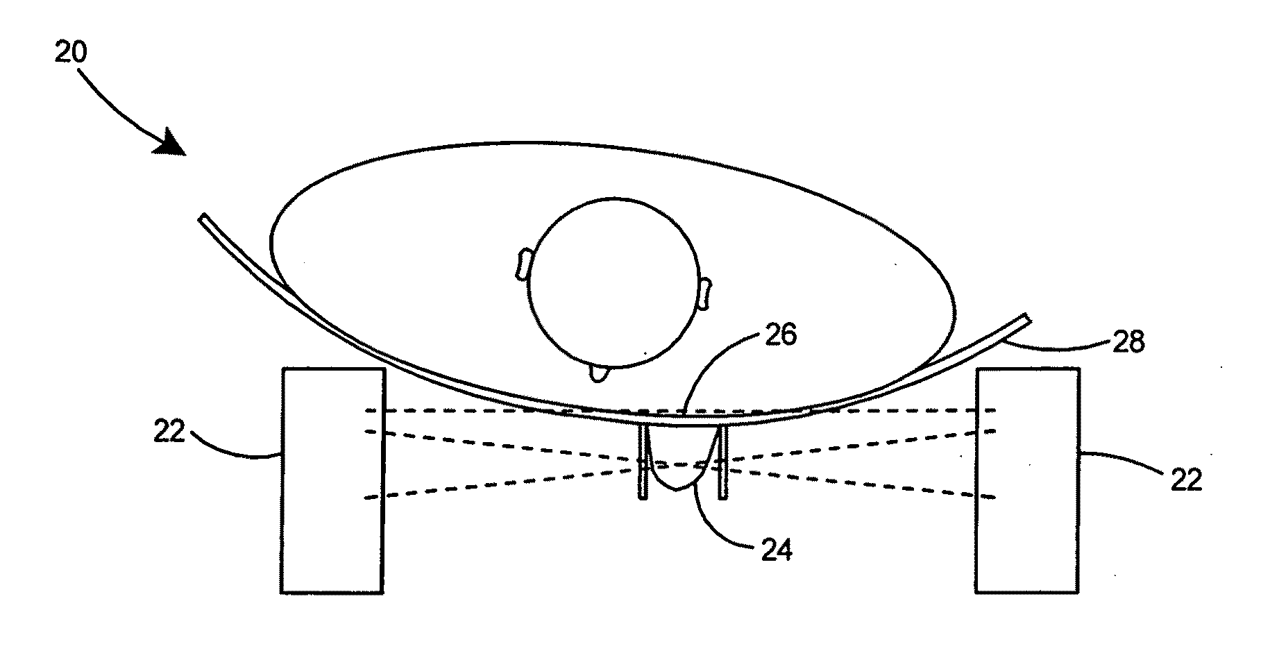
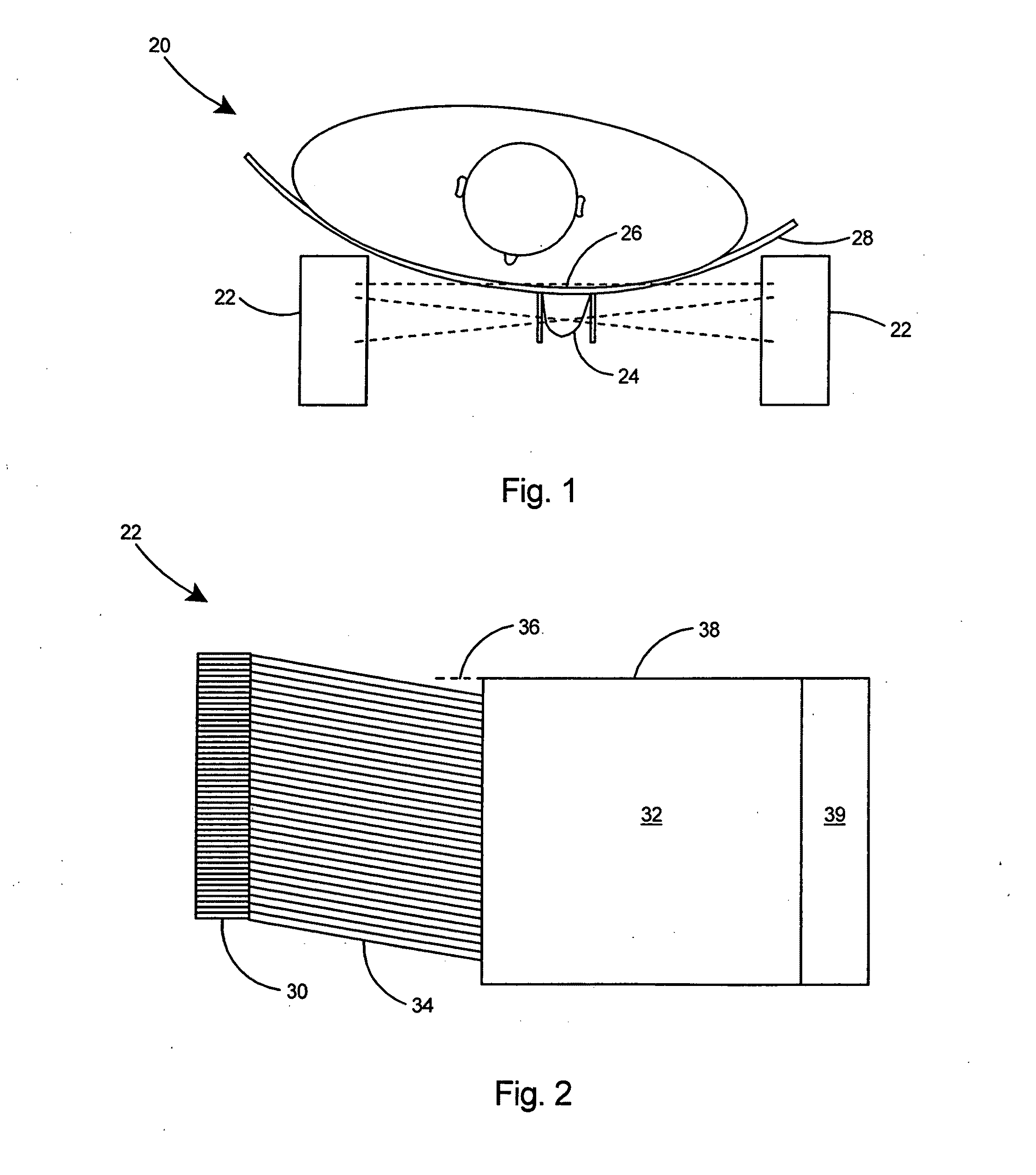
[0080]With reference to FIG. 1 there is shown a breast imaging system 20 according to the present invention including a set of photodetector modules 22 viewing the breast 24 in a lateral position and with the detector heads raised past the edge of the chest wall 26 to permit efficient detection of lesions in the breast region close to the chest wall. The imaging system includes lateral imaging geometry with the detector heads 22 put further apart with one or two detector heads 22 raised past the edge of the chest wall 26 to permit efficient detection of lesions in the breast region close to the chest wall 26. The light-weight patient bed 28, which is preferably constructed of hard plastic or a carbon-fiber matrix, has a shape approximating a fraction of a cylinder to facilitate placement of the detector heads 22 higher than the chest wall 26.

[0081]The second and preferred embodiment of the imaging system of the present invention includes a ring of detection modules surrounding the i...

second embodiment

[0084]the breast imaging system 60 is depicted in FIG. 5. In the embodiment depicted in FIG. 5, two rings of imaging modules 46 are provided, including a top imaging ring 62 and a bottom imaging ring 64, and the whole set of imaging rings 62 and 64 can be moved vertically to cover the whole volume of the breast 24. In this particular embodiment the dead edge at the PMT perimeter 66 results in a small physical gap 68 between the two imaging rings 62 and 64. The impact of the gap 68 is minimal on the final quality of the reconstructed images. If desired, additional detector rings could be added to create a breast imaging system with three or more rings (not shown) to eliminate the need for vertical scanning and cover the whole breast volume without requiring movement of the detector rings.

[0085]In the breast imaging systems of the present invention, the breast to be imaged will be pending through the inner tube 48 in the patient bed 44 as shown in FIG. 5. An alternative embodiment for...

third embodiment

[0087]FIG. 6B depicts a top view of an imaging system 78 similar to the imaging system of FIG. 6A but in which the imaging modules 72 are in two imaging ring sectors 79A and 79B. The imaging modules 72 are rotated around the breast (not shown) to provide all the angular views that are necessary for 3D tomographic reconstruction. This embodiment of the breast imager 78 is an economical arrangement composed of two imaging ring sectors 79A and 79B only. This embodiment of the breast imager 78 offers convenient side ports 80 for installing a biopsy device. The imager 78 provides the convenience of performing the biopsy procedure while imaging the breast. By using a radioactive tip biopsy needle (not shown), the needle tip is guided to the lesion by the imaging system 78.

[0088]Referring to FIG. 2, a dedicated breast PET imager according to the present invention could include several imaging technologies. The preferred general type of a solution will have a scintillator sensor 30 as a sen...


PUM


Abstract

A highly efficient PET breast imager for detecting lesions in the entire breast including those located close to the patient's chest wall. The breast imager includes a ring of imaging modules surrounding the imaged breast. Each imaging module includes a slant imaging light guide inserted between a gamma radiation sensor and a photodetector. The slant light guide permits the gamma radiation sensors to be placed in close proximity to the skin of the chest wall thereby extending the sensitive region of the imager to the base of the breast. Several types of photodetectors are proposed for use in the detector modules, with compact silicon photomultipliers as the preferred choice, due to its high compactness. The geometry of the detector heads and the arrangement of the detector ring significantly reduce dead regions thereby improving detection efficiency for lesions located close to the chest wall.

Description

[0001]The United States of America may have certain rights to this invention under Management and Operating contract No. DE-AC05-06OR23177 from the Department of Energy.FIELD OF THE INVENTION[0002]This invention relates to breast imaging and more particularly to a high resolution PET breast imaging system with improved detection efficiency for lesions located close to the chest wall of a patient.BACKGROUND OF THE INVENTION[0003]Breast cancer biology seems to indicate that breast cancer metastasis starts at the lesion size of about 2 mm. While X-ray mammography offers much better intrinsic spatial resolution, on the order of 50 micrometers, this anatomical imaging modality suffers from poor specificity to the type (cancerous or benign, etc.) of the suspicious structure seen on the mammogram. In some patients this X-ray imaging technique is not useful at all due to dense breast tissue, implants, or scars as the result of previously performed surgery. Breast MRI can provide more struct...

Claims


Application Information

Patent Timeline
Patent Type & Authority Applications(United States)
IPC IPC(8): A61B6/04G01T1/164
CPCA61B6/037G01T1/00G01T1/2008
Inventor MAJEWSKI, STANISLAW
Owner JEFFERSON SCI ASSOCS LLC
Why Eureka
Try Eureka