Radiographic imaging device and radiographic imaging system

Inactive Publication Date: 2010-04-01
KONICA MINOLTA MEDICAL & GRAPHICS INC
View PDF8 Cites 31 Cited by
  • Summary
  • Abstract
  • Description
  • Claims
  • Application Information

AI Technical Summary

Benefits of technology

[0028]According to the present invention, it becomes possible to fully exhibit a Talbot effect and to detect the configuration of a photographic object with sufficient accuracy in a moire stripe image. At this time, since X rays having an average energy of 15 to 60 keV are irradiated from the X-ray tube for a short time period of 1/several seconds or less, an X-ray image can be obtained without blurring due to motion of a human body being a photographic object and a burden to a human body can reduced. Further, with a setup to make a distance between an X-ray tube and an X-ray detector, a distance between an X-ray tube and a first diffraction grating, and the focal size of an X-ray tube appropriate respectively, it

Problems solved by technology

However, it is very difficult to detect the above-mentioned early symptoms of rheumatic disease from an X-ray radiograph which is an easy inspection method, and it is difficult to judge easily whether or not symptoms have been developed.
However, the photography by MRI causes a heavy burden on a person to be photographed from the viewpoints of expenses and time necessary for the diagnosis.
Therefore

Method used


Image

  • Radiographic imaging device and radiographic imaging system
  • Radiographic imaging device and radiographic imaging system
  • Radiographic imaging device and radiographic imaging system

Examples


Example

EXPLANATION OF SYMBOL

[0049]1 radiation image radiographing apparatus[0050]8 X-ray tube[0051]15 first diffraction grating[0052]16 second diffraction grating[0053]15a, 16a temperature sensors[0054]152, 162 diffractive members[0055]17 X-ray detector[0056]20 control device[0057]30 image processing apparatus[0058]50 image output apparatus[0059]100 radiation image radiographing system[0060]a focal size of an X-ray tube[0061]H a photographic object[0062]L distance between an X-ray tube and a first diffraction grating[0063]M moire stripes[0064]Z1 distance between the first diffraction grating and the second diffraction grating

BEST MODE FOR CARRYING OUT THE INVENTION

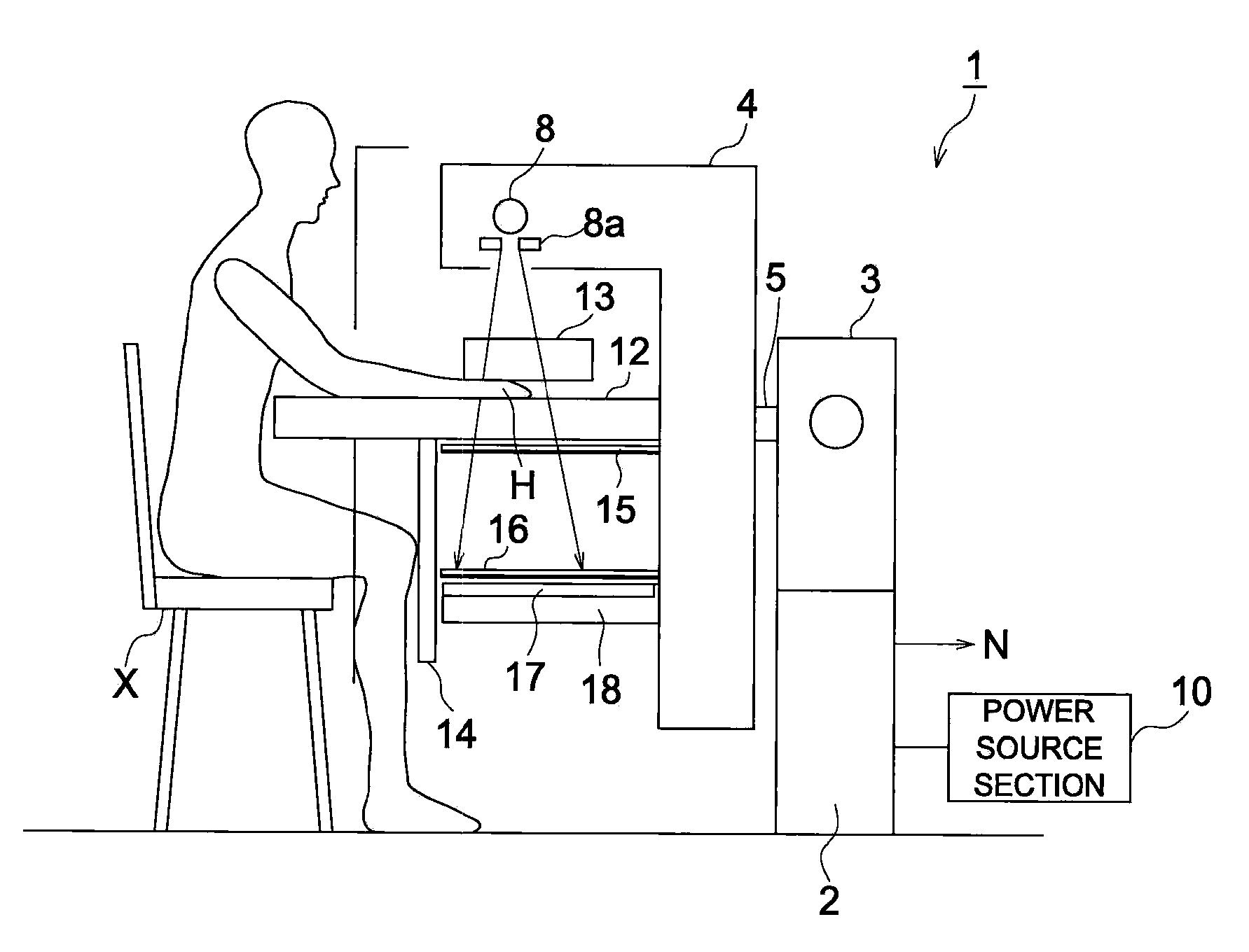
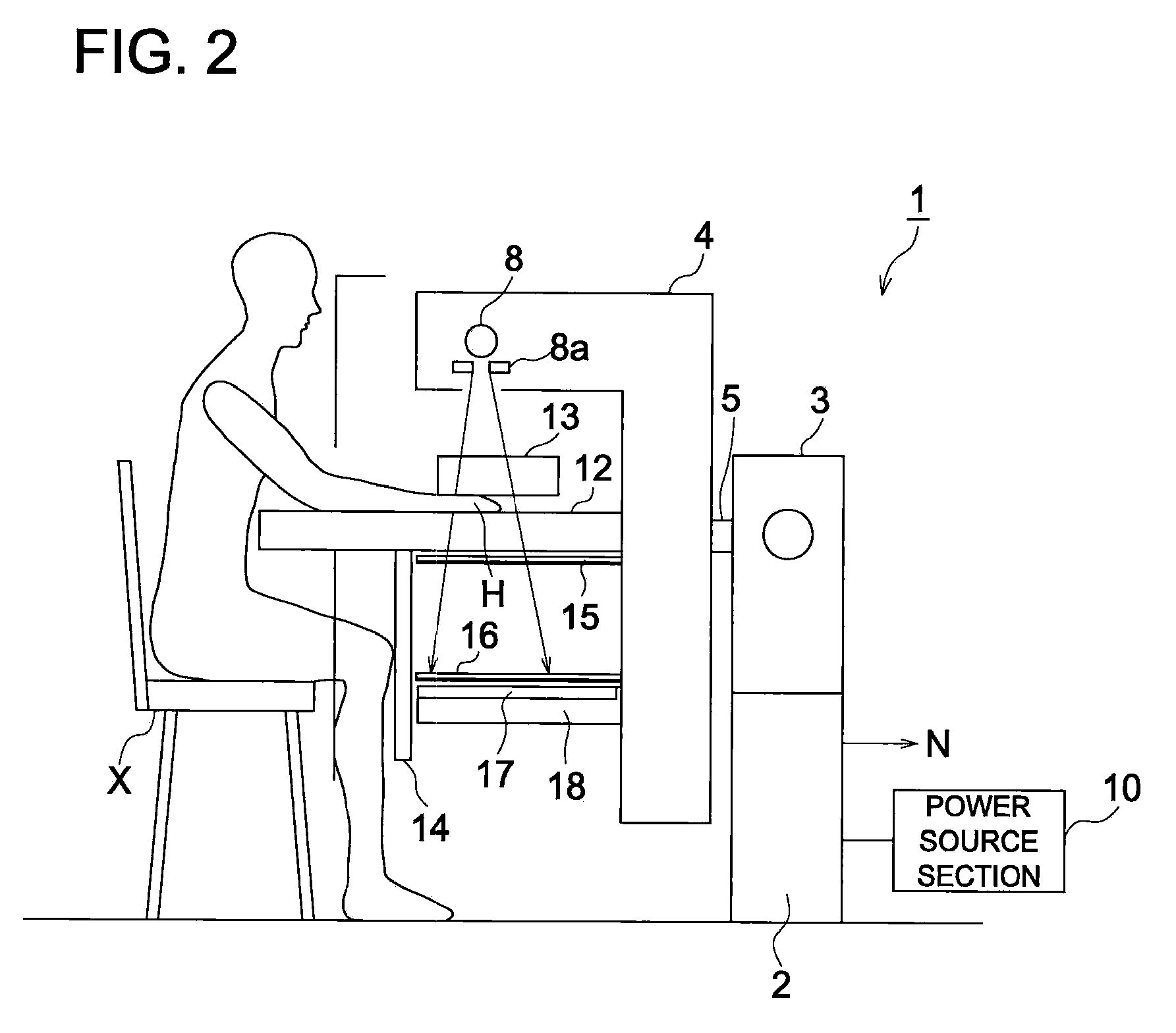
[0065]Hereafter, embodiments of a radiation image radiographing apparatus and a radiation image radiographing system according to the present invention will be described with reference to drawings. However, the scope of the invention is not limited to the examples shown in the drawings.

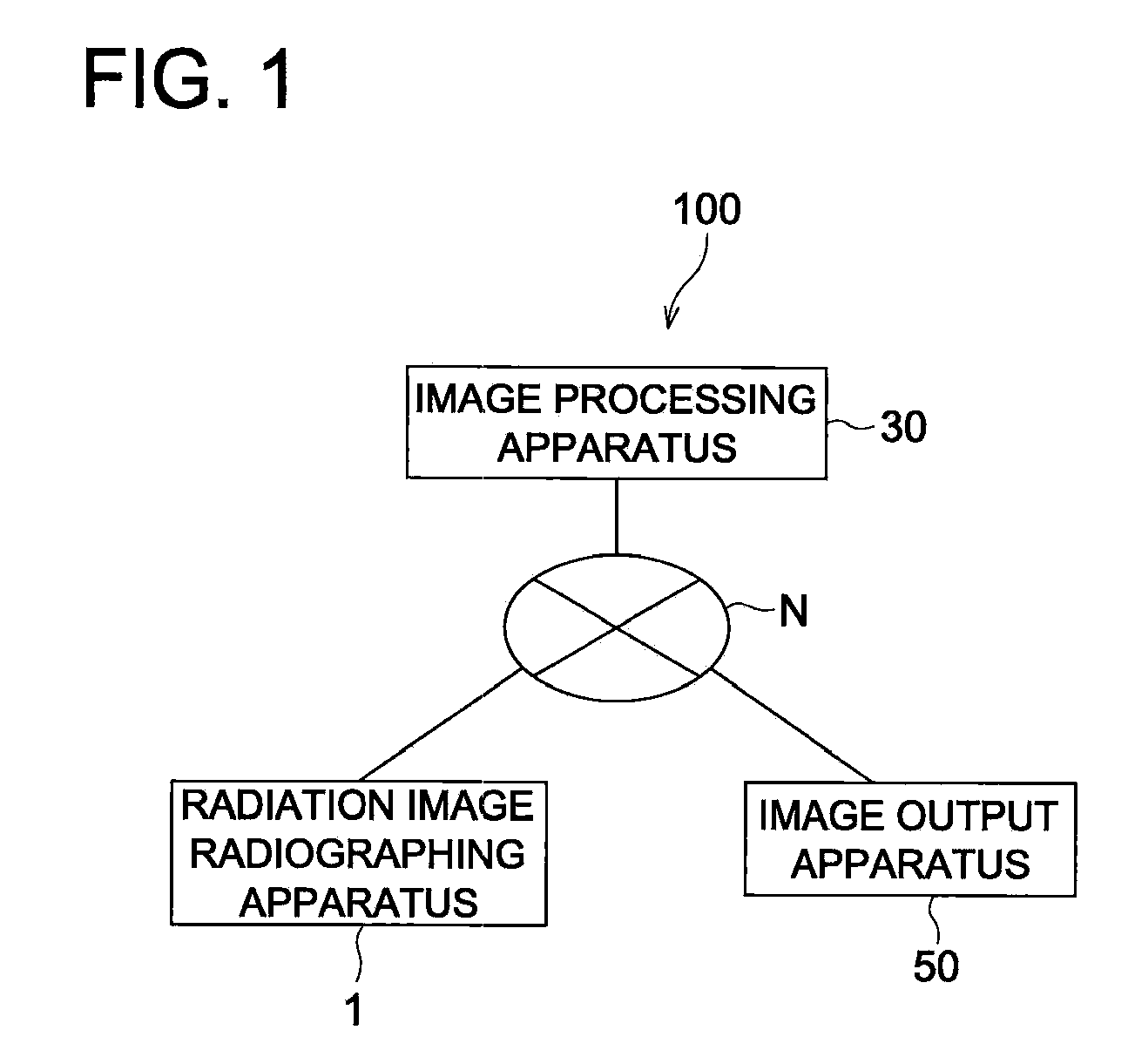
[0066]In the present embodiment, as shown in...


PUM


Abstract

A radiographic imaging device for capturing an excellent X-ray image in which the contrast of edge portions such as a cartilage tissue of a human body is enhanced by using a Talbot interferometer, and a radiographic imaging system for processing the captured image. The radiographic imaging device comprises an X-ray tube for applying X-rays having an average energy of 15 to 60 keV, a first diffraction grating for diffracting X-rays transmitted through a subject (H) to produce the Talbot effect, a second diffraction grating for diffracting X-rays diffracted by the first diffraction grating, and an X-ray detector for detecting X-rays diffracted by the second diffraction grating. The second diffraction grating is disposed in contact with the X-ray detector. The distance L between the X-ray tube and the first diffraction grating is 0.5 m or more. The distance Z1 between the first and second diffraction gratings is 0.05 m or more. The focus diameter a of the X-ray tube is 1 μm or more.

Description

TECHNICAL FIELD[0001]The present invention relates to a radiation image radiographing apparatus (radiographic imaging device) and a radiation image radiographing system (radiation imaging system), and especially relates to a radiation image radiographing apparatus employing a Talbot interferometer system and a radiation image radiographing system to conduct a processing for an image captured by the radiation image radiographing apparatus.BACKGROUND ART[0002]The disease rate of rheumatic disease in Japan has reached to even 1%, and, now, the rheumatic disease is made into a folk disease. As its early symptoms, the abrasion of cartilaginous portions (cartilage destruction) and some changes in minute bone geometries and bone trabeculae are observed, and at the stage where the symptom advanced, big changes in configurations of a bony part are observed. Therefore, since it is possible to diagnose the disease condition of a rheumatic disease by observing changes in the configurations of c...

Claims


Application Information

Patent Timeline
IPC IPC(8): G06K9/00G01N23/04
CPCA61B5/4514A61B5/4528A61B6/06A61B6/4488A61B6/4291A61B6/484A61B6/502A61B6/505A61B6/483
Inventor OHARA, HIROMU
Owner KONICA MINOLTA MEDICAL & GRAPHICS INC
Why Eureka
Try Eureka