

Use of crystal location in nuclear imaging apparatus to minimize timing degradation in a photodetector array
a nuclear imaging and crystal location technology, applied in the field of nuclear medical imaging, can solve the problems of long-felt and unresolved problems such as the inability to differentiate random events from true coincidence events, the inability to optimally couplage the scintillation crystals of the the inability to optimally coupl the photo detectors in a 1, so as to improve the overall prompt, improve the quality of images, and reduce the random rate
- Summary
- Abstract
- Description
- Claims
- Application Information
AI Technical Summary
Benefits of technology
Problems solved by technology
Method used
Image

Examples
Embodiment Construction
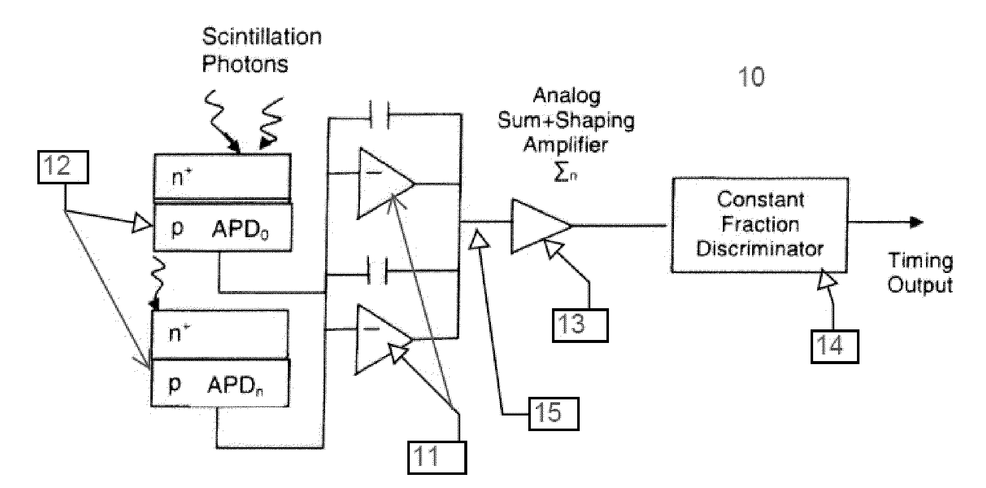
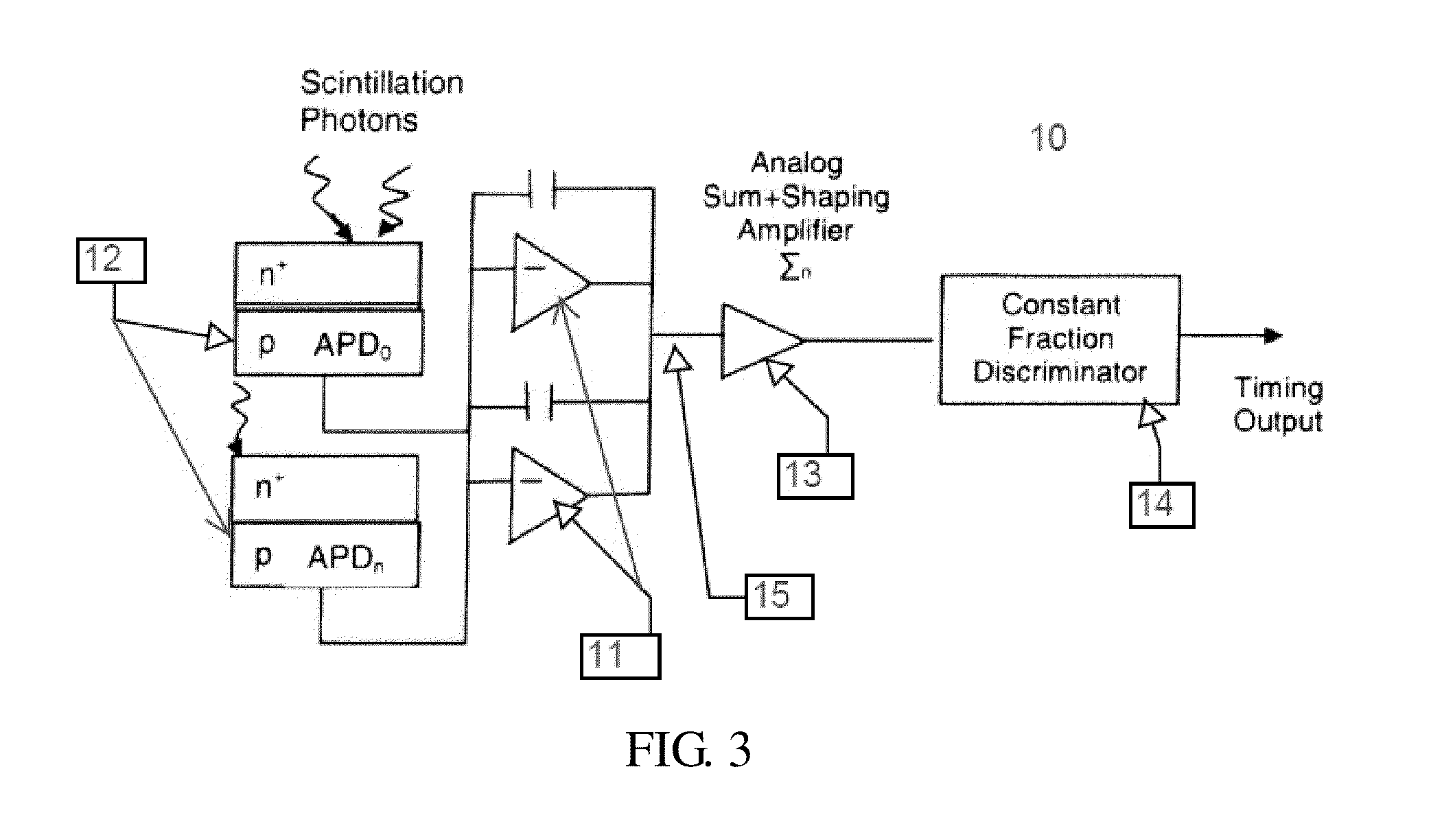
[0022]FIG. 3 illustrates an exemplary embodiment of an APD-based block detector timing signal architecture 10 of a PET scanner. Timing signal architecture 10 comprises low-noise charge-sensitive preamplifiers 11, which can be used with APDs 12, with moderate gain of about 100, to achieve timing coincidence window resolution values that are comparable to PMT-based detector blocks. A method is described which improves timing coincidence window resolution using a variable number of APDs to derive the timing signal based on the current detected photon's crystal location.
[0023]The charge liberated from each individual APD (APDn=9 in the example given), when a gamma photon is detected by a scintillation crystal, can be integrated by a charge-sensitive preamplifier 11. N detector block APDs, for example 9, are summed into a single timing channel 15 and then filtered in a time shaping amplifier 13 (TSA) before being sent to a constant fraction discriminator 14 (CFD) for determining the dete...
PUM
 Abstract
Description
Claims
Application Information