Method and device to determine an inversion time value of tissue by means of magnetic resonance technology

a tissue inversion and value technology, applied in the field of tissue inversion time value determination, can solve the problem that the patient is not able to remain in the magnetic resonance apparatus for a long time due to multiple measurements, and achieve the effect of reducing the amount of measurement time, stably estimating the relaxation response, and reducing the patient's risk of having to remain in the magnetic resonance apparatus

Inactive Publication Date: 2010-04-08
SIEMENS HEALTHCARE GMBH
View PDF11 Cites 10 Cited by
  • Summary
  • Abstract
  • Description
  • Claims
  • Application Information

AI Technical Summary

Benefits of technology

[0022]In a further embodiment, the series of magnetic resonance images is acquired by using a late enhancement measurement. The inversion times are established by the interval of the inversion module from the actual image acquisition sequence. Since the late enhancement measurement always ensues in the same cardiac phase, no significant heart movement is contained in the image data. The segmentation of the structure to be examined is therefore particularly simple.
[0023]An additional advantageous embodiment is characterized in that a parameterizable model of the time response of the relaxation of the longitudinal magnetization of one of the different tissues (advantageously the healthy tissue) is used for segmentation. The model is adapted to the current temporal signal curve in that the free parameter of the model is determined so that, for example, the difference between the observed signal intensity and the signal intensity of the parameterized relaxation model is minimal. In this way it is possible to stably estimate the relaxation response based on only a few acquired images, such that a considerable amount of measurement time can be saved.
[0024]In a further advantageous embodiment, the imaging area is a short-axis section of the heart. The short-axis section shows simple characteristic geometric structures that can be segmented well.

Problems solved by technology

Repeat measurements due to incorrect first measurements are no longer required, and the danger to the patient of having to remain in the magnetic resonance apparatus longer due to multiple measurements no longer exists.

Method used


Image

  • Method and device to determine an inversion time value of tissue by means of magnetic resonance technology
  • Method and device to determine an inversion time value of tissue by means of magnetic resonance technology
  • Method and device to determine an inversion time value of tissue by means of magnetic resonance technology

Examples


Embodiment Construction

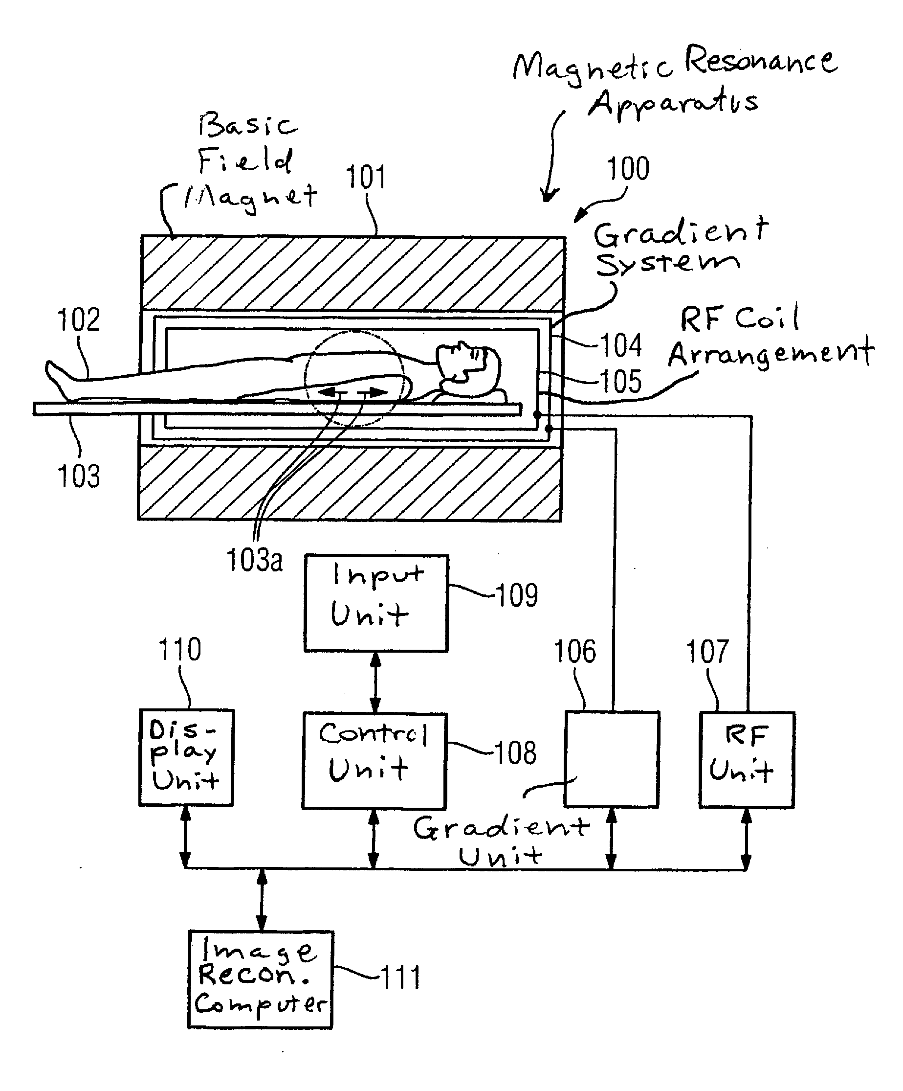
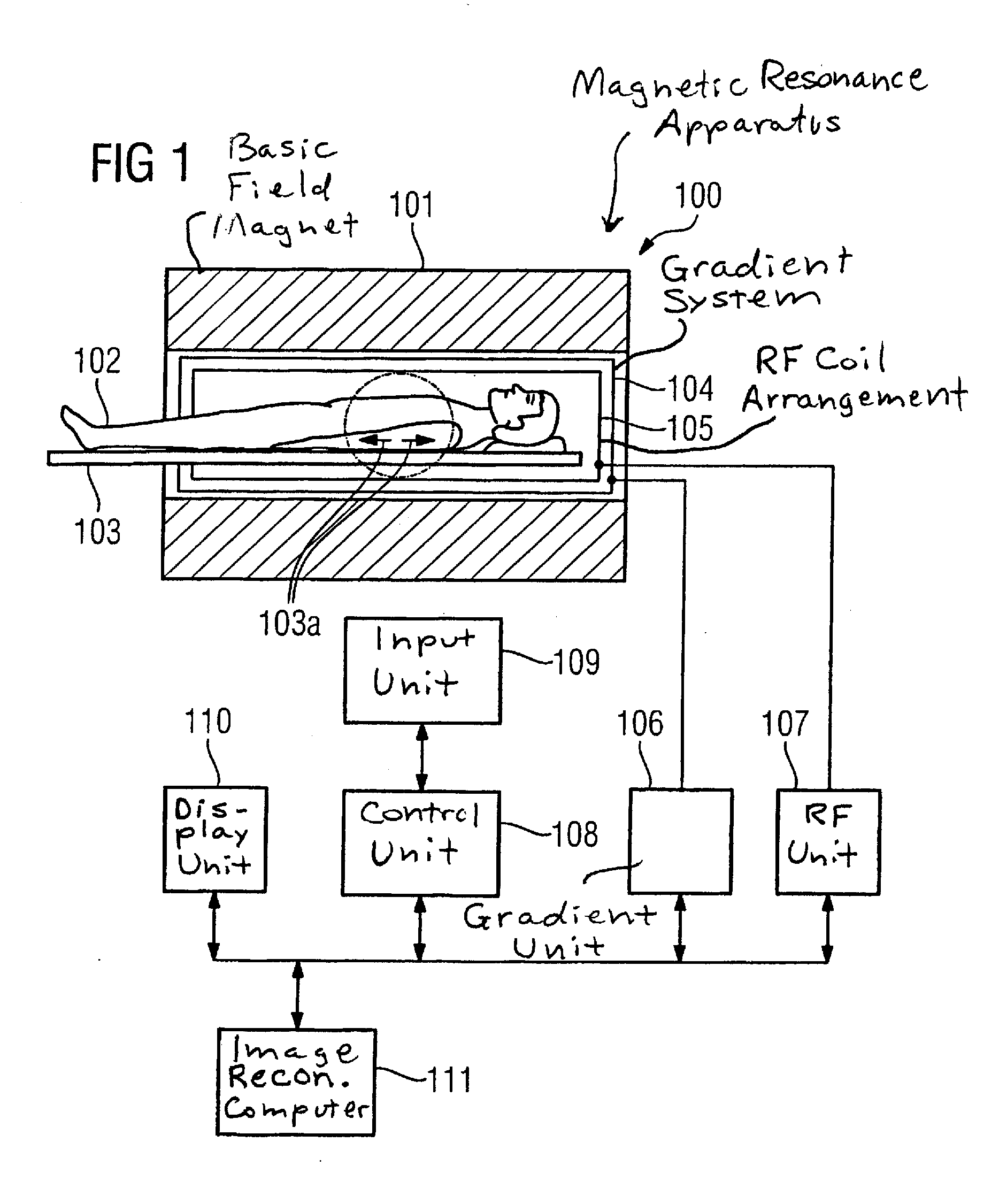
[0029]FIG. 1 schematically shows a magnetic resonance apparatus 100 with which an inversion time value for contrast improvement between different tissue can be determined in a contrast agent-supported magnetic resonance image acquisition, for example for a following vitality examination of the heart muscle. Such a magnetic resonance apparatus 100 has a magnet 101 to generate a polarization field or basic magnetic field B0. In the example shown here, the examination subject is an examination person 10 on a bed 103. As is schematically represented by arrows 103a, the bed 103 can be driven into the magnet 101 and be positioned therein for the examination of an organ. Furthermore, the magnetic resonance apparatus 100 has a gradient system 104 to generate magnetic field gradients that are used for imaging and spatial coding. To excite the polarization resulting in the basic magnetic field B0, a radio-frequency (RF) coil arrangement 105 is provided that radiates a radio-frequency field in...


PUM


Abstract

In a method to determine an inversion time value for contrast improvement between different tissue in a contrast agent-supported magnetic resonance imaging, a series of magnetic resonance images of an imaging area is acquired using an inversion recovery sequence with different inversion times. A structure in the magnetic resonance images is segmented and a time response of the signal intensity of image elements corresponding to one another in the magnetic resonance images of the segmented structure is automatically determined. Minima of the signal intensity in the segmented structure are determined automatically and associated with the associated inversion time values. The optimal inversion time value for contrast improvement is automatically determined from the inversion time values that have been associated with the minima of the signal intensity in the segmented structure.

Description

BACKGROUND OF THE INVENTION[0001]1. Field of the Invention[0002]The present invention relates to a method for determining an inversion time value of tissue using magnetic resonance technology, as well as to a magnetic resonance apparatus for implementing such a method.[0003]2. Description of the Prior Art[0004]In recent years, contrast agent-supported tissue characterization by means of magnetic resonance technology has found increasing distribution in clinical practice. Conclusions about the extent and degree of damage in undernourished tissue or, respectively, after infarcts can be obtained with the aid of contrast agent-supported magnetic resonance examinations.[0005]The basic principle of contrast agent-supported magnetic resonance imaging for tissue characterization utilizes the kinetics of a contrast agent based on gadolinium in tissue. In infracted, necrotic or scarred tissue, the contrast agent enrichment takes significantly longer than in healthy tissue due to the poor perf...

Claims


Application Information

Patent Timeline
Patent Type & Authority Applications(United States)
IPC IPC(8): G01R33/48
CPCA61B5/055A61B5/7285G01R33/281G01R33/56G01R33/56366G01R33/5602G01R33/5608G01R33/563G01R33/56325G01R33/5601
Inventor LITTMANN, ARNESPEIER, PETERSPRUNG, KATRIN CHRISTEL
Owner SIEMENS HEALTHCARE GMBH
Why Eureka
Try Eureka