Assay Membrane and Method of Use Thereof

a technology of assay membrane and assay method, which is applied in the field of assay membrane, can solve the problems of high cost and cumbersomeness of available screening technologies for large number of samples from subjects, and the technology has yet to be adopted as a routine method for diagnostic testing

Active Publication Date: 2010-04-15
PICTOR
View PDF13 Cites 32 Cited by
  • Summary
  • Abstract
  • Description
  • Claims
  • Application Information

AI Technical Summary

Benefits of technology

[0031]In one embodiment the fiduciary marker is a dye, dye-conjugated protein or chromogenic protein, hapten-conjugated protein or enzyme-conjugated protein; for example, Coomassie Blue, colloidal gold, Ponceau S, a peroxidase enzyme such as horseradish peroxidase (HRP), or a dyed molecular weight marker. Preferably the fiduciary marker permits orientation and gridding of the array.

Problems solved by technology

Currently available technologies for screening large numbers of samples from subjects are expensive and cumbersome.
However, this technology has yet to be adopted as a routine method for diagnostic testing, due to technical challenges surrounding the sensitivity, specificity and cross-reactivity of the assay reagents and the need for expensive instrumentation.

Method used


Image

  • Assay Membrane and Method of Use Thereof
  • Assay Membrane and Method of Use Thereof
  • Assay Membrane and Method of Use Thereof

Examples


example 1

General Procedures for Array Manufacture

[0187]Membranes or films attached to a bottomless 96-well polystyrene plate (such as nylon, Nalge Nunc International, USA,) were used for printing microarrays. Various methods may be used to attach the membranes to the 96-well bottomless plates. In this example, a rubber sheet with dimensions of 128 mm length and 86 mm and 1 mm thickness was used. 96 round holes were stamped on the sheet with a diameter of 6.35 mm and a center to center distance of 9 mm. The sheet was then coated with adhesive on both sides and glued to one side of the 96-well bottomless plate such that the second adhesive layer was still available for binding to a membrane. A nylon membrane was then cut to the dimensions of 128 mm length and 86 mm width and attached to the other side of the rubber sheet to create a gasket that creates leak proof wells.

[0188]Another method for attaching the membranes to the 96-well bottomless plates involves use of an adhesive that is applied ...

example 2

General Procedure for Array Processing and Analysis

[0195]The arrays were incubated at 37° C. for 60 min after adding 100 μl of Blocker [1% casein (Vector Labs, USA) in phosphate buffered saline containing 0.1% Tween 20 (PBS-T)] to each well. The Blocker was then aspirated off.

[0196]Samples containing analytes were added by diluting in Blocker at a volume of 50 μl in each well and the membrane incubated at 37° C. for 60 min. The membrane was washed 3× with PBS-T to remove excess non-bound analytes.

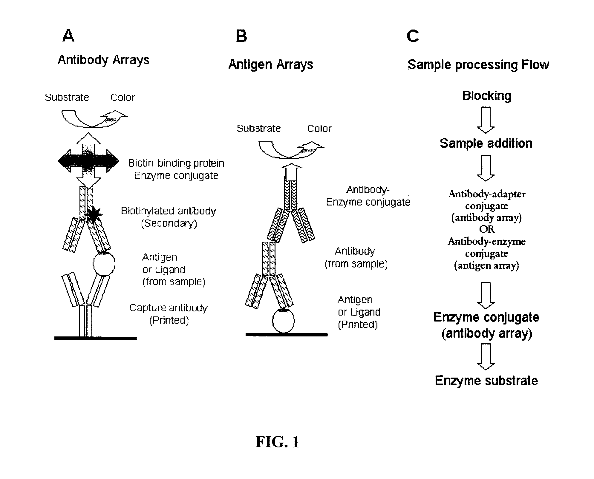
[0197]For antibody arrays (for antigen detection), adapter-conjugated detection antibodies were added to the wells at concentrations either recommended by the manufacturer or empirically determined by experimentation. Examples of adapter-conjugated antibodies include hapten-conjugated antibodies such as biotin-conjugated antibodies. The membrane was incubated at 37° C. for 60 min and washed 3× with PBS-T. The membrane was then incubated with an anti-adapter antibody (such as an anti-biotin ...

example 3

Antibody Arrays

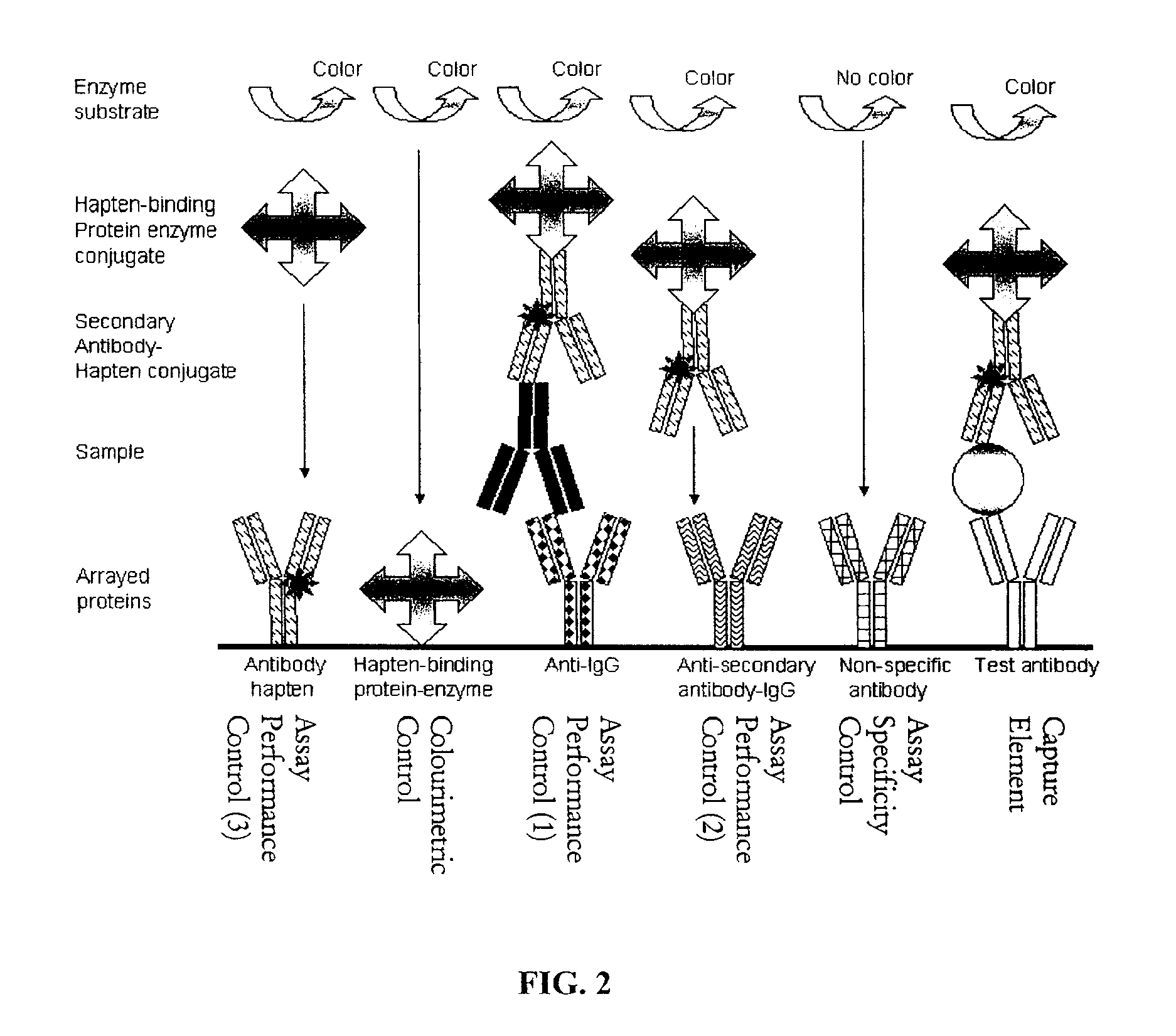
[0202]Arrays for detection of antigens such as protein markers of autoimmune diseases, cardiovascular diseases, cancer and infectious agents, or ligands such as growth factors, hormones, cytokines and chemokines are created by printing panels of antibodies as capture elements for specific capture of the antigen. A series of control antibodies and control proteins are also printed. These controls serve a variety of functions including controls for monitoring assay performance including performance of individual reagents, controls for monitoring the specificity of the capture antibodies and fiduciary markers for gridding the arrays after sample processing for determination of signal intensity at each spot in the array. Table 4 summarises the reagents that may be used to print and process antibody arrays. The assay performance control numbering relates to the numbering in FIG. 2.

TABLE 4Antibody array reagentsReagentFunctionExampleCommentsProtein or antibodyFiduciary mark...


PUM

PropertyMeasurementUnit
diameteraaaaaaaaaa
diameteraaaaaaaaaa
temperatureaaaaaaaaaa

Abstract

The present invention provides a microporous membrane for detecting at least one target analyte in a sample. The membrane includes an array that comprises at least one capture element and the at least one control element printed on the membrane surface, the at least one capture element corresponding to and being able to bind a target analyte, the plurality of control elements, when present including: i) at least one fiduciary marker, ii) at least one negative control to monitor background signal, iii) at least one negative control to monitor assay specificity, iv) at least one positive colorimetric control, v) at least one positive control to monitor assay performance or any combination thereof.

Description

BACKGROUND OF THE INVENTION[0001]1. Field of the Invention[0002]The present invention relates to a method for the colorimetric detection of at least one analyte in a sample, preferably multiple analytes in a sample, and membranes for use in the method.[0003]2. Background Information[0004]Biomarkers can identify disease prior to exhibition of clinical symptoms by a subject, and therefore provide the ability to treat the molecular basis of disease using targeted therapies. Thus, biomarkers allow the pharmacodynamic effects of targeted therapeutics to be evaluated before clinical signs and symptoms become evident.[0005]New biomarkers are being discovered by means of a large number of proteomic and genomic studies using low throughput, labour intensive technologies such as mass spectrometry and high performance liquid chromatography. These technologies are great discovery tools for identifying novel biomarkers in small numbers of samples from subjects. However, biomarkers must be qualif...

Claims


Application Information

Patent Timeline
Patent Type & Authority Applications(United States)
IPC IPC(8): C40B30/04C40B60/12
CPCG01N2800/52G01N33/54306B01L3/5025B01L3/5085B01L3/50855G01N33/543
Inventor KUMBLE, SARITA
Owner PICTOR
Why Eureka
Try Eureka