Method and apparatus for medical imaging using near-infrared optical tomography combined with photoacoustic and ultrasound guidance

Inactive Publication Date: 2010-04-15
UNIV OF CONNECTICUT
View PDF25 Cites 114 Cited by
  • Summary
  • Abstract
  • Description
  • Claims
  • Application Information

AI Technical Summary

Problems solved by technology

Although diffusive optical tomography is a promising medical imaging technique, DOT imaging methods have yet to yield high quality reconstructions of inclusions due to fundamental issues with intense light scattering.
The primary limitation of DOT is related to the intense light scattering in tissues that dominates near infrared light propagation and makes three-dimensional localization of lesions and accurate quantification of lesion optical properties difficult.
Ultrasound can detect breast lesions that are a few millimeters in size; however, its specificity in breast cancer detection is not high as a result of the overlapping characteristi

Method used


Image

  • Method and apparatus for medical imaging using near-infrared optical tomography combined with photoacoustic and ultrasound guidance
  • Method and apparatus for medical imaging using near-infrared optical tomography combined with photoacoustic and ultrasound guidance
  • Method and apparatus for medical imaging using near-infrared optical tomography combined with photoacoustic and ultrasound guidance

Examples


example 1

[0081]This example was conducted to demonstrate the synergistic role of PAT and DOT in detection and characterization of deep, closely spaced targets. Because both PAT and DOT utilize optical contrast, this guidance can be more specific than with non-optical modalities to improve reconstruction accuracy and robustness.

[0082]Three types of spherical targets embedded in turbid liquid mediums were used to simulate mechanical and / or optical contrast. Hard spherical resin balls of 1 cm diameter with (μa=0.07 cm−1, μ′s=5.5 cm−1) and higher (μa=0.23 cm−1, μ′s=5.5 cm−1) absorption provided a high contrast ratio of 3.3. μα is the coefficient of absorption while μ′s is a reduced scattering coefficient. A pair of 1 cm-diameter near-spherical soft-gelatin absorbers of higher (μa=0.14 cm−1, μ′s=4.3 cm−1) and lower (μa=0.08 cm−1, μ′s=6.32 cm−1) optical contrast representative of malignant and benign lesion optical properties are used as soft tissue targets with moderate contrast (ratio=1.75). Fin...

example 2

[0099]This example was conducted to demonstrate the synergistic role of PAT and DOT in detection and characterization of deep, closely spaced targets. In this example, both single-lobed and multi-lobed polyvinylchloride (PVC) plastisol absorbers were used in separate measurements to stimulate a tumor. The PVC absorbers were disposed in Intralipid (a material that is representative of human breast tissue). The PVC absorbers were cube shaped having each side equal to 1 centimeter and had absorption coefficients of 0.075 cm−1 to 0.23 cm−1. The PVC absorbers were imaged at depths of up to 2.5 centimeters in the Intralipid. As will be seen in the experiment below one of the absorbers was a high contrast target and the other a low contrast target.

[0100]From the results detailed below, it can be seen that without PAT guidance the absorber location was not clear and lower contrast targets in the two-absorber configurations were not distinguishable. With PAT guidance, the two targets were we...


PUM


Abstract

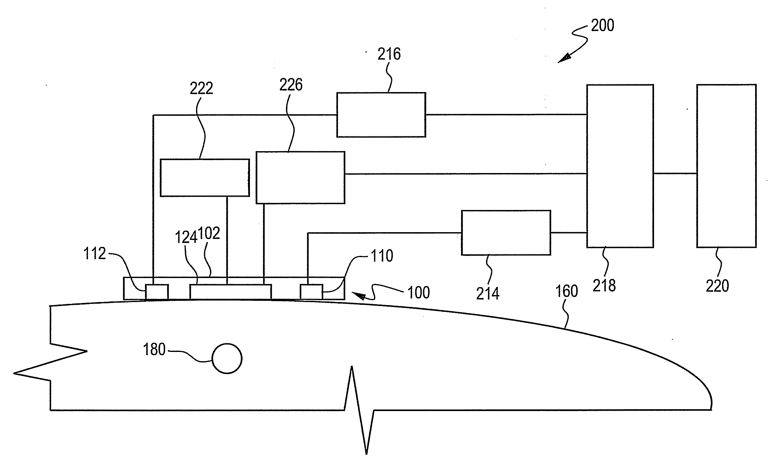
Disclosed herein is an apparatus for biological imaging comprising a probe comprising an emitter and a detector; a source circuit connected in operational communication to the emitter; a detector circuit connected in operational communication to the detector; a central processing unit connected to the source circuit and the detector circuit; a display operably connected to the central processing unit; and wherein the apparatus is capable of photoacoustic tomography and diffusive optical tomography and/or ultrasound tomography.

Description

CROSS-REFERENCE TO RELATED APPLICATION[0001]This application claims priority to provisional application 61 / 105,174 filed on Oct. 14, 2009, the entire contents of which are hereby incorporated by reference.TECHNICAL FIELD[0002]This disclosure relates primarily to the field of biological imaging, particularly to medical imaging. More specifically, this disclosure relates to medical imaging equipment and methods for medical imaging using combined near-infrared optical tomography with photoacoustic and ultrasound guidance.BACKGROUND OF THE INVENTION[0003]Diffusive optical tomography (DOT) is a form of computer-generated tomography wherein near-infrared light (NIR) is directed at a biological object (e.g., a inclusion, tumor, and so forth) and the amount of light transmitted and / or diffused through the object, and / or reflected from the object, is detected and utilized to reconstruct a digital image of the target area (e.g., the object can exhibit a differential in optical absorption and ...

Claims


Application Information

Patent Timeline
IPC IPC(8): A61B6/00
CPCA61B5/0073A61B5/0095A61B8/4416A61B5/418A61B8/08A61B5/415A61B5/0035
Inventor ZHU, QUINGGAMELIN, JOHN
Owner UNIV OF CONNECTICUT
Why Eureka
Try Eureka