Biomarkers and methods for detecting and treating spinal and joint pain

a biomarker and spinal joint technology, applied in the direction of dna/rna fragmentation, peptide/protein ingredients, depsipeptides, etc., can solve the problems of difficult identification difficult treatment of spinal and joint pain, joint injury, etc., to reduce the expression of fibronectin, inhibit or reduce one or more biological activities, and reduce the formation

Active Publication Date: 2010-04-22
CYTONICS CORP
View PDF37 Cites 13 Cited by
  • Summary
  • Abstract
  • Description
  • Claims
  • Application Information

AI Technical Summary

Benefits of technology

[0022]Other methods for treating spinal or joint pain include administering antagonists of fibronectin-aggrecan complexes. Suitable fibronectin-aggrecan complex antagonists inhibit or reduce one or more biological activities of fibronectin-aggrecan complexes, inhibit or reduce the formation of fibronectin-aggrecan complexes from fibronectin and aggrecan and / or their fragments, cause the dissociation of fibronectin-aggrecan complexes, reduce the expression of fibronectin and / or aggrecan, or reduce the degradation and the breakdown of aggrecan from cartilage. Exemplary fibronectin-aggrecan complex antagonists include, but are not limited to, antibodies and other polypeptides, peptidomimetics, small organic molecules, and inhibitory RNAs, such as ribozymes, triplex-forming oligonucleotides, antisense DNA, siRNA and miRNA.

Problems solved by technology

Spinal and joint pain can be difficult to treat.
In particular the cause of spinal and joint pain can be difficult to identify.
Increasing degrees of force applied to joints result in joint injury.
Physicians currently do not have a system or method available to differentiate between acute injury due to trauma and age related joint deteriorations.
It is presently difficult to determine the appropriate course of treatment for a given patient since it is frequently unclear whether the particular condition the patient suffers from is acute or chronic.
This problem is exacerbated by the low specificity of MRI, currently a mainstay of diagnosing this pathology.
It has been shown that MRI will identify a meniscal injury in as many as 65% of asymptomatic people making MRI a questionable diagnostic tool and highlighting the lack of correlation between abnormal meniscal anatomy and knee pain.
Lack of a clear correlation between abnormal meniscus anatomy and knee pain is particularly problematic in the elderly patient population many of whom develop osteoarthritis.
It has been demonstrated that the application of nucleus pulposus to the spinal nerve root can result in axonal damage and functional changes to nerve root micro-anatomy, resulting in pain-related behaviors.
Thus, it has been theorized that a mechanical defect releasing the nucleus pulposus into the epidural space may cause nerve root damage resulting in radicular pain.
However, no clear causal pathway leading from injury or degeneration to the painful state has been confirmed.
Although some studies have provided evidence that the epidural space may be affected by an intervertebral disk herniation, none has measured concentrations of biomolecules in the epidural space in an attempt to detect the differences between affected and non-affected persons.

Method used


Image

  • Biomarkers and methods for detecting and treating spinal and joint pain
  • Biomarkers and methods for detecting and treating spinal and joint pain
  • Biomarkers and methods for detecting and treating spinal and joint pain

Examples


example 1

Application of Chromatography for the Purification of Fibronectin-Aggrecan Complexes from Human Samples

[0235]To purify fibronectin-aggrecan complexes from a disc lavage sample or synovial fluid, size exclusion chromatography (SEC) followed by anion-exchange chromatography was performed. SEC and anion-exchange chromatography were carried out using the Bio-Rad BioLogic DuoFlow® computer controlled HPLC system equipped with BioLogic QuadTec® UV / VIS detector and conductivity monitor.

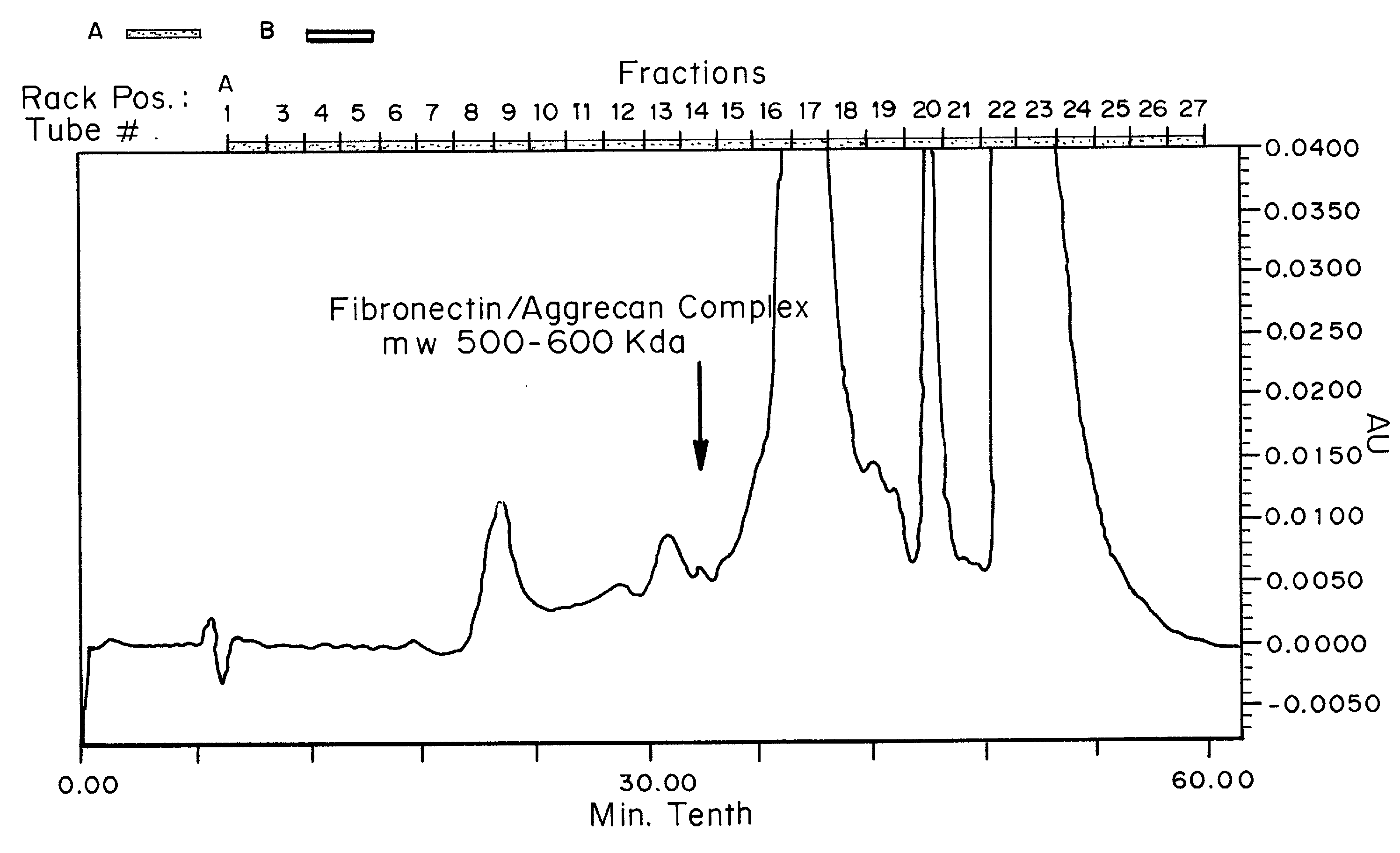
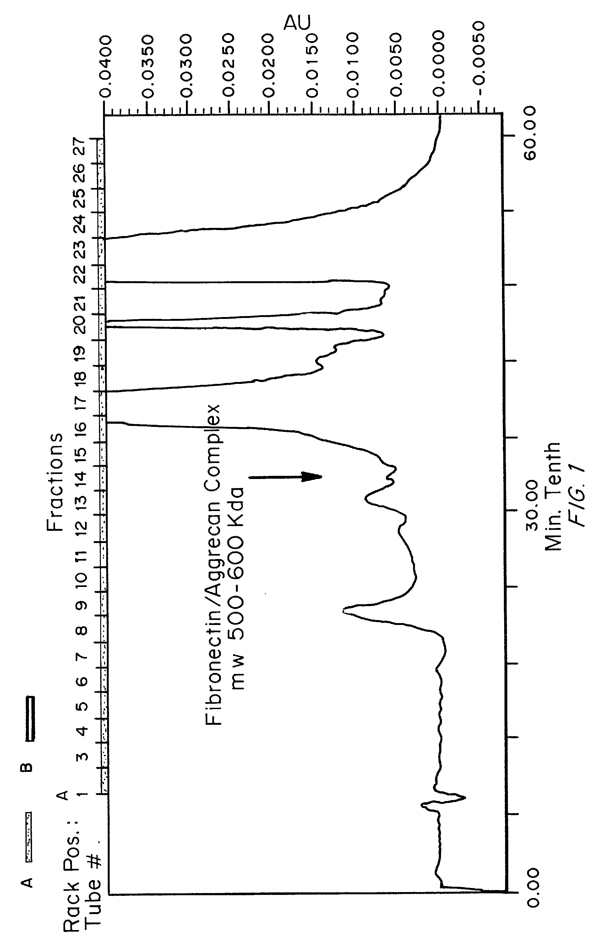
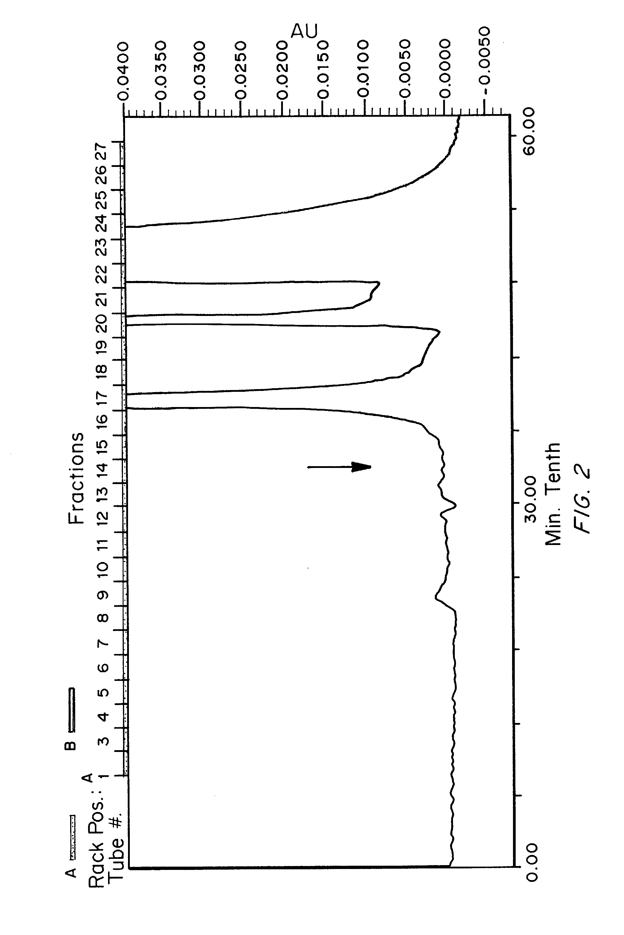
[0236]SEC was performed using two Bio-Rad® SEC-400-5 columns in series using isocratic elution with 50 mM Tris / HCl, 100 mM NaCl, pH 8.0. The resulting fractions were analyzed for total protein content (A280nm and A215nm) and for the presence of the discovered biomarker consisting of Fibronectin complexes with aggrecan fragment. FIG. 1 shows the SEC-HPLC elution profile (A215nm) for a symptomatic sample and FIG. 2 shows the SEC-HPLC elution profile for an asymptomatic sample under the same conditions. Subsequ...

example 2

Silver Stain and Western Blot Analysis Using Anti-Fibronectin, and Anti-Aggrecan (G3 Domain) Specific Antibodies

[0239]Example 1 demonstrates the fractionation of a human sample by SEC-HPLC. Fractions 14 and 15 from the SEC analysis were subjected to SDS-PAGE separation followed by silver staining or Western blotting to determine the size of the proteins in the fractions and to probe their identity. Western blotting was conducted using standard methods using anti-fibronectin monoclonal (DiaPharma Group, Inc. detection antibody from DPGR028A fibronectin ELISA kit), and anti-aggrecan monoclonal (G3 domain) (Santa Cruz Biotechnology, Aggrecan C20, sc-16493) specific antibodies.

[0240]Silver staining analysis showed the presence of proteins in the range of greater than 250 Kd in both fractions. Western blotting using the anti-fibronectin monoclonal and anti-aggrecan monoclonal (G3 domain) antibodies revealed immunoreactive band at a molecular weight range of 400-600 Kd, indicating that th...

example 3

Analysis of Aggrecan Domains that Co-Migrate with Fibronectin Through Polyacrylamide Gels

[0241]Size exclusion chromatography of a single human sample was conducted as described in Example 1 and eluted fractions 8 through 20 from the SEC were subjected to separation by SDS-PAGE and then transferred to membranes for Western blotting using polyclonal anti-aggrecan G1 (Santa Cruz Biotechnology, Aggrecan D20, se-16492), G2 (Santa Cruz Biotechnology, Aggrecan E12, se-67513) or G3 (Santa Cruz Biotechnology, Aggrecan C20, sc-16493) antibodies. Western blots using the anti-aggrecan G3 domain antibody revealed immunoreactive bands of different molecular sizes in fractions 8-20, corresponding to various proteolytic fragments of aggrecan containing the G3 domain. Western blots using the anti-aggrecan G1 domain antibody revealed immunoreactive bands in fractions 8-20 that exhibited a different banding pattern than that observed using the anti-aggrecan G3 domain antibody.

[0242]Western blots using...


PUM

PropertyMeasurementUnit
dissociation constantaaaaaaaaaa
volumeaaaaaaaaaa
pHaaaaaaaaaa

Abstract

Biomarkers, including isolated fibronectin-aggrecan complexes that correlate with spinal or joint pain and inflammation, and methods for their detection are provided. Also provided are methods for identifying treatment sites in the spine or joint for treatment of pain and inflammation by detecting the presence of, or increased levels of, fibronectin-aggrecan complexes. Methods for treating spinal or joint pain and inflammation are also provided.

Description

CROSS-REFERENCE TO RELATED APPLICATIONS[0001]This application claims benefit of and priority to U.S. Provisional Patent Application Nos. 61 / 106,059 filed on Oct. 16, 2008, and 61 / 118,401 filed on Nov. 26, 2008, and 61 / 122,045 filed on Dec. 12, 2008 all of which are by Gaetano J. Scuderi, Lewis S. Hanna, and Robert Bowser, and are incorporated by reference in their entirety.FIELD OF THE INVENTION[0002]The invention is generally related to methods for diagnosing pathologies associated with spinal- or joint-related pain as well as compositions and methods for treating spinal- or joint-related pain.BACKGROUND OF THE INVENTION[0003]Spinal and joint pain can be difficult to treat. In particular the cause of spinal and joint pain can be difficult to identify. Increasing degrees of force applied to joints result in joint injury. Abnormal joint anatomy is frequently a hallmark of ageing, but joint injury is also frequently seen as a result of trauma. For instance, chondral lesions are often ...

Claims


Application Information

Patent Timeline
Patent Type & Authority Applications(United States)
IPC IPC(8): A61K39/395C07K14/78G01N33/53A61K38/02A61K31/711A61K31/7105A61P19/02A61P37/06
CPCG01N33/6887G01N2333/4722G01N33/68G01N2800/102G01N2800/52G01N2333/78A61P19/02A61P29/00A61P37/06A61P43/00A61K38/39C07K14/78G01N33/53
Inventor SCUDERI, GAETANO J.HANNA, LEWIS S.BOWSER, ROBERT
Owner CYTONICS CORP
Why Eureka
Try Eureka