
Minimally-Invasive Method and Device for Permanently Compressing Tissues within the Body

a tissue and minimally invasive technology, applied in the field of surgery methods and devices, can solve the problems of limiting the ability of the heart to pump blood effectively, affecting the function of the heart, so as to prevent clot formation and circulating, and prevent strokes. , the effect of minimal invasiveness

Inactive Publication Date: 2010-05-06
SHUKLA HIMANSHU
View PDF16 Cites 13 Cited by
  • Summary
  • Abstract
  • Description
  • Claims
  • Application Information

AI Technical Summary

Benefits of technology

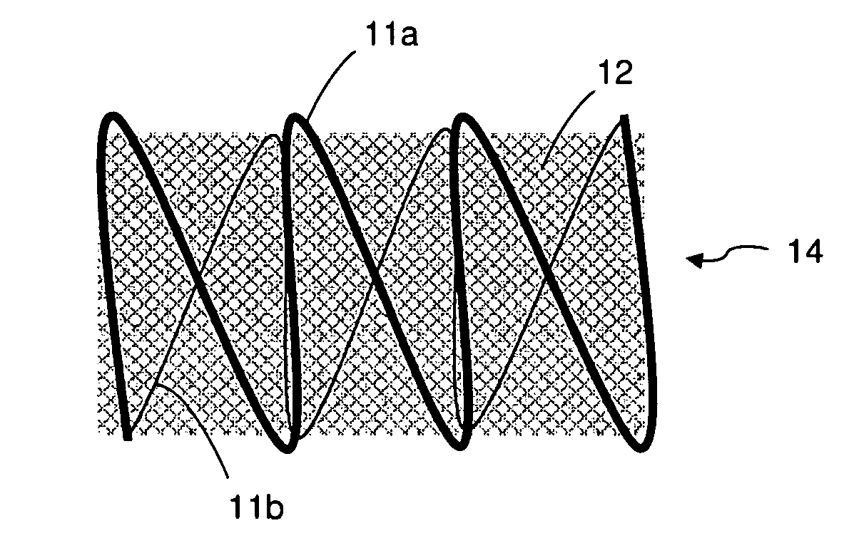
[0009]The present invention is method for permanently compressing tissues in the body. The method employs a compression device made of a spring and a flexible sheet that cooperate to form a compressive envelope around the desired tissue. The spring is preferably Z-shaped or a coil. The sheet is made of a flexible material and the material is preferably elastic. The sheet is preferably a biocompatible elastic material, such as a mesh made of stainless steel or a woven or non-woven elastomer. The method is minimally invasive because it deploys the compression device through the patient's skin directly to the tissue, as opposed to through catheterization or open invasive surgery, such as open-heart surgery. The preferred use is for compressing the left atrial appendage to prevent clots from forming and circulating, thereby preventing strokes. The device is deployed by making an incision in a patient's chest, inserting the compression device through the incision into the pericardium without piercing the heart, and deploying it around the entire appendage. The device remains in place by its own compressive nature: either the spring, the sheet, or both components compress the appendage and cause the device to stay in place due to friction. Preferably the entire compression device is left in place, but the spring or the sheet may be removed, leaving the remaining component to compress the appendage.

Problems solved by technology

An arrhythmia is the irregular beating of the heart which limits the ability of the heart to pump blood effectively.
It is caused when the natural rhythm of the heart becomes abnormal, the regular pathways for electrical signals in the heart are interrupted, or parts of the heart improperly emit electrical signals.
Atrial fibrillation is a serious disease which can lead to chronic fatigue, congestive heart failure, and stroke.
Atrial fibrillation occurs when the atria pump quickly and unevenly.
This quivering creates a serious risk that blood will collect and pool in the left atrial appendage.
If this pooled blood forms clots that leave the heart, a stroke can occur.
However, medication must be taken regularly and frequently is accompanied by unwanted side effects.
This often involves surgically opening the chest cavity, which significantly increases the risk to the patient and adds significant recovery time.
While reducing the patient's recovery time, open surgery and entering the heart from within still creates significant risk to the patient.

Method used


Image

  • Minimally-Invasive Method and Device for Permanently Compressing Tissues within the Body
  • Minimally-Invasive Method and Device for Permanently Compressing Tissues within the Body
  • Minimally-Invasive Method and Device for Permanently Compressing Tissues within the Body

Examples


Embodiment Construction

[0020]The present invention is method for permanently compressing tissues within the body so that they no longer function or no longer cause dysfunction. The method employs a compression device 10 that envelopes substantially the entire tissue. The method may be applied to tissues in the body such the appendix, gallbladder and stomach, and will be described with particular reference to the left atrial appendage to prevent blots clots from forming inside it.

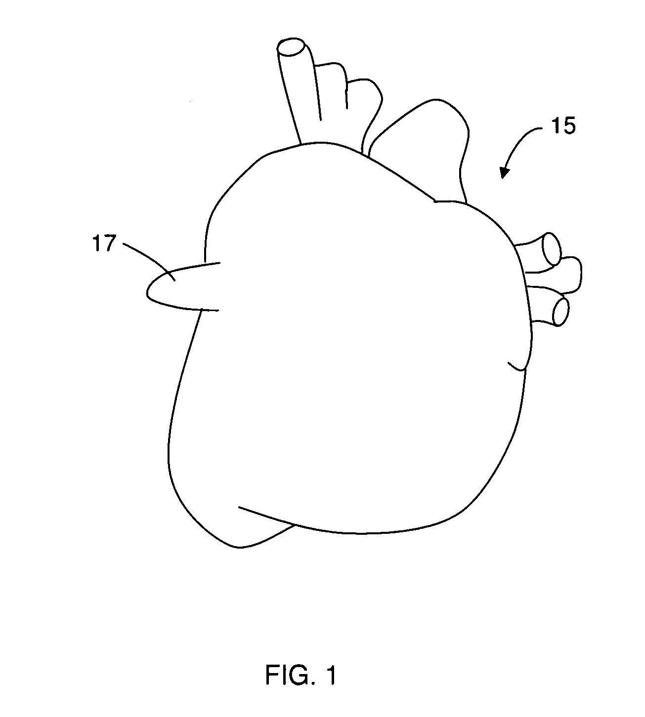
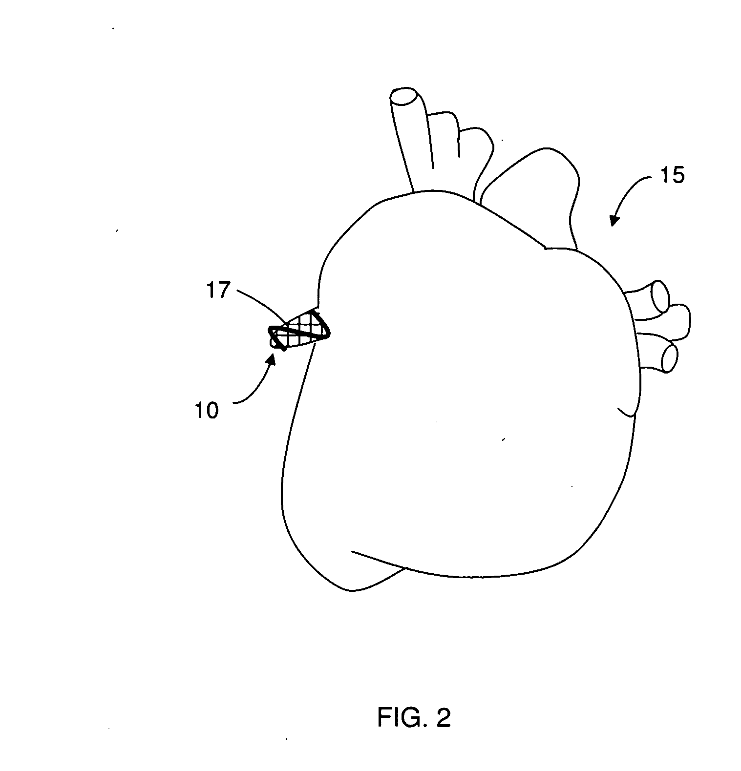
[0021]FIG. 1 shows an untreated heart 15 and left atrial appendage 17 (referred to hereafter as “LAA”). FIG. 2 shows a heart 15 after being treated with the method herein, namely with the preferred embodiment of the compression device 10 disposed around the LAA 17.

[0022]FIGS. 3-6 show several embodiments of the compression device 10. Each compression device 10 comprises a spring 11 and a flexible sheet 12. The spring 11 can take any form provided that in its relaxed state the opposing sides of the spring are biased towards each ot...


PUM


Abstract

The present invention is method for permanently compressing tissues in the body. The method employs a compression device made of a spring and a flexible sheet that cooperate to form a compressive envelope around the desired tissue. The spring is preferably Z-shaped or a coil. The sheet is made of a flexible material and the material is preferably elastic. The sheet is preferably a biocompatible elastic material, such as a mesh made of stainless steel or a woven or non-woven elastomer. The method is minimally invasive because it deploys the compression device through the patient's skin directly to the tissue, as opposed to through catheterization or open invasive surgery, such as open-heart surgery. The preferred use is for compressing the left atrial appendage to prevent clots from forming and circulating, thereby preventing strokes. The device is deployed by making an incision in a patient's chest, inserting the compression device through the incision into the pericardium without piercing the heart, and deploying it around the entire appendage. The device remains in place by its own compressive nature: either the spring, the sheet, or both components compress the appendage and cause the device to stay in place due to friction. Preferably the entire compression device is left in place, but the spring or the sheet may be removed, leaving the remaining component to compress the appendage.

Description

CROSS-REFERENCE TO RELATED APPLICATIONS[0001]This application claims the benefit of co-pending provisional application No. 61 / 198,545 filed Nov. 6, 2008.FIELD OF INVENTION[0002]This invention relates to surgery methods and devices. This invention relates particularly to methods and devices for collapsing and occluding appendages and tissues within the body, including the left atrial appendage.BACKGROUND[0003]An arrhythmia is the irregular beating of the heart which limits the ability of the heart to pump blood effectively. It is caused when the natural rhythm of the heart becomes abnormal, the regular pathways for electrical signals in the heart are interrupted, or parts of the heart improperly emit electrical signals. Arrhythmias vary in seriousness, from brief, almost imperceptible events, to life-threatening conditions.[0004]Atrial fibrillation is one form of arrhythmia which affects over two million people. Atrial fibrillation is a serious disease which can lead to chronic fatig...

Claims


Application Information

Patent Timeline
IPC IPC(8): A61M29/00
CPCA61B17/12013A61B2017/00243A61B2017/00867A61F2/2481A61F2002/2484
Inventor SHUKLA, HIMANSHU
Owner SHUKLA HIMANSHU
Learn More