Medical image-processing device, medical image-processing method, medical image-processing system, and medical image-acquiring device

Inactive Publication Date: 2010-05-27
CANON MEDICAL SYST COPRPORATION
View PDF17 Cites 19 Cited by
  • Summary
  • Abstract
  • Description
  • Claims
  • Application Information

AI Technical Summary

Benefits of technology

[0030]The inventor of the present invention has confirmed that the opacity curve set by the medical image-processing device according to the present invention is effective for setting the opacity in three-dimensional images. Namely, in the present invention, effective presetting of the opacity curve is done by performing gray-scale processing tasks such as window conversion, gain adjustment, and STC adjustment. Therefore, the display-adju

Problems solved by technology

In addition, in MR images and ultrasound images, a 256 gray-scale is insufficient to express all pixel intensities.
Thus, the image viewer must set a display condition of an image, requ

Method used


Image

  • Medical image-processing device, medical image-processing method, medical image-processing system, and medical image-acquiring device
  • Medical image-processing device, medical image-processing method, medical image-processing system, and medical image-acquiring device
  • Medical image-processing device, medical image-processing method, medical image-processing system, and medical image-acquiring device

Examples


first embodiment

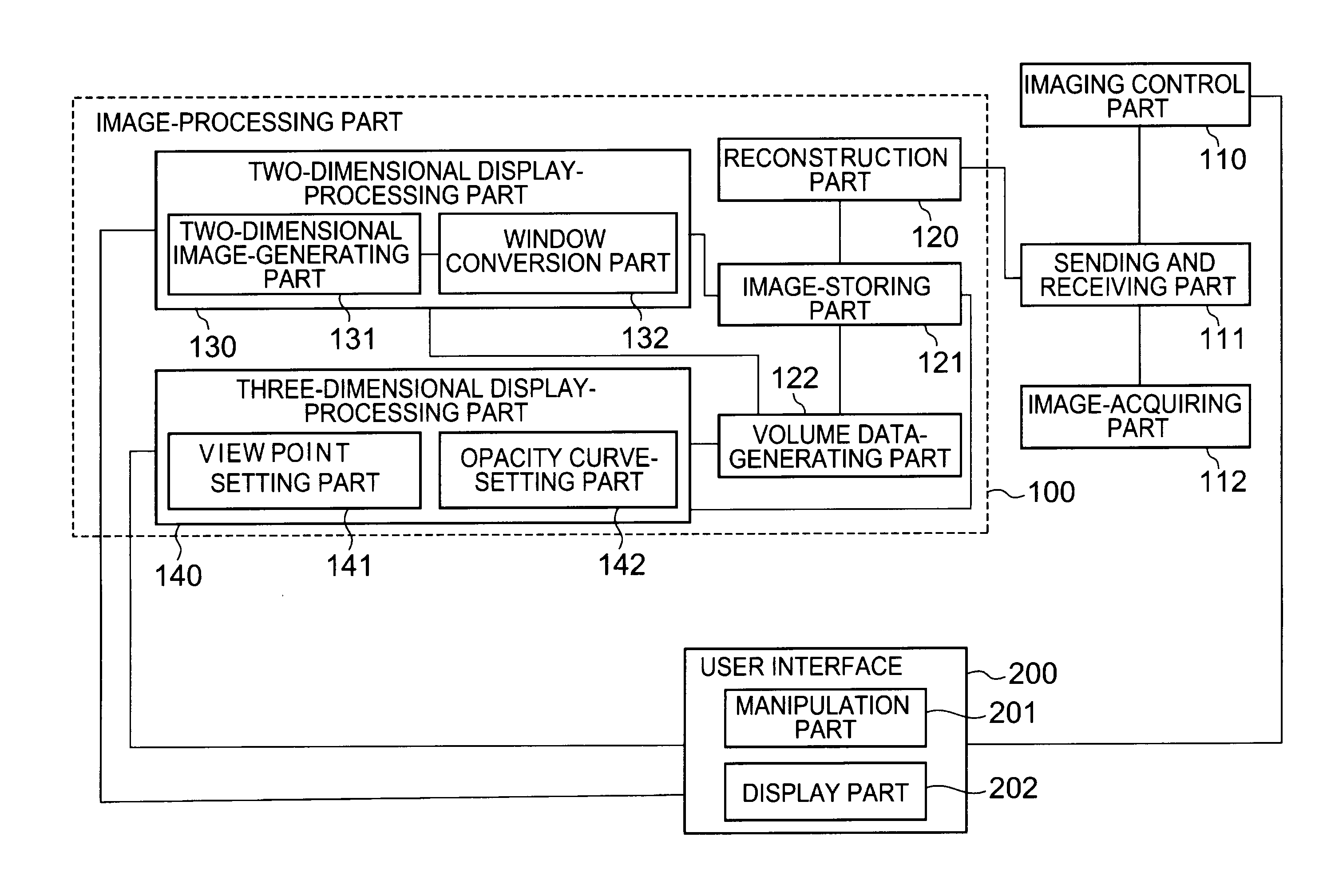
[0041]The medical image-processing device according to the first embodiment of the present invention is described as follows with reference to FIGS. 1-5. FIG. 1 is a schematic block diagram showing the schematic conformation of the medical image-processing device according to the first embodiment of the present invention. The first embodiment performs both two-dimensional display processing and three-dimensional display processing using the medical image-processing device.

[0042]In addition, the medical image-processing device according to the present embodiment performs not only display processing of image data but also imaging of a subject, reconstruction processing, and volume data generation. On the other hand, the medical image-processing device according to the present invention is not necessarily limited to a device performing these processes. The medical image-processing device according to the present invention may be one that performs only display processing, such as image ...

second embodiment

[0108]Next, the medical image-processing system according to the second embodiment of the present invention is described with reference to FIGS. 6 and 7. FIG. 6 is a block diagram showing the schematic conformation of a medical image-processing system according to the second embodiment of the present invention.

[0109]In the medical image-processing system according to the second embodiment, image data is generated by the image-acquiring part 112 in the medical image-processing device. The generated image data is sent to an image server 400 by the sending and receiving part 111 in the medical image-processing device and stored in the image server 400. Moreover, the image data stored in the image server 400 may be read from the image server and displayed on an image display terminal 500.

[0110]An example of an operation in a case where the medical image-processing device in the medical image-processing system of the present embodiment is applied to an ultrasound image-acquiring image is...

third embodiment

[0121]Next, an X-ray CT device as the medical image-processing device according to the third embodiment of the present invention will be described. With the X-ray CT device for performing medical image processing according to the third embodiment, image data is generated by the image-acquiring part 112 in a similar manner to the volume data generation process described above. Moreover, this image data undergoes window conversion as two-dimensional display processing, but for this X-ray CT device, window conversion is done as follows.

[0122]For the X-ray CT device, before scanning by the image acquiring part 112, parameters of various conditions such as scanning condition, reconstruction condition, and display condition are set via the manipulation part 201 in order to acquire the X-ray CT images. For example, for the X-ray CT device of the present embodiment, before acquiring the X-ray CT images, parameters such as slicing positions, a scope of imaging, a tube voltage, and a tube cur...


PUM


Abstract

A medical image-processing device stores the parameters of window level and window width set by a window conversion part. An opacity curve-setting part sets the settings of the OWL and OWW in an opacity curve based on the stored parameters during volume rendering for volume data. In this way, the medical image-processing device sets the opacity of a three-dimensional image.

Description

BACKGROUND OF THE INVENTION[0001]1. Field of the Invention[0002]The present invention relates to techniques regarding display condition settings for imaged medical image data.[0003]2. Description of the Related Art[0004]In medical institutions, after acquiring information of tissue within a subject—such as perspective images, tomographic images, and blood flow within the subject—using a medical image-acquiring device, the acquired tissue information is converted into medical images. Moreover, in medical institutions, examinations and diagnoses are conducted with such medical images. There are various types of this medical image-processing device. For example, there are X-ray CT (computed tomography) devices, MRI (magnetic resonance imaging) devices, ultrasound image-acquiring devices (ultrasound diagnostic equipment), nuclear medicine diagnostic devices (NM: nuclear medicine), PET-CT (positron emission tomography-computed tomography) devices, and others.[0005]Moreover, these medical...

Claims


Application Information

Patent Timeline
IPC IPC(8): A61B8/14G06K9/00A61B5/00A61B5/055A61B6/03A61B8/00G06T1/00
CPCA61B5/055A61B6/03A61B8/483G06T15/08A61B8/13
Inventor YAMAGATA, HITOSHI
Owner CANON MEDICAL SYST COPRPORATION
Why Eureka
Try Eureka