Method and apparatus for delivery of agents across the blood brain barrier

a blood brain barrier and agent technology, applied in the field of active staining, can solve the problems of limited bbb region, difficult to extend to mice, limited use of diagnostic and therapeutic agents in the brain, etc., and achieve the effects of enhancing brain imaging, rapid administration, and opening of blood-brain barrier

Inactive Publication Date: 2010-06-10
DUKE UNIV
View PDF3 Cites 49 Cited by
  • Summary
  • Abstract
  • Description
  • Claims
  • Application Information

AI Technical Summary

Benefits of technology

[0066]1) Blood-brain barrier disruption using unfocused ultrasound: Previous work has been with mechanically focused ultrasound transducers which only allow single spots of disruption. Using an unfocused transducer allows the opening of much larger areas of the blood-brain barrier, including the whole brain.
[0069]3c) Wide availability: clinical diagnostic scanners are ubiquitous and require no special engineering skills to set up and use.
[0070]Our invention insonifies the entire brain to achieve a global opening of the blood-brain barrier. To the best of our knowledge, no technique has been demonstrated that achieves global opening of the blood-brain barrier. While the existing osmotic technique allow for hemispheric opening of the blood-brain barrier, it is invasive while our invention is not invasive.
[0071]Utilizing this global blood-brain barrier opening, our invention rapidly administers a contrast agent to enhance brain imaging. No other technique exists for rapidly administering contrast agents to the whole brain. While some agents can be administered to the whole brain slowly (e.g., divalent manganese will diffuse slowly into the brain), there are significant scientific and practical advantages to rapid administration. While some blood-brain barrier disruption techniques (e.g., mechanically focused ultrasound) can administer contrast agents to small portions of the brain, they have not been implemented to rapidly administer contrast to the whole brain. While some other blood-brain barrier disruption techniques (e.g., osmotic opening) can administer contrast agents to a whole hemisphere of the brain, they suffer from significant technical and scientific drawbacks (detailed above).
[0072]By choosing agents that are indicators for specific biological phenomena (such as neuronal activity), this invention can be used to identify non-morphological brain features. For example this invention can administer Mn2+ (a contrast agent that highlights active neurons) to the whole brain, allowing the detection of neuronal activity anywhere in the brain. No other technique of which we are aware can do this. This invention could administer any number of agents which have specificity for tissues of a certain type or nature.
[0073]Obviously, this invention could be used to administer other diagnostic and therapeutic agents, not just contrast agents. Our work to date has focused on the use for administering agents to enhance both sensitivity and specificity in imaging studies. But there is enormous potential in the use of this method to introduce and monitor the administration of a wide range of molecules for both diagnostic and therapeutic purposes.

Problems solved by technology

The use of diagnostic and therapeutic agents in the brain is limited by the blood-brain barrier (BBB), which restricts entry into the brain.
However, this technically challenging approach is invasive, opens only a limited region of the BBB, and is difficult to extend to mice.

Method used


Image

  • Method and apparatus for delivery of agents across the blood brain barrier
  • Method and apparatus for delivery of agents across the blood brain barrier
  • Method and apparatus for delivery of agents across the blood brain barrier

Examples


example 1

In Vivo Magnetic Resonance Microscopy by BOMUS

Methods

Microbubbles

[0096]Prior to opening the BBB, perflutren lipid microspheres (Definity, Lantheus, N. Billerica, Mass.) were produced by “activating” the vial (i.e., shaking it in the manufacturer-supplied device for 45 seconds) according to the prescribing information sheet. Immediately prior to microbubble administration, the vial was agitated by hand for 1 minute.

Ultrasound System

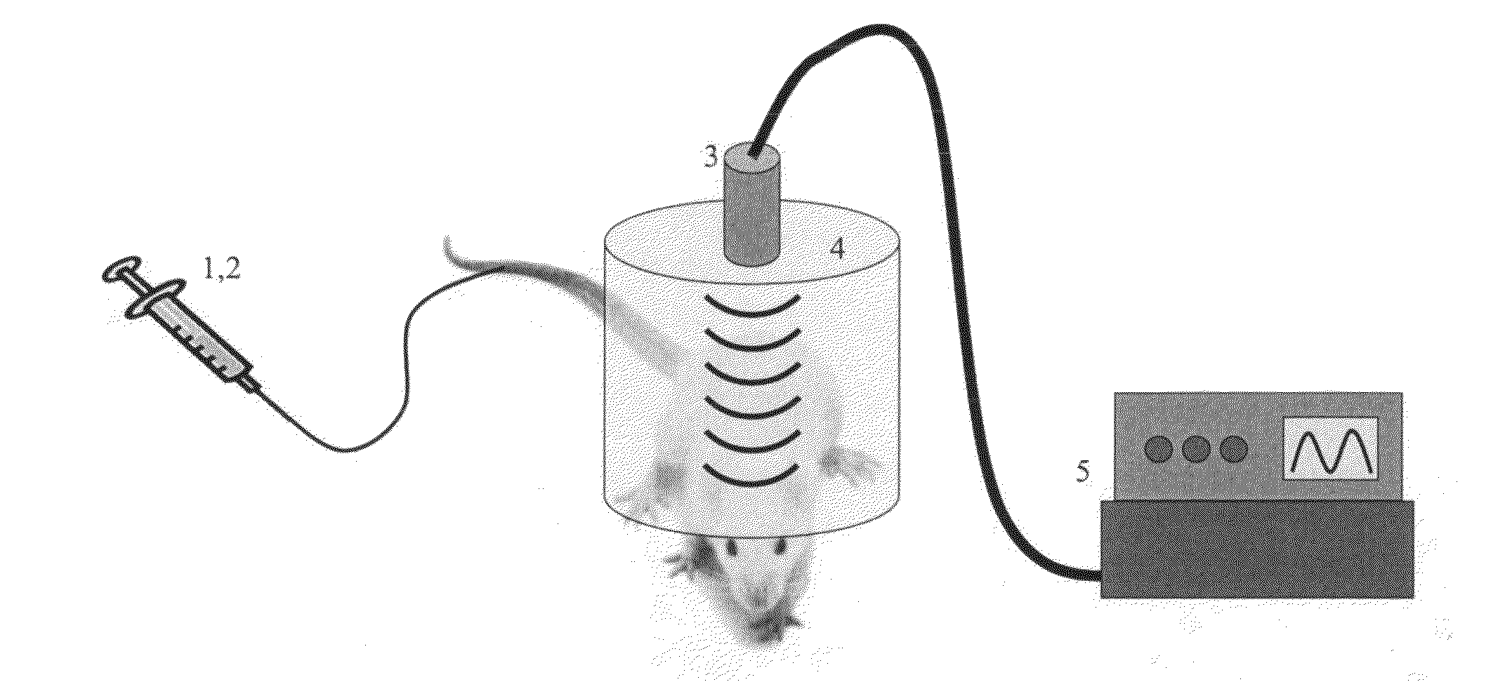
[0097]For insonification a circular single-element ultrasound transducer (model A382S-SU, Olympus NDT) was used, which had a diameter of 13 mm and a center frequency of 2.15 MHz. See, FIG. 14 where panel a depicts the BOMUS setup and panel b depicts the experimental timeline. The transducer was positioned using a 3-axis frame (VisualSonics, Toronto, ON) at its natural focal distance (58 mm) in the water column directly over the mouse brain. The natural focus distance (i.e., the Rayleigh distance) was estimated as d2 / 4 lambda, where d is the element diamete...

example 2

Blood-Brain Barrier (BBB) Disruption Using a Diagnostic Ultrasound Scanner

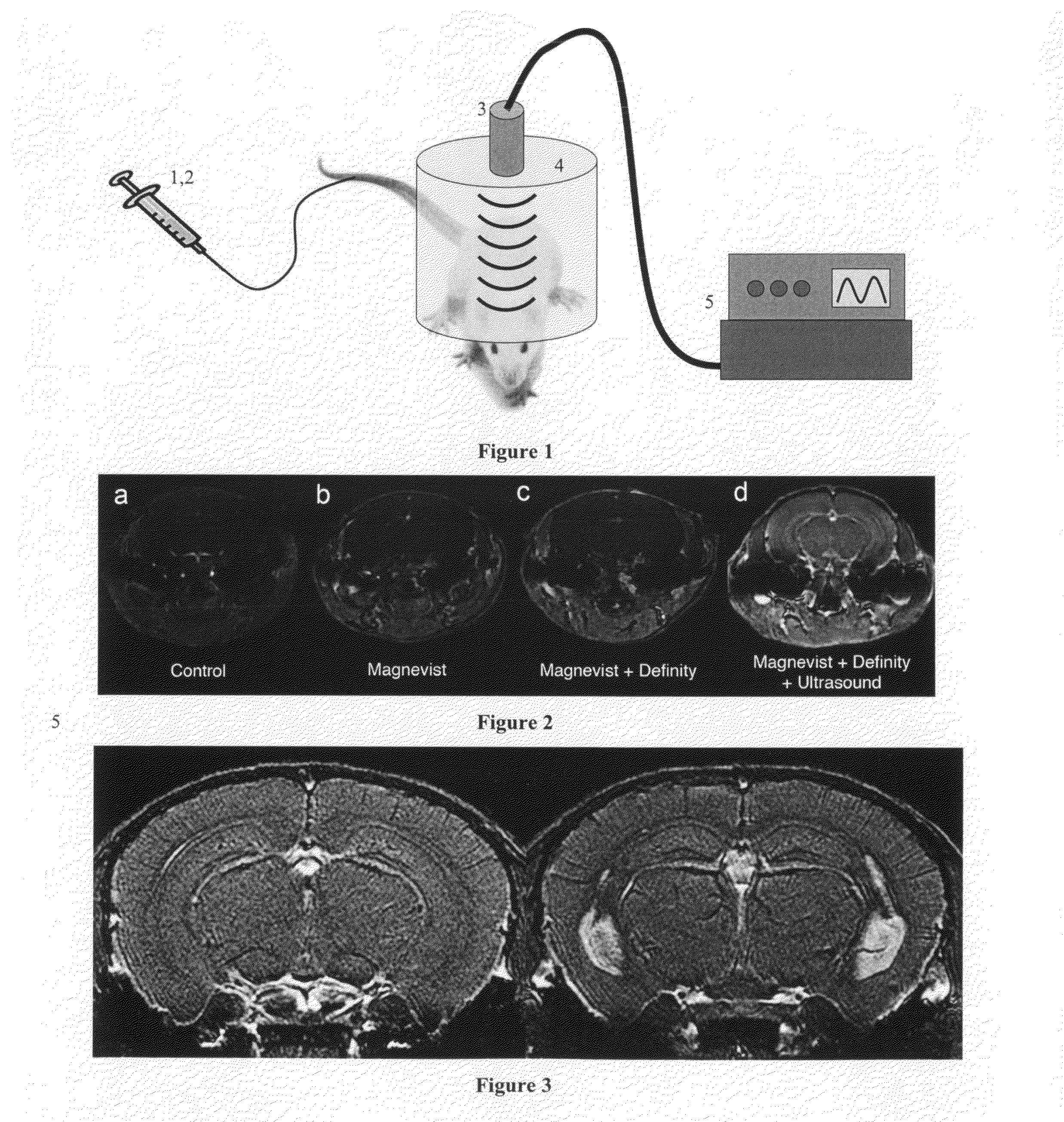
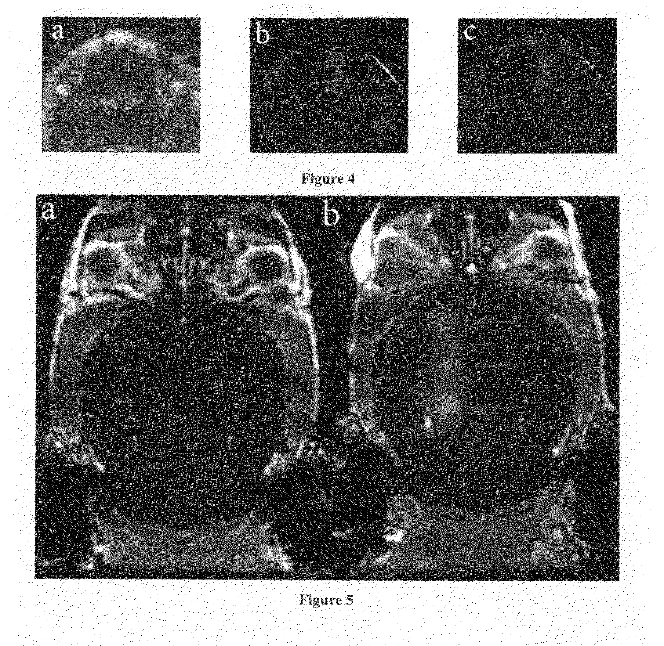
[0132]The objective of this example was to transcranially and nondestructively disrupt the BBB in the mouse using focused, diagnostic ultrasound and contrast agent, and to quantify that disruption using MRI and MR contrast agent. Each mouse was placed under isoflurane anesthesia and the hair on top of its skull was removed before treatment. A diagnostic ultrasound transducer was placed in a water bag coupled with gel to the mouse skull. Definity (US contrast) and Magnevist (MR contrast) were injected concurrent with the start of a custom ultrasound transmission sequence. The transducer was translated along the rostral-caudal axis to insonify three spatial locations (2 mm apart) along one half of the brain for each sequence. T1-weighted MR images were used to quantify the volume of tissue over which the BBB disruption allowed Magnevist to enter the brain, based upon increases in MR contrast-to-noise ratio (CNR)...


PUM


Abstract

We describe a method for opening the blood-brain barrier (BBB) using ultrasound and preformed microbubbles. With this method, diagnostic or therapeutic agents may be administered to the brain. This method can open a focal region of the BBB and administer agents in a targeted fashion or the method can open large regions (or the entirety) of the brain for more global administration of agents. In one embodiment, the method can be used to administer contrast agents (e.g., agents that increase or decrease the magnetic resonance imaging signal) to the brain and thereby improve the quality or information content of imaging data. In another embodiment, a standard clinical diagnostic ultrasound scanner can be used to open specific regions of the BBB and administer diagnostic or therapeutic agents. Importantly, this invention can open the BBB in a non-destructive/non-invasive fashion, allowing the subject to be awake and suffer no detectable side effects.

Description

RELATED APPLICATIONS[0001]This Application claims the benefit of priority from U.S. Provisional Application Ser. No. 61 / 193,006 filed on Oct. 22, 2008. This provisional application is hereby incorporated in its entirety by reference.FEDERALLY-SPONSORED RESEARCH OR DEVELOPMENT[0002]The U.S. Government has certain rights in this invention as provided for by the terms of NIH / NCRR: 5P41-RR005959-18, 2U24CA092656-07, 5R01CA114075, 2R01EB002132-05 and NSF 2003014921 awarded by the Department of Health and Human Services.BACKGROUND OF THE INVENTIONActive Staining[0003]In magnetic resonance imaging, the term “active staining” refers to the use of contrast agents to selectively enhance specific tissue properties to improve the MRI images. Active staining was introduced by the Center for In Vivo Microscopy in 2002 for perfusion-fixed specimens (Johnson 2002). A patent was subsequently issued for active staining of fixed specimens (U.S. Pat. No. 6,023,162). Contrast agents are routinely used i...

Claims


Application Information

Patent Timeline
Patent Type & Authority Applications(United States)
IPC IPC(8): A61K51/00A61B8/00A61K49/06A61K49/04A61K49/22
CPCA61K41/0028A61K49/105A61K49/223A61B8/0816A61N2007/0039A61N2007/0078A61N2007/0082A61M37/0092
Inventor JOHNSON, G. ALLANHOWLES-BANERJI, GABRIELBING, KRISTIN FRINKLEYNIGHTINGALE, KATHRYNPALMERI, MARK
Owner DUKE UNIV
Why Eureka
Try Eureka