Method and Apparatus for Quantification of Optical Properties of Superficial Volumes Using Small Source-to-Detector Separations

a technology of optical properties and source-to-detector separation, which is applied in the field of methods and probe designs, can solve the problems of insufficient model, limited applicability of biopsies of subjects, and inability to take biopsies from subjects

Active Publication Date: 2010-06-24
RGT UNIV OF CALIFORNIA
View PDF9 Cites 29 Cited by
  • Summary
  • Abstract
  • Description
  • Claims
  • Application Information

AI Technical Summary

Benefits of technology

[0010]The illustrated embodiments of the invention are directed to a probe for obtaining quantitative optical properties and chromophore concentrations of tissue components in tissue in-vivo at superficial depths and at source-detector separations of 5 mm or less, and typically with source-detector separations of 3 mm or less. One embodiment of the invention comprises a source fiber providing light to expose the tissue, a diffuser layer into which light from the source fiber is directed and then from the diffuser layer to and / or into the tissue, and a detector fiber arranged relative to the diffuser layer for detecting backscattered and / or reflected light returned from the tissue without transmission through the diffuser layer.

Problems solved by technology

Although the integrating sphere based techniques can be used to investigate ex vivo optical properties of epidermis, dermis, and subcutaneous tissue, the need to take biopsies from subjects limits their applicability in the clinic.
Furthermore, once tissue has been excised, it is no longer part of the living organism; oxy and dexoyhemoglobin concentrations begin to diverge from physiologic quantities and if not carefully handled, the hydration of the tissue begins to change.
It has been proven to be a not adequate model because boundary conditions and the assumption of multiple scattering in a turbid medium cannot be satisfied.
In-vivo techniques which are capable of measuring optical properties of skin do exist, but have some important limitations.
In addition, for the case where the reduced scattering coefficient does not have a linear dependence on wavelength, the empirical mathematical model will not recover tissue optical properties properly.
Probes for use in free space or for quantitative measurements of chromophores in tissues that can be reached by an endoscope or similar instrument in which the source and detector are in relatively close proximity with one another has been a significant challenge for quantitative optical methods.
In each of these cases, a simple application of diffusion approximation based modeling will yield inaccurate tissue optical properties and chromophore concentrations.
The problem with this approach is that the calibration samples have to be very similar to the target (unknown) sample set of interest.
Clearly this is impractical when thinking in terms of a probe for interrogating, for example, oral tissues.

Method used


Image

  • Method and Apparatus for Quantification of Optical Properties of Superficial Volumes Using Small Source-to-Detector Separations
  • Method and Apparatus for Quantification of Optical Properties of Superficial Volumes Using Small Source-to-Detector Separations
  • Method and Apparatus for Quantification of Optical Properties of Superficial Volumes Using Small Source-to-Detector Separations

Examples


Embodiment Construction

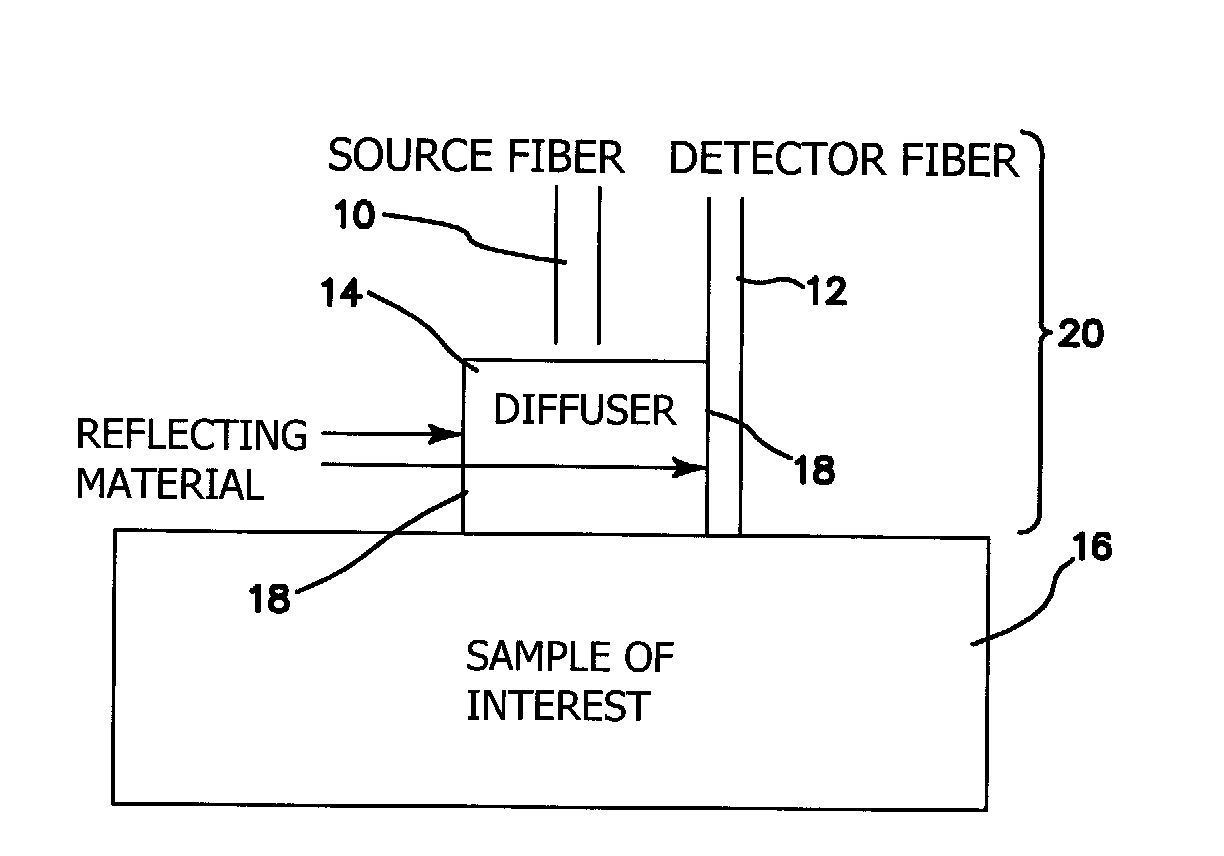
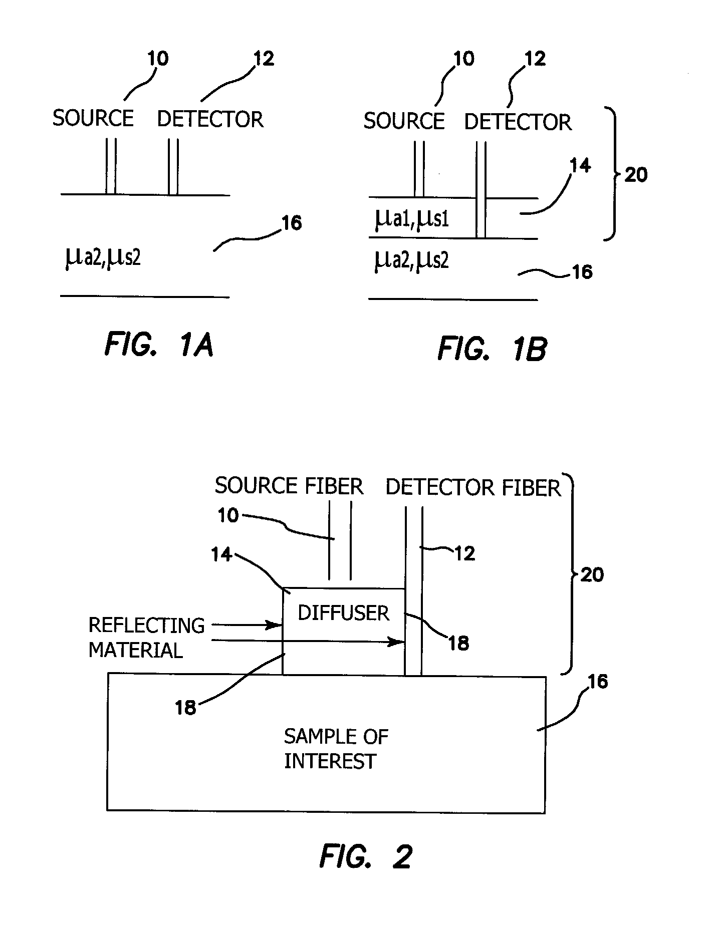
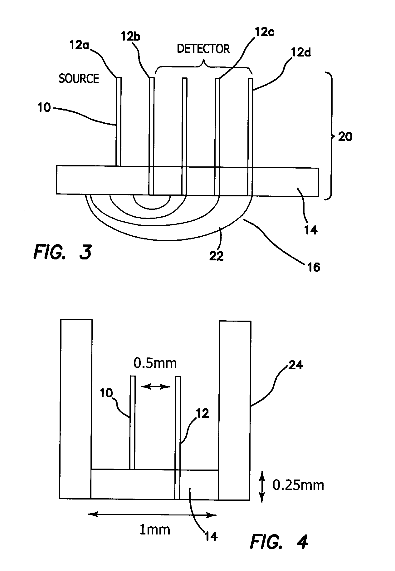
[0038]The probe of the illustrated embodiments is amenable to use in free space or for quantitative measurements of chromophores in tissues that can be reached by an endoscope or similar instrument. We present a method to reduce source-detector separation while maintaining the validity of the diffusion approximation, which employs a high scattering, low absorption layer (μs′=9 mm−1 and μa=0.0015 mm−1 at 661 nm) placed on the surface of the tissue under investigation. This effectively increases the photon path length and allows the source-detector separation to be made arbitrarily small. In order to demonstrate feasibility, we have carried out frequency domain measurements at several wavelengths to recover the optical properties of tissue phantoms, using a two-layer model for which the optical properties and thickness of the upper, highly scattering layer are known.

[0039]In a sense, the approach of the illustrated embodiments of the invention “force” diffusive light propagation on th...


PUM


Abstract

A probe for obtaining quantitative optical properties and chromophore concentrations of tissue components in tissue in-vivo at superficial depths and at source-detector separations of 5 mm or less includes a source fiber providing light to expose the tissue, a diffuser layer into which light from the source fiber is directed and then from the diffuser layer to and/or into the tissue, and a detector fiber arranged relative to the diffuser layer for detecting backscattered and/or reflected light returned from the tissue without transmission through the diffuser layer.

Description

RELATED APPLICATIONS[0001]The present application is related to U.S. Provisional Patent Application Ser. No. 61 / 140,323, filed on Dec. 23, 2008, which is incorporated herein by reference and to which priority is claimed pursuant to 35 USC 119.BACKGROUND OF THE INVENTION[0002]The invention relates to the field of methods and probe designs for obtaining quantitative optical properties and chromophore concentrations of tissue components in-vivo at superficial depths and “short” source-detector separations.[0003]The absorption coefficient μa, the scattering coefficient μs′, and chromophore concentrations of skin are fundamental properties of tissue that can provide essential information for many aesthetic, therapeutic, and diagnostic applications such as monitoring of skin blood oxygenation, melanin concentration, detection of cancer with fluorescence, laser surgery, and photodynamic therapy. Many researchers have quantified the optical properties of skin tissue and most of them used ex...

Claims


Application Information

Patent Timeline
Patent Type & Authority Applications(United States)
IPC IPC(8): A61B5/1459
CPCA61B5/0075A61B5/0086A61B5/14532A61B2562/0242A61B5/4312A61B5/443A61B5/1459
Inventor DURKIN, ANTHONY J.TSENG, SHENG-HAO
Owner RGT UNIV OF CALIFORNIA
Why Eureka
Try Eureka