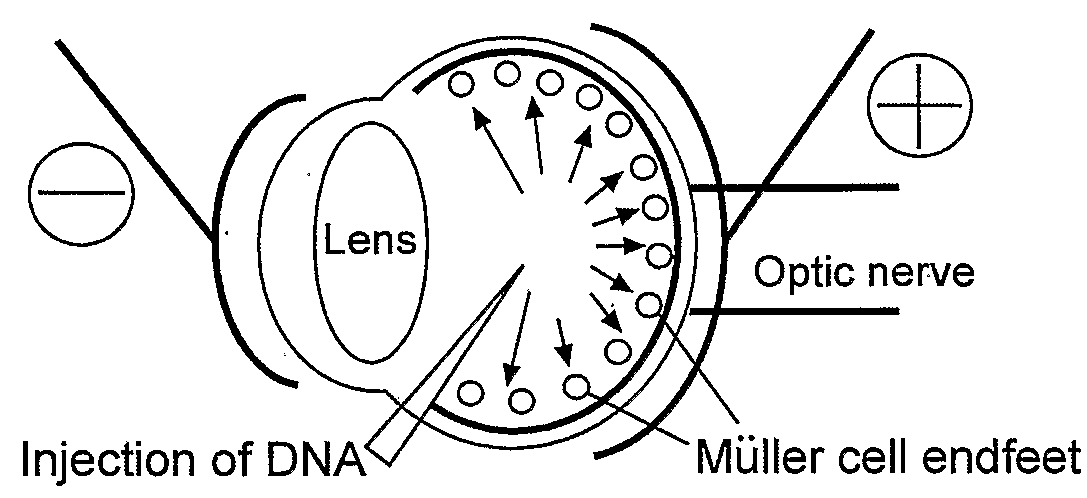
Muller Cell Specific Gene Therapy
a gene therapy and muller cell technology, applied in the field of muller cell specific gene therapy, can solve the problems of increased intraocular pressure, visual loss or complete blindness, eye diseases that represent a significant health problem, etc., and achieve the effect of treating or preventing
- Summary
- Abstract
- Description
- Claims
- Application Information
AI Technical Summary
Benefits of technology
Problems solved by technology
Method used
Image

Examples
example 1
In Vitro and In Vivo Characterization of RRV Pseudotyped LV Vectors
[0144]Lentiviral vector particles were constructed as described above to contain envelope glycoproteins (pseudotyped) derived from the Ross River Virus (RRV). RRV is an enveloped retrovirus that was first isolated from mosquitoes in the Ross River, Australia. It exhibits an extremely broad host range and RRV infection leads to epidemic polyarthritis in humans.
[0145]RRV pseudotyped LV vector particles were packaged as described above and concentrated to high titer (108-109 TU / mL). Titer was determined by Q-PCR and direct GFP visualization as described above.
[0146]In vitro characterization was performed as follows. RRV-LV (CMV-GFP) vector particles were added to in vitro cultures of 293T, primary Müller, and a rat Müller cell line rMC-1 (Sarthy et al. IOVS V39 212-215 1998) along with polybrene (8 μg / mL). These Müller cell lines were stained with antibodies to GFAP and Vimentin and exhibit an expression profile similar...
example 2
CD44, GFAP, and Vim Promoters Drive GFP Expression in Müller Cells In Vivo
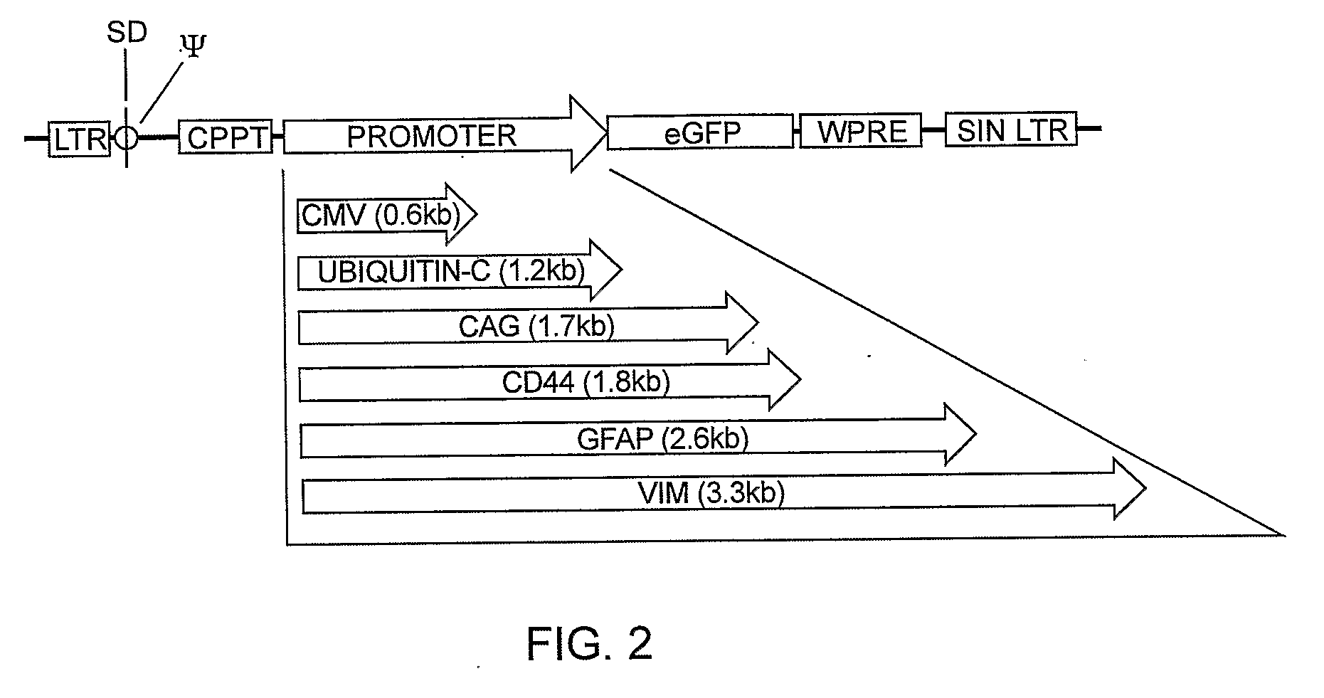
[0151]High titer vectors or PBS controls were injected subretinally or intravitreally into SD and S334Ter+ / −rat eyes. GFP expression was evaluated by fluorescent fundus imaging 2-180 days following subretinal injection of 3 μl LV vector. GFP was observed over a 6 month period, showing persistent transgene expression and stable proviral integration. After subretinal injection of CD44, GFAP and Vim promoted vectors, high level GFP was consistently seen by fundus imaging (FIG. 16), and confocal microscopy revealed Müller cells were transduced with an efficiency approaching 95% in the subretinal bleb area (FIG. 17).
[0152]Overall fluorescence intensity viewed by fundus imaging consistently appeared highest with the CD44 promoter, followed by GFAP, and finally Vim promoted vectors. VSV.CD44.GFP vector injected retinas exhibited GFP expression in Müller cell processes spanning the entire retinal thickness (FIG. 18, p...
example 3
Photoreceptor Rescue by GDNF Secretion from LV Transduced Müller Cells
[0153]The present invention is used for treatment of multiple neurodegenerative diseases of the retina (i.e. RP, AMD, glaucoma). The neurotrophin GDNF has significant application in the treatment of RP and has been shown to delay photoreceptor degeneration when expressed in photoreceptors of the S334Ter-4 transgenic rat model for RP (Sanfter et al. Molec Ther, 4, 1-9, 2001).
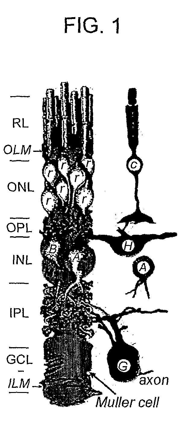
[0154]The secretion of neurotrophins from Müller cells directly to rescue degenerating photoreceptors has advantages over previous methods for neurotrophin rescue; Müller cells are not directly affected by known gene defects resulting in retinal degenerations and are therefore healthy reservoirs capable of secreting protective factors. Their unique retinal anatomy permits vector access from either intravitreal or subretinal injection. Furthermore, their close association with all other classes or retinal neurons insures the secreted factor will...
PUM
| Property | Measurement | Unit |
|---|---|---|
| thick | aaaaa | aaaaa |
| thickness | aaaaa | aaaaa |
| volume | aaaaa | aaaaa |
Abstract
Description
Claims
Application Information
 - R&D
- Intellectual Property
- Life Sciences
- Materials
- Tech Scout
- Unparalleled Data Quality
- Higher Quality Content
- 60% Fewer Hallucinations
Browse by: Latest US Patents, China's latest patents, Technical Efficacy Thesaurus, Application Domain, Technology Topic, Popular Technical Reports.
© 2025 PatSnap. All rights reserved.Legal|Privacy policy|Modern Slavery Act Transparency Statement|Sitemap|About US| Contact US: help@patsnap.com