Surgical Guide for use during sinus elevation surgery utilizing the caldwell-luc osteotomy

a surgical guide and caldwell-luc technology, applied in the field oforal implantology, can solve the problems of negating the purpose, inability of the operator to precisely locate the floor of the sinus, and no bony support around the implant portion, so as to prevent any damage of overcutting and accurately prepare the caldwell-luc osteotomy

Inactive Publication Date: 2010-07-29
MASSOUD YEHIA A
View PDF15 Cites 31 Cited by
  • Summary
  • Abstract
  • Description
  • Claims
  • Application Information

AI Technical Summary

Benefits of technology

[0015]The invention described discloses a surgical guide and accompanying surgical bur with a ledge for depth control. The guide is fabricated from acrylic and is based on the treatment plan set forth from the diagnostic information provided by 3-D imaging software from a CT scan taken of a patient. The surgical guide is shaped and dimensioned (i.e., customized) based on the CT scan and 3-D imaging software results of the sinus area for each patient and accordingly, allows the surgeon to accurately prepare a Caldwell-Luc osteotomy in all three planes. The outline of the osteotomy, as well as the depth of the bone to be removed without damaging the underlying the Schneiderian Membrane is created utilizing the surgical guide and proper depth bur. This permits open access to the maxillary sinus cavity within the dimensions required for the sinus elevation and enables the surgeon easy access to the Schneiderian Membrane without anatomical obstructions.
[0016]Previously, this procedure was performed merely with an approximation as to where the floor of the sinus was, where the superior portion was, as well as where the anterior wall and the posterior wall are located. In addition, the variable depth of the lateral wall of the sinus was accessed only with the experience and visual sense of the clinician without exact measurements as to the varying thickness of the osteotomy as it moved along the x-y axis. The proposed surgical guide eliminates all approximations of the osteotomy in the x-y axis as to the outline of the osteotomy, as well as along the Z axis as to the depth of the osteotomy so as to prevent any damage of overcutting into the Schneiderian Membrane, and thus enabling easy access into the sinus cavity as outlined by the treatment plan set forth utilizing 3-D imaging software from a CT scan of the patient's maxillary sinus.

Problems solved by technology

This would result in no bony support around that portion of the implant and negate the purpose for dental implant placement as a means of tooth replacement in the posterior maxilla.
One of the technical difficulties encountered during this procedure is the inability of the operator to precisely locate the floor of the sinus as he prepares the osteotomy from an antero-posterior direction (along the X-Y axis).
Since the floor of the sinus can elevate and descend variably as the osteotomy moves antero-posteriorly, it is impossible to visualize this course.
Obviously, the additional trimming of bone is traumatic and removes bone unnecessarily.
Another error occurs if the osteotomy is placed too superior to the floor of the sinus.
This technically difficult maneuvering of the instruments along two planes increases the risk of tearing the membrane and thus compromising the outcome of the graft.
This would lead to additional trimming of bone and increase the risk of tearing the membrane during the expansion of the osteotomy.
It is nearly impossible to visualize the variable course of the sinus floor as the osteotomy progresses antero-posteriorly.
This inability to visualize the course of the sinus floor is the first difficulty encountered in the procedure.
Another difficulty encountered is the varying thickness of the lateral wall of the sinus as the osteotomy penetrates it to expose the underlying Schneiderian membrane.
However, if the osteotomy is prepared too deep, it can tear through the fragile membrane.
A further difficulty encountered is the anterior wall of the sinus.
This would require manipulation anteriorly and then laterally to dissect the membrane from the lateral and anterior walls thus increasing the risk of tearing the membrane from the difficult manipulation in two planes.
Again, any additional adjustments to the osteotomy would cause unnecessary bone removal and trauma as well as increase the risk of tearing the membrane.
Since it is nearly impossible to accurately follow the varying course of the sinus during the osteotomy, inevitably there would be areas that are not exposed by the osteotomy.
Even with the information provided by the CT scan utilizing the 3-D imaging software as to the outline of the sinus in the X, Y and Z planes and all other parameters, there has been no mechanism to accurately transfer this highly precise information to the surgical field.
Meticulous planning of the parameters of the sinus to be elevated and augmented has been thwarted by the inaccurate approximation in the transfer of this information during the surgical procedure.

Method used


Image

  • Surgical Guide for use during sinus elevation surgery utilizing the caldwell-luc osteotomy
  • Surgical Guide for use during sinus elevation surgery utilizing the caldwell-luc osteotomy
  • Surgical Guide for use during sinus elevation surgery utilizing the caldwell-luc osteotomy

Examples


Embodiment Construction

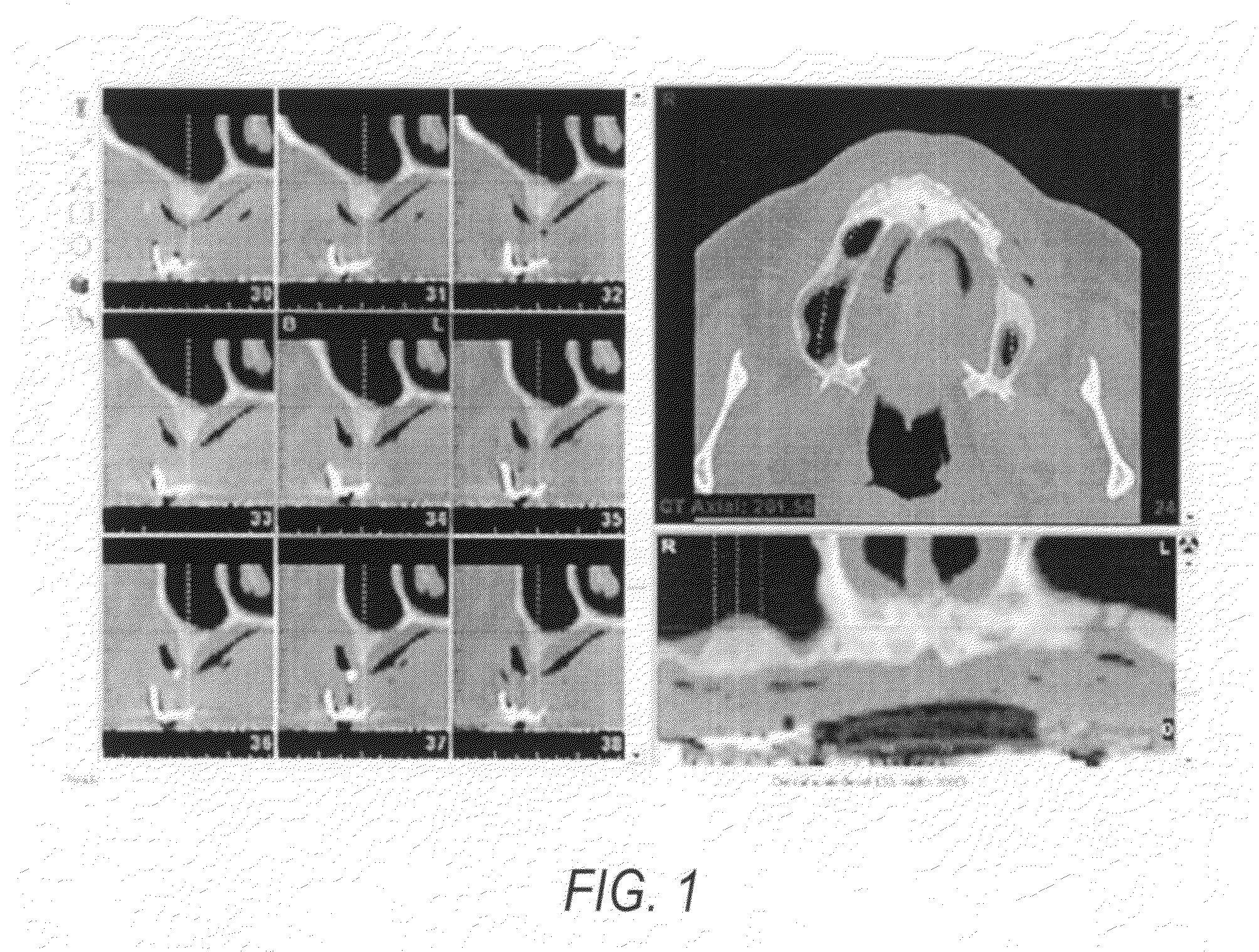
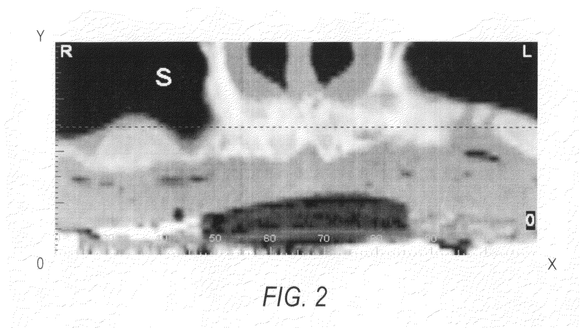
[0031]In the oral cavity, when there is tooth loss, the maxillary sinus pneumatisizes, meaning that there is resorption in a three dimensional plane. The floor of the sinus drops towards the oral cavity, as well as resulting in expansion of the lateral walls. This leaves less maxillary bone for placement of an implant to replace the missing teeth. If the remaining maxillary bone is insufficient to support an implant in terms of height and width, then sinus elevation and bone grafting is required in order to regain the resorbed bone. The treatment planning for the sinus elevation involves the patient to receive a CT scan which provides different views of the sinus and maxillary bone (FIG. 1). This diagnostic information allows the surgeon to prepare a treatment plan which outlines the volume and borders of the area of the sinus to be grafted (FIGS. 2A and 3A).

[0032]Referring to FIG. 1, nine sagittal cross-sectional CT scan views along the Y-Z axes are illustratively shown for a patie...


PUM


Abstract

The present invention discloses a surgical guide and accompanying depth bur with a method for its use during sinus elevation surgery utilizing the Caldwell-Luc Osteotomy procedure. The surgical guide of the present invention provides accurate transfer of the parameters of the surgical osteotomy to be performed during sinus elevation surgery. It bridges the gap between the precise information obtained from the CT scan and the surgical field on which this surgical plan must be placed. This is accomplished by the use of the surgical guide which is constructed based on the information in the CT scan and the treatment plan prepared utilizing 3-D imaging software.

Description

CROSS REFERENCE TO RELATED APPLICATION[0001]This patent application is a Continuation-in-Part of U.S. patent application Ser. No. 10 / 810,553, filed Mar. 29, 2004, the contents of which are incorporated hereine by reference in its entirety.FIELD OF THE INVENTION[0002]The present invention relates generally to the field of oral implantology in the surgical reconstruction of the maxillary sinus in preparation for dental implant placement. More particularly, the present invention relates to a surgical guide and accompanying bur utilized to guide the surgeon in preparation of the osteotomy to enter the maxillary sinus as part of the procedure of sinus elevation and grafting.BACKGROUND OF THE INVENTION[0003]Following maxillary posterior tooth loss, the maxillary sinus pneumatisizes or expands in every dimension towards the maxilla. As the maxillary bone resorbs, the sinus is enlarged in a coronal, lateral, anterior and posterior direction. This pneumatization, or expansion of the maxillar...

Claims


Application Information

Patent Timeline
Patent Type & Authority Applications(United States)
IPC IPC(8): A61B17/56
CPCA61B17/1615A61B17/1688A61B17/176A61B2017/1785A61C1/084A61C8/0092A61B2019/304A61B2090/034A61B17/1785
Inventor MASSOUD, YEHIA A.
Owner MASSOUD YEHIA A
Why Eureka
Try Eureka