Eureka AIR delivers breakthrough ideas for toughest innovation challenges, trusted by R&D personnel around the world.

Enzyme disruption of bacterial biofilms

a technology of enzymes and biofilms, applied in the direction of prosthesis, catheters, peptides/protein ingredients, etc., can solve the problems of completely destroying the biofilm matrix and eradicating it from the surfa

Inactive Publication Date: 2010-09-02
BIOSYNEXUS INC
View PDF18 Cites 11 Cited by
  • Summary
  • Abstract
  • Description
  • Claims
  • Application Information

AI Technical Summary

Benefits of technology

[0024]It has now been discovered that antibacterial enzymes such as lysostaphin unexpectedly not only kill all bacteria in a biofilm, they also disrupt the biofilm matrix completely, eradicating it from the surface on which it has formed. This makes possible the treatment of biofilm-related infections, especially those that form on damaged tissue or on the surfaces of indwelling prosthetic devices and catheters, without resorting to surgical removal.
[0025]Therefore, according to one aspect of the present invention, a method is provided for treating a patient in whom damaged tissue or an indwelling prosthetic device or catheter has a bacterial biofilm growing thereon, to at least partially disrupt said biofilm thereon, comprising administering to said patient at least one antibacterial enzyme that is lethal or damaging to the biofilm-forming bacteria in an amount that is effective to at least partially disrupt the biofilm upon contact therewith. For staphylococcal and other bacterial-based biofilms, lysostaphin and lysostaphin analogues have proven to be particularly effective in both preventing biofilm growth and eradicating biofilms that are already established.
[0027]The present invention also includes the disinfection or sterilization of ex-vivo surfaces not necessarily intended for patient contact. That is, the method of the present invention is suitable for disinfecting or sterilizing essentially any surface, including anything implantable into the body such as polymers and metals such as titanium, on which the growth of a biofilm has occurred, or on which the growth is possible but undesirable. For practical purposes, the inventive method will be primarily used in those circumstances where more rigorous sterilization or disinfection conditions used for biofilm removal or prevention are unsuitable, including situations where residual traces of the harsh chemicals employed would be harmful. Thus, the method of the present invention is particularly useful for preventing biofilm growth on a surface intended for medical implants in a patient or eliminating contamination before biofilm formation begins.
[0028]Therefore, according to another aspect of the present invention a method is provided for disinfecting or sterilizing a surface ex-vivo, with a bacterial biofilm growing thereon, to at least partially remove the biofilm therefrom, in which the surface is contacted with at least one antibacterial enzyme that is lethal or damaging to the biofilm-forming bacteria in an amount that is effective to at least partially disrupt the biofilm upon contact therewith. This aspect of the present invention is particularly effective for disinfecting or sterilizing surfaces to prevent or remove the growth of a biofilm.
[0029]The present invention also includes ex-vivo methods for preventing the growth of a biofilm on a susceptible surface. Therefore, according to another aspect of the present invention, a method is provided for disinfecting, protecting or sterilizing a surface ex-vivo to prevent biofilm-forming bacteria from growing thereon, by contacting the surface with a prophylactically effective amount of at least one antibacterial enzyme that is lethal or damaging to a biofilm-forming bacteria. The aspect of the present invention is particularly effective for disinfecting, protecting or sterilizing surfaces susceptible to biofilm growth and intended for medical implantation into a patient, such as catheters and prosthetic devices.
[0031]The coating may be physically retained by the ionic charge of the enzyme. For a polymeric surface, the coating may be retained by covalent attachment of the enzyme to the polymeric surface, or it may be blended with a surface polymer by techniques that result in presentation of the enzyme at the polymer surface without substantial release therefrom. The present invention thus further includes methods for preparing polymer compositions resistant to the growth of a bacteria biofilm on a surface formed therefrom by blending the polymer with an effective amount of at least one antibacterial enzyme that is lethal to a biofilm-forming bacteria. The invention also includes polymer compositions for fabrication of a prosthetic device or catheter in which the polymer is blended with at least one antibacterial enzyme that is lethal to a biofilm-forming bacteria in an amount that is effective to prevent biofilm formation on a surface formed therefrom.

Problems solved by technology

It has now been discovered that antibacterial enzymes such as lysostaphin unexpectedly not only kill all bacteria in a biofilm, they also disrupt the biofilm matrix completely, eradicating it from the surface on which it has formed.

Method used


Image

  • Enzyme disruption of bacterial biofilms
  • Enzyme disruption of bacterial biofilms
  • Enzyme disruption of bacterial biofilms

Examples


example 1

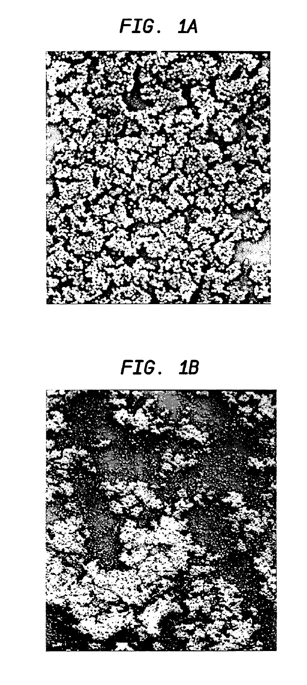
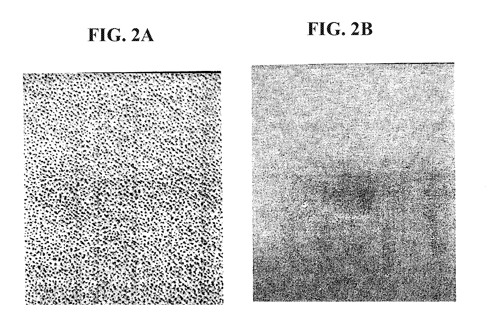
Disruption of S. aureus Biofilms with 100 .mu.g / 1 ml Lysostaphin In Vitro

[0058]Staphylococcal strains were stored in .about. 0.5 mL Tryptic Soy Broth (TSB, Difco Bacto) aliquots at −70° C. Prior to each experiment, an aliquot was taken from the freezer, plated on sheep's blood agar (Remel), and incubated at 37° C. overnight.

TABLE 1Bacteria strains used:SpeciesDesignationInformationS. aureusATCC 49521 (SA5)type 5 capsuleS. aureusColMRSAS. aureusCol-lysoRLysostaphin-resistant variant ofaboveS. aureusMBT 5040MRSAS. aureusMBT 5040 lysoRLysostaphin-resistant variant ofaboveS. aureusATCC 35556wild type for belowS. aureusdltA negativedoes not make biofilmS. epidermidisSE 380Clinical IsolateS. epidermidisHAYClinical IsolateS. epidermidisSE 1175Clinical IsolateS. epidermidisATCC 35984High slime producer

[0059]Biofilm Assay 20

[0060]Five ml of TSB supplemented with 0.25% glucose (Sigma-Aldrich) was inoculated with five isolated staphylococcal colonies. The cultures were incubated at 37° C. over...

example 2

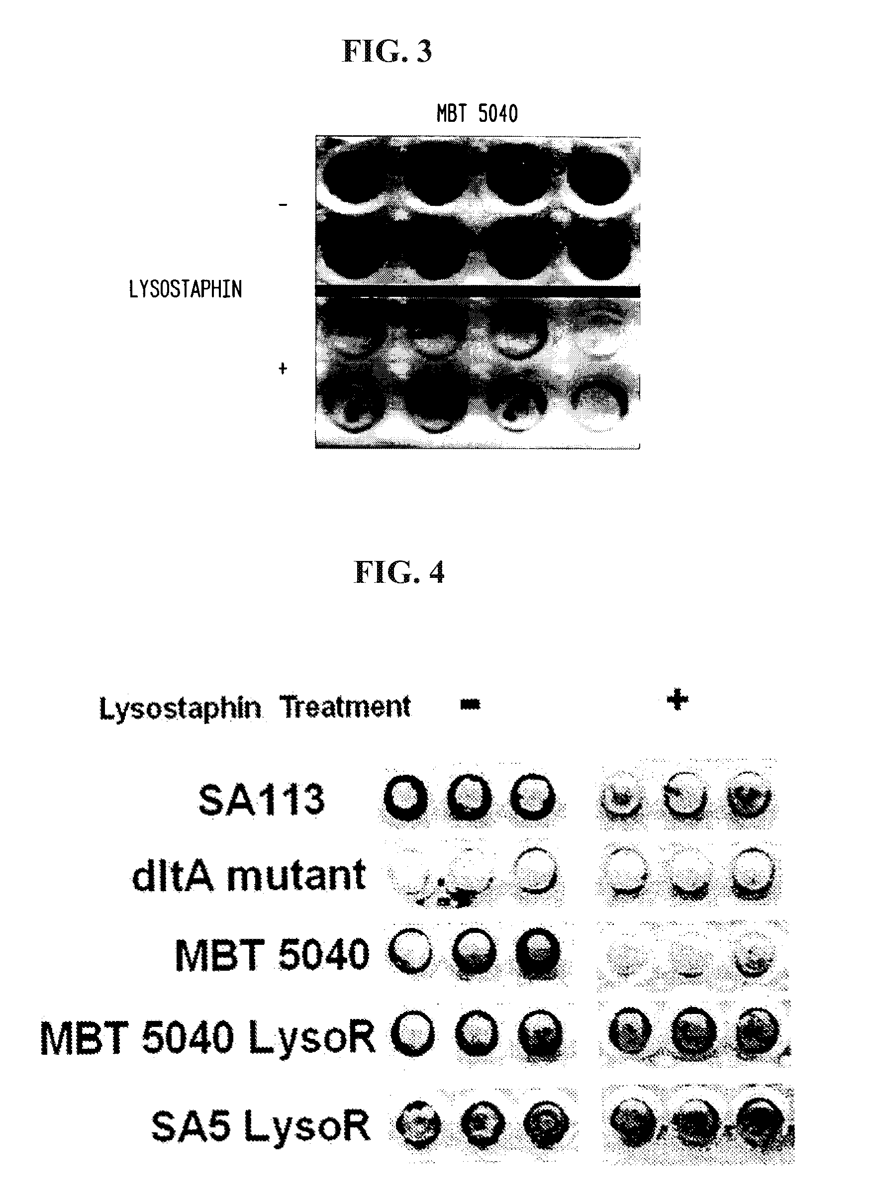
Disruption of S. aureus Biofilms With 50 μg / ml Lysostaphin In Vitro

[0068]Methicillin-resistant S. aureus strain MBT 5040 was grown overnight in TSB plus glucose as in Example 1. Twenty four hours later, a 96 well tissue culture plate containing 200 μl TSB plus glucose was inoculated with a 1:200 dilution of the overnight culture, also as in Example 1. The 96 well plate was incubated overnight at 37° C. with shaking and transferred to a stationary 37° C. incubator for an additional 24 hours. After the second incubation, the wells were washed twice with PBS to remove planktonic cells and incubated for three hours at room temperature with either PBS without lysostaphin (−) or PBS containing 50 μg / ml lysostaphin (+). Following another three hour incubation, the wells were washed twice with PBS and then fixed in Bouin's solution (Sigma-Aldrich) for five minutes. The wells were stained with safranin for one minute and washed again with PBS. The results are depicted in FIG. 3, which demons...

example 3

Preparation of Biofilm-Formation Resistant Lysostaphin-Coated Catheters

[0069]Six wells were incubated with 300 μl of either 10 mg / ml, 1 mg / ml or 100 μg / ml of lysostaphin diluted in PBS. All the samples were done in duplicates. The plate was allowed to incubate overnight at 4° C. The following morning the wells were washed with 1 ml of PBS ten times, using vacuum suction to clean out the wells. S. aureus strain SA5 was diluted in PBS to a percent transmittance of 40. A 1:10,000 dilution of this solution was made, and 300 μl was added to each well. The plates were put in a shaking incubator at 75 rpm for two hours at 37° C. After two hours, 40 μl from each well was taken out and plated onto a blood agar plate and put in the incubator overnight at 37° C. The colonies on the plates were counted the following morning.

[0070]Two Angiocath catheters (Becton Dickinson) were incubated with 200 PI of a 100 μg / mL solution of lysostaphin, while two others were incubated in PBS. The catheters wer...


PUM

PropertyMeasurementUnit
molecular weightaaaaaaaaaa
body weightaaaaaaaaaa
concentrationaaaaaaaaaa

Abstract

Methods for treating patients in which damaged tissue or an indwelling prosthetic device or catheter has a bacterial biofilm growing thereon, to at least partially disrupt said biofilm, by administering at least one antibacterial enzyme that is lethal or damaging to the biofilm-forming bacteria in an amount that is effective to at least partially disrupt the biofilm upon contact therewith. Methods for prophylactically treating a patient, and methods for disinfecting or sterilizing a surface ex-vivo to remove a biofilm or prevent biofilm growth are also disclosed, as well as implantable articles susceptible to biofilm growth to which a prophylactic coating of an antibacterial enzyme has been applied.

Description

CROSS-REFERENCE TO RELATED APPLICATION[0001]The present application is a divisional of U.S. patent application Ser. No. 10 / 401,342 filed on Mar. 26, 2003, which claims priority benefit under 35 U.S.C. §119(e) of U.S. Provisional Application Ser. No. 60 / 367,189 filed on Mar. 26, 2002, the disclosure of which is incorporated herein by reference.BACKGROUND OF THE INVENTION[0002]1. Field of the Invention[0003]This invention pertains to the disruption of bacterial biofilms with antibacterial enzymes. More specifically, this invention relates to the disruption of staphylococcal biofilms with lysostaphin.[0004]2. Background Art[0005]A. Biofilms[0006]Bacteria that adhere to implanted medical devices or damaged tissue can encase themselves in a hydrated matrix of polysaccharide and protein and form a slime layer also known as a biofilm. Biofilms pose a serious problem for public health because of the increased resistance of biofilm-associated organisms to antimicrobial agents and the associa...

Claims


Application Information

Patent Timeline
Patent Type & Authority Applications(United States)
IPC IPC(8): A61K38/48A61P31/04A01N63/50A61L27/54A61L29/16
CPCA01N63/00A61K38/482A61L27/54A61L29/16A61L2300/254A61L2300/45A61L2300/404A61L2300/406A61K2300/00A01N63/50
Inventor KOKAI-KUN, JOHN F.WU, JULIE ADAMSMOND, JAMES J.WALSH, SCOTT M.SHAH, ANJALI G.CHANTURIYA, TATYANA I.
Owner BIOSYNEXUS INC
Why Eureka
Eureka Blog
Learn More