Detection and tracking of interventional tools

a tracking and interventional technology, applied in the field of minimally invasive xray guided interventions, can solve the problems of difficult subtraction with reference images and inability to always provide reference images, and achieve the effects of enhancing the contrast of those patterns, reducing the contrast of common patterns, and improving visibility

Inactive Publication Date: 2010-09-09
KONINKLIJKE PHILIPS ELECTRONICS NV
View PDF5 Cites 23 Cited by
  • Summary
  • Abstract
  • Description
  • Claims
  • Application Information

AI Technical Summary

Benefits of technology

[0020]It may thus be an object of the present invention to improve the visibility as well as detection and tracking of interventional tools which are applied in the scope of minimally invasive interventions under X-ray fluoroscopic guidance.
[0021]A first exemplary embodiment of the present invention is directed to a method which addresses this object by calculating differences between 2D projected image data of a preoperatively acquired 3D voxel volume showing an anatomical structure or region of interest, pathological abnormalities (such as e.g. an intracranial arterial stenosis, an aneurysm of a cerebral, pulmonary or coronary artery branch, a gastric carcinoma or sarcoma, etc.), pacemakers and/or other medical implants in a tissue of a patient's body to be examined or in a blood vessel segment of said patient's vascular system and intraoperatively recorded 2D fluoroscopic images showing the aforementioned objects in the interior of said patient's body, wherein said 2D fluoroscopic images have been co-registered with the 2D projected image data, and using the resulting difference image that is generated based on said calculation step for enhancing the contrast and visibility of interventional tools inserted into said tissue region or said blood vessel segment by removing common patterns detrimental to the visibility of the interventional tools, said patterns being contained both in the 2D projection image of the preoperatively acquired 3D voxel volume and in the intraoperatively recorded 2D fluoroscopic images.
[0022]After registration of the aforementioned 2D projected image data of said preoperatively acquired 3D voxel volume with said intraoperatively recorded 2D fluoroscopic images, the registered 2D projection image may be used to calculate a discrepancy map which can either be realized as a difference image—obtained by subtracting a representative of said 2D fluoroscopic images from the 2D projection image that has been co-registered with said 2D fluoroscopic image—or as the result of any image processing operator that may be specially designed to reduce the contrast of common patterns and enhance the contrast of those patterns which are visible only in one of the aforementioned two 2D images. Said discrepancy map can be displayed to a radiologist, which is especially useful as interventional tools, which are present in the intraoperatively recorded 2D fluoroscopic images but not in the 2D projected image data of the preoperatively acquired 3D voxel volume, are thus better visible and not hidden by other objects with high Hounsfield density values (such as e.g. bones), which supports the radiologist to detect, guide and navigate these interventi...

Problems solved by technology

Nevertheless, such a reference image is not always available since most of the time the patient already has ECG electrodes or ...

Method used


Image

  • Detection and tracking of interventional tools
  • Detection and tracking of interventional tools
  • Detection and tracking of interventional tools

Examples


Embodiment Construction

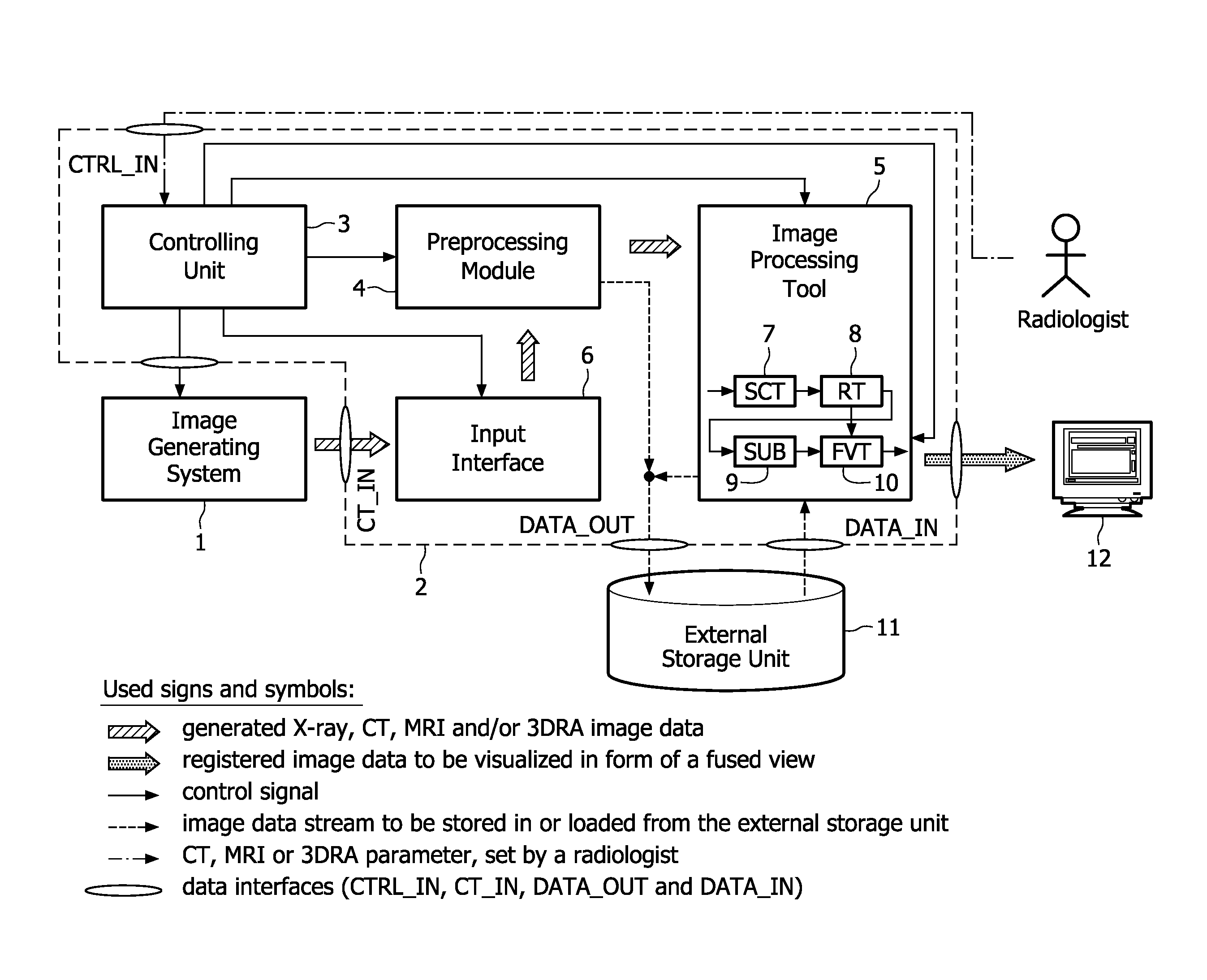
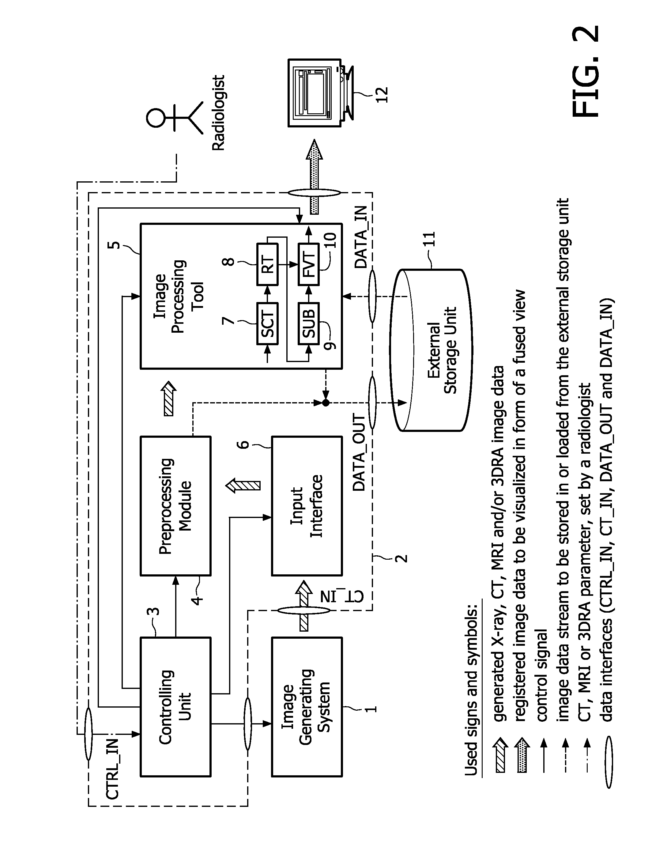
[0029]In the following, the aforementioned image processing method and system will be explained in more detail with respect to special refinements and referring to the accompanying drawings.

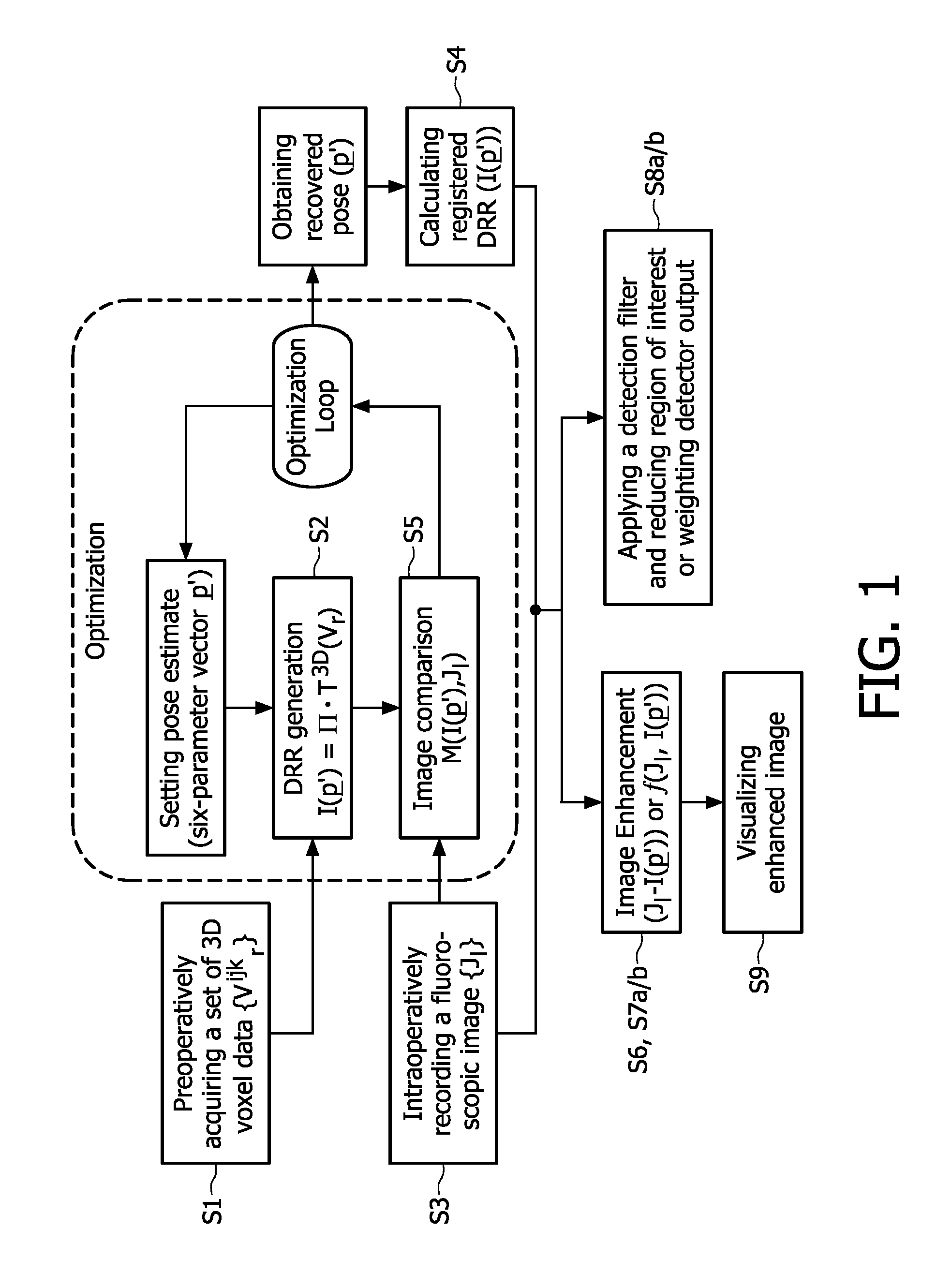
[0030]As shown in the flowchart depicted in FIG. 1, it is proposed to use a preoperatively acquired 3D voxel data showing an anatomical structure or a pathological tissue region in the interior of a patient's body, which has e.g. been recorded (S1) with a 3D imaging device, such as e.g. a CT, MRI or rotational angiography device, etc., and register (S4) this voxel volume with acquired 2D data of intraoperatively recorded (S3) fluoroscopic X-ray images. This may be done in such a way that a digitally reconstructed radiograph (DRR), that may have been generated (S2) from the 3D voxel data by means of a ray cast algorithm which comprises the step of transforming a voxel volume into a 2D image through central projection, matches the 2D fluoroscopic images as closely as possible. To be more precisely,...


PUM


Abstract

The present invention relates to minimally invasive X-ray guided interventions, in particular to an image processing and rendering system and a method for improving visibility and supporting automatic detection and tracking of interventional tools that are used in electrophysiological procedures. According to the invention, this is accomplished by calculating differences between 2D projected image data of a preoperatively acquired 3D voxel volume showing a specific anatomical region of interest or a pathological abnormality (e.g. an intracranial arterial stenosis, an aneurysm of a cerebral, pulmonary or coronary artery branch, a gastric carcinoma or sarcoma, etc.) in a tissue of a patient's body and intraoperatively recorded 2D fluoroscopic images showing the aforementioned objects in the interior of said patient's body, wherein said 3D voxel volume has been generated in the scope of a computed tomography, magnet resonance imaging or 3D rotational angiography based image acquisition procedure and said 2D fluoroscopic images have been co-registered with the 2D projected image data. After registration of the projected 3D data with each of said X-ray images, comparison of the 2D projected image data with the 2D fluoroscopic images—based on the resulting difference images—allows removing common patterns and thus enhancing the visibility of interventional instruments which are inserted into a pathological tissue region, a blood vessel segment or any other region of interest in the interior of the patient's body. Automatic image processing methods to detect and track those instruments are also made easier and more robust by this invention. Once the 2D-3D registration is completed for a given view, all the changes in the system geometry of an X-ray system used for generating said fluoroscopic images can be applied to a registration matrix. Hence, use of said method as claimed is not limited to the same X-ray view during the whole procedure.

Description

FIELD OF THE INVENTION[0001]The present invention relates to minimally invasive X-ray guided interventions, in particular to an image processing and rendering system and a method for improving visibility and supporting automatic detection and tracking of interventional tools that are used in electrophysiological procedures. According to the invention, this is achieved by calculating differences between co-registered X-ray images and 2D projected image data of a preoperatively acquired 3D voxel volume and using these differences for enhancing the contrast and visibility of the interventional tools.BACKGROUND OF THE INVENTION[0002]Cardiovascular diseases (CVD), such as e.g. atherosclerosis, hypertension and ischemia, remain the leading cause of death in most developed countries as they cause permanent damage to the heart and blood vessels, which may lead to chronic heart failure, angina, or myocardial infarction (heart attack). For a patient showing symptoms of a cardiovascular diseas...

Claims


Application Information

Patent Timeline
IPC IPC(8): G06K9/00G06T17/05
CPCG06T5/50G06T2207/10072G06T2207/30101G06T2207/30021G06T2207/10116G06T7/32
Inventor VILLAIN, NICOLAS F.PICARD, CECILE A.M.GOGIN, NICOLAS P.B
Owner KONINKLIJKE PHILIPS ELECTRONICS NV
Why Eureka
Try Eureka